Search Results for: vegetarian

The Benefits of Near Infrared Light

We have a great respect for the influence of light upon health. We’ve blogged and spoken about the importance of blue light for circadian rhythm entrainment, and ultraviolet light for production of vitamin D and nitric oxide (for example, in “Nitric Oxide and AO+Mist Skin Probiotic at the Perfect Health Retreat”; use the coupon code phd25 for 25% off AOBiome’s nitric oxide enhancing skin probiotic).

However, red and near infrared light are healthful too. Vladimir Heiskanen (also known as “Valtsu”) is a Finnish blogger and intelligent young scholar who has been researching the effects of red and near infrared light, and he wrote a post on his blog which deserves greater exposure. Vladimir has graciously allowed me to revise his post for PHD readers. – Paul Jaminet

I (Valtsu) used to spend a lot of time reading Ray Peat’s articles, trying to make sense of his ideas. In many of his articles, Peat praised red light. For example (from here):

Old observations such as Warburg’s, that visible light can restore the activity of the “respiratory pigments,” showed without doubt that visible light is biochemically active. By the 1960s, several studies had been published showing the inhibition of respiratory enzymes by blue light, and their activation by red light.

Peat didn’t give many references to justify his claims, but after doing some searches on PubMed, I realized that there are thousands of papers supporting his views.

Mechanisms

Activation of cytochrome c oxidase (Cox), the mitochondrial respiratory enzyme discovered by Nobel laureate Otto Warburg, seems to be the primary mechanism by which red light enhances mitochondrial function. [1] [2] [3] [4] [5] [6]

Cox is the centerpiece of the last stage of mitochondrial energy production, Complex IV. Cox utilizes energetic electrons and protons from opposite sides of the inner mitochondrial membrane to turn one molecule of oxygen (O2) into two molecules of water (H2O), in the process contributing the energy required to form ATP.

But a number of small molecules can displace the O2, spoiling the reaction and acting as inhibitors of ATP synthesis. These include cyanide (HCN), carbon monoxide (CO), hydrogen sulfide (H2S), and nitric oxide (NO). If too much oxygen is displaced, as in cyanide or carbon monoxide poisoning, cells die from chemical asphyxiation.

Nitric oxide is a native molecule of crucial importance for health, especially cardiovascular health. Nitric oxide generation by ultraviolet light (UV-A) is probably a major reason for the healthfulness of sunshine. Nitric oxide is commonly generated by stressed cells (for example, during the heat shock response) to support cellular health, in part by increasing blood flow.

However, the binding of NO to Cox, inhibiting mitochondrial respiration, can be an unwanted side effect. It turns out, fortunately, that red and near infrared light photodissociate NO from Cox, leading to its release from mitochondria back into circulation with beneficial effects on blood flow.

Removal of NO from mitochondria appears to be the mechanism by which red light phototherapy enhances mitochondrial respiration [2] [7a] [7b] [8] [9] [10] [11]. Supporting evidence: provision of NO abolishes the cellular effects of red and near infrared light.

Visible light doesn’t penetrate the body well, but infrared light does. Near infrared light is only reduced in intensity by about half after passage through 2 mm of tissue, and in daytime outdoor environments may dissociate NO and promote mitochondrial respiration at a depth of 2-3 cm beneath the skin. [13] [14] [15]

This image, from here, is illuminating:

Valtsu image 01

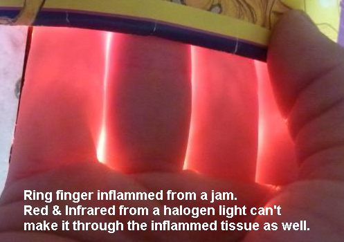
First, it illustrates the relative transparency of human tissue in the red (and even greater transparency in the infrared). Transparency in this frequency range appears to have been evolutionarily selected, to the point that one molecule – cytochrome c oxidase in mitochondria – absorbs 35% or more of red light. If it was so important to let red light reach mitochondria that other human molecules had to evolve transparency in the red, then it is surely important for us to provide our mitochondria with red light.

Second, when tissues are injured, they release extra nitric oxide, and NO bound to Cox absorbs red light, making the injured tissue more opaque in the red. In this case the middle finger had been jammed; due to the injury it passes significantly less red light than the fingers on either side.

The History of Red Light Therapy

There is nothing new under the sun, and when a simple activity is beneficial for health, we often find that somebody discovered the effect long ago. So it is with red light.

Niels Ryberg Finsen won the Nobel Prize for Medicine in 1903 for his explorations of the therapeutic effects of light, notably the use of ultraviolet light to treat lupus and other diseases; but he had also found that red light could be beneficial, and had published an article titled “The Red Light Treatment of Smallpox” in 1895.

The idea of using red light for therapy was picked up by John Harvey Kellogg, who published a 200-page book titled Light Therapeutics in 1910. Kellogg had long been famous as one of the first vegetarian doctors, leader of the Battle Creek Sanitarium for the Seventh Day Adventist Church from 1876 until 1933, and as the inventor of corn flakes breakfast cereals (in 1878). Kellogg recommended light therapy for diabetes, obesity, chronic fatigue, insomnia, baldness, and cachexia. [18]

After the invention of lasers, it was found that red laser light could accelerate wound healing in animals, and in the 1970s similar results were obtained in humans. [2]

The most interesting work on phototherapy, however, has been conducted recently. It is becoming a hot field: a Pubmed search for LLLT (an acronym for “low-level laser therapy” or “low-level light therapy”) generates about 10 papers per year in the 1990s, 100 per year in the 2000s, and 400 per year in the 2010s.

Therapeutic Benefits from Local Application of Red Light

Therapeutic benefits from local application of red or near infrared light to injured tissues have been reported for several conditions:

  • Age-related macular degeneration. The eyes of 200 elderly subjects with age-related macular degeneration were exposed to near infrared light of wavelength 780 nm. Visual acuity was improved in 95% of the subjects; most were able to see two rows lower on an eye chart. Results achieved in two weeks of treatment were maintained three to thirty-six months. [65]
  • Knee osteoarthritis. Application of near infrared (830 nm) light to the knees of osteoarthritis patients dramatically reduced knee pain scores. [73]
  • Herpes labialis. Cold sores around the lips caused by herpes simplex virus 1 were treated with red laser light. Time to recurrence was a median 37.5 weeks in the treatment group, 3 weeks in the placebo group. (Subjects wore masks and couldn’t tell which group they were in.) [61]
  • Hypothyroidism. Hashimoto’s hypothyroidism patients were exposed to near infrared (830 nm) radiation of the skin over the thyroid gland. Nine months later, 48% of the treatment group had been able to stop taking thyroid hormone, and the average T4 dose had dropped from 93 mcg to 39 mcg. In the control group, the average T4 dose had increased from 90 mcg to 107 mcg. [37] Similar results have been reported in other studies. [36] [37] [38] [40]  [41] [42] [43]
  • Cognitive dysfunction following traumatic brain injury. Eleven patients with continuing cognitive dysfunction following traumatic brain injury (from motor vehicle accidents, sports injuries, and an improvised explosive device detonation) were treated with red and near-infrared light to the scalp. They experienced improvements in executive function, learning, and memory, as well as improved sleep and fewer post-traumatic stress disorder symptoms.
  • Cellulite. This is more speculative, but there are indications that red and near infrared light can help reduce cellulite.
  • Hair loss. Use of a laser hair comb led to fuller and thicker hair in hair loss patients.

Systemic Benefits of Phototherapy

Even when light is applied locally, some of the benefits may be shared systemically.

For instance, exposure to light causes release of NO from mitochondria and also an increase in NO levels due to photoactivation of nitric oxide synthase. Elevation of NO anywhere increases blood flow throughout the body. In one experiment, one hand was irradiated with white light; blood flow rate increased 45% in the irradiated hand and 39% in the non-irradiated hand.

Irradiation with white light has been found to increase antibody production, presumably improving immune function.

Light exposure has also been found to have anti-inflammatory effects. For example, white light exposure reduces levels of the pro-inflammatory cytokines TNF- α, IL-6, interferon-gamma, and interleukin-12 and increases levels of anti-inflammatory cytokines interleukin-10 and TGF-beta.

Other studies have found that UV radiation increases TNF-α, IL-6 and other pro-inflammatory cytokines. [90] [91] So it is likely that the anti-inflammatory effects of the white light were chiefly due to its red and near infrared components.

These anti-inflammatory effects may shed light on the improvements hypothyroid subjects experienced from near infrared phototherapy. TNF-α and IL-6 suppress peripheral thyroid hormone metabolism by decreasing T3 and increasing rT3. [92] [93] Inflammation seems to commonly trigger hypothyroidism, while anti-inflammatory strategies are almost always therapeutic for hypothyroidism.

Epidemiological evidence suggests that light exposure improves serum lipid profiles. At mid-latitudes, serum cholesterol levels typically rise 5% to 10%, but HDL cholesterol levels decrease, in winter. Blood pressure is also higher in winter. [97] [98] [99] [100] [101] [102] Lack of red light may be the reason. In a pilot study, red light exposure reduced serum cholesterol levels in 84% of subjects.

Widespread Deficiencies in Light Exposure

It’s likely that our modern environments lead to systemic deficiencies in light exposure. It’s common for health to worsen in low-light locations or seasons, as Ray Peat observed:

Many people who came to cloudy Eugene to study, and who often lived in cheap basement apartments, would develop chronic health problems within a few months. Women who had been healthy when they arrived would often develop premenstrual syndrome or arthritis or colitis during their first winter in Eugene.

Since the last ice age ended, humanity has populated more northerly latitudes and moved indoors. We are getting far less light than our ancestors. The evolutionary mismatch principle suggests that humans will be optimized for ancestral light levels, and that we moderns can improve our health by getting more light.

Health Risks of Blue-Only Light

While red light tends to enhance mitochondrial function, high intensities of blue light can damage mitochondria by triggering oxidative stress. Blue-only light can kill retinal cells. Exposing rats to blue-only light or to “white” LED light that peaks unnaturally strongly in the blue led to retinal damage. LED lights are the worst, due to their concentration at single frequencies, but compact fluorescent lights which have peaks in the blue can also generate mild to moderate retinal damage. A commentary on the research is here.

Reactive oxygen species are generated in mitochondria when they can’t dispose of electrons in the manufacture of ATP. For this reason, NO binding to Cox promotes oxidative stress, and release of NO from Cox by red and infrared light reduces oxidative stress. It is probably by this mechanism that red and near infrared light protects against retinal injury.

Because of this research, we don’t recommend using blue-only light boxes for circadian rhythm entrainment; rather, use full spectrum white light.

Incandescent lights, which produce a smooth spectrum including red and near-infrared wavelengths, are probably safest for eye health. Some researchers, such as Richard Funk and Alexander Wunsch, who appeared in the Bulb Fiction documentary, assert that increased CFL usage may be harmful to eyes. Of course, governments are here to help us, and have banned incandescent lighting.

Circadian Rhythm Considerations

There are hints that bright red and near infrared light exposure should occur in the daytime.

Night-time levels of melatonin, but not day-time levels, have been shown to abolish the effects of red and infrared light on cellular function, just as nitric oxide does.

This shows that melatonin, a circadian rhythm hormone, evolved to inhibit red light influences at night. If our “night hormone” is trying to block the influence of red light, we probably shouldn’t willfully expose ourselves to bright red light at night.

What to Do

If fluorescent lights are problematic, how should we get adequate red and near infrared light, while getting sufficient blue in the daytime to entrain circadian rhythms?

The general lighting types that provide the most red and near-infrared are incandescent lights, heat lamps, and LEDs.

Heat lamps by Philips or Osram  generate little blue light but, thanks in part to their high power levels (up to 250 W), provide significant red and near-infrared light. However, only ~12% of the power is emitted at therapeutic wavelengths (600-1070 nm); most of their power is emitted at the warming IR-B wavelengths. In fact, these lamps emit so much heat that they can substitute for central heating in the winter. Most people will find they produce too much heat for summer use. Although we recommend using red or orange lighting at night, heat lamps are not really suitable for night use; ambient temperature is a zeitgeber and our exposure to warmth should occur in the daytime. Moreover, as noted above, it might be best to get red light exposure in the daytime, when melatonin levels are low. So heat lamps are best used in the daytime as a complement to other broad-spectrum lighting.

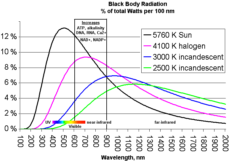
Incandescent lights (including halogen lights), which produce a blackbody spectrum, can generate a significant amount of red and near infrared and are excellent daytime light sources. A color temperature of 5500 K mimics the sun and provides substantial amounts of red and near infrared; a color temperature of 4100 K is also excellent in the red and near infrared, but is rather weak in the blue and ultraviolet for daytime circadian rhythm entrainment. A look at blackbody spectra as a function of color temperature:

Valtsu image 02

LEDs are another possibility. One of their virtues is that it’s possible to obtain monochromatic LEDs with frequencies optimized for Cox-NO photodissociation (680 and 820 nm work best; inexpensive LEDs are available at 630, 660, 850, and 880 nm. Here is a video of a fellow who created a homemade LED helmet:

A variety of commercial light devices have been used in LLLT/light therapy studies: AnodyneBioptronHairMax LaserCombOmniluxNoveon NaiLaserBiolightQuantum WarpSyrolight BioBeamHIRO 3.0Picasso LiteHELBO® TheraLite Laser and Mustang 2000.

Conclusion

Exposure to sunshine on bare skin, which was common in our ancestral environment, is something we need to obtain or mimic if we want optimal health.

Unfortunately, it’s hard to reproduce the many facets of sunshine in indoor environments. Blue light (for daytime circadian rhythm entrainment), UV-A light (for nitric oxide), UV-B light (for vitamin D), red light (for nighttime circadian rhythm entrainment), and now red and near infrared light in the daytime (for mitochondrial respiration) all seem to be important.

Awareness is the first step toward optimization. Thank you, Vladimir, for sorting through this research for us!

Examine.com Supplement Stack Guides

A PHD reader and friend of the blog, Sol Orwell, and colleagues have compiled a comprehensive database of free information about nutritional supplements at Examine.com. Our own Kamal Patel is their nutrition director.

Examine.com supplement recommendations are research based, everything is cited to the literature, and recommendations are in line with PHD recommendations. Think of it like a super-PHD.

Although all the content is freely available, a huge online database is not easy to navigate. For example, their vitamin D page has 322 unique citations, and that’s far from the biggest page on the site. They have therefore worked to create paid products which distill the information down to more digestible pieces. I’ve previously recommended their Supplement Goals Reference Guide, but even that is over 1,000 pages.

So now they’ve come out with even more distilled set of guides, that they call “supplement stacks.” These are practical and actionable summaries of the evidence for how supplements can be used to address major health goals. The health goals are:

  • Testosterone Enhancement
  • Fat Loss
  • Muscle Gain & Exercise Performance
  • Mood and Depression
  • Heart Health
  • Sleep Quality
  • Insulin Sensitivity
  • Memory and Focus
  • Skin and Hair Quality
  • Libido and Sexual Enhancement
  • Liver Health
  • Allergies and Immunity
  • Bone Health
  • Joint Health
  • Vegetarianism/Veganism
  • Seniors

In each guide, supplements are classed according to the evidence for them. Base supplements are generally safe and often synergistic with other supplements, proven supplements have a good deal of evidence whether it’s meta-analyses or solid trials, unproven supplements show promise but may have caveats or not enough extended research in humans, and cautionary supplements are either overhyped or downright dangerous. Each guide wraps up with steps to assembling your stack depending on who you are and your goals.

Kamal tells me, “The Examine.com team has learned much from the Perfect Health Diet, and in addition to me (PHD resident blogger) our team includes a variety of health researchers, medical doctors, and other clinical practitioners. Examine also brought in specialists, including a PhD in toxicology and a specialist in pyschiatric pharmacology, to check over nutrient-supplement-medication interactions for each guide.”

The guides are updated for life. So when new studies come out, the evidence is re-assessed and new supplements may be introduced or lacklustre supplements may be shifted down in recommendation.

If you spend a lot of money on supplements, these guides can save you a lot of money as well as improve your health by steering you to beneficial, and only beneficial, supplements. I’ve reviewed a number of guides and they are excellent – also they get right to the point and are easy to read.

To find out more, visit the Examine.com Supplement Stack Guides. There is an introductory sale through midnight tonight.

The Case of the Killer Protein

Earlier this week a paper was released to much fanfare, claiming that diets with over 20% of energy as animal protein might be as life-threatening as smoking.

  • The Huffington Post said, “Atkins aficionados, Paleo enthusiasts, and Dukan devotees, you may want to reconsider what’s on your plate. While high-protein diets have been all the rage over the last few years for their waist-whittling goodness, a new study says they could be as bad for you as smoking.”
  • Scientific American said “People who eat a high-protein diet during middle age are more likely to die of cancer than those who eat less protein, a new study finds.”
  • NPR said, “Americans who ate a diet rich in animal protein during middle age were significantly more likely to die from cancer and other causes.” They added, “In an age when advocates of the Paleo Diet and other low-carb eating plans such as Atkins talk up the virtues of protein because of its satiating effects, expect plenty of people to be skeptical of the new findings.” A sound prognostication!

Ray, Alex, Navy87Guy, Kat, Sam, and others asked for my thoughts.

What the Researchers Did

The article appeared in Cell Metabolism, a high-impact journal which likes long complex papers reporting years of work. [1] A common strategy for getting into such journals is to piece together a great variety of work into one article, weaving a narrative theme to unite them. That’s what this article did, using the theme “high protein diets may shorten lifespan” to link several relatively disconnected projects.

The NHANES Findings

The work that generated most of the buzz was an analysis of data from the National Health and Nutrition Examination Survey (NHANES). They looked at a group of 6,381 NHANES respondents and found, “Respondents aged 50–65 reporting high protein intake had a 75% increase in overall mortality and a 4-fold increase in cancer death risk during the following 18 years. These associations were either abolished or attenuated if the proteins were plant derived.”

Here’s their Figure 1:

Longo et al Figure 1

Two oddities in this result raise red flags:

  • First, protein appears harmful at age 50, neutral at age 65, and beneficial at age 80. This reversal of effects is incompatible with most mechanisms by which protein could affect aging or disease risk. In animal studies, we see the opposite: protein restriction extends maximum lifespan, which means that at high ages, mortality is lower, but increases risk of early death, which means that in middle age mortality is higher.
  • Second, they report that the effect was specific to animal protein: “[W]hen the percent calories from animal protein was controlled for, the association between total protein and all-cause or cancer mortality was eliminated or significantly reduced, respectively, suggesting animal proteins are responsible for a significant portion of these relationships. When we controlled for the effect of plant-based protein, there was no change in the association between protein intake and mortality, indicating that high levels of animal proteins promote mortality.” Yet, plant and animal proteins are biologically similar.

These two oddities strongly suggest that the appearance of negative health outcomes from protein is due to confounding factors – behaviors or foods associated with animal protein consumption in middle age, rather than effects caused by the protein itself.

When we look at how the analysis was performed, we find more reasons to doubt that protein is at fault. All of this data was found using a model which adjusted for the following covariates:

Model 1 (baseline model): Adjusted for age, sex, race/ethnicity, education, waist circumference, smoking, chronic conditions (diabetes, cancer, myocardial infarction), trying to lose weight in the last year, diet changed in the last year, reported intake representative of typical diet, and total calories.

Adjustment for a host of health-related conditions – waist circumference, diabetes, cancer, myocardian infarction, and even total calories which is effectively a proxy for obesity – can radically distort results, and even transform effects from positive to negative. I’ve discussed this issue previously in The Case of the Killer Vitamins.

In practice, many factors are highly correlated. The variables being studied – protein intake, waist circumference, total calorie intake, and others – are beset by the problem of collinearity. Attempting multiple regression analysis on collinear variables can generate very peculiar results. The more the number of adjustment factors grows, the more strange things tend to happen to data.

If they wanted us to understand whether their results are trustworthy, authors would present raw data, and then a sensitivity analysis that shows how introducing each covariate individually affects the results, then showing how including combinations of two covariates affects the results, and so forth. This would help us judge how robust the results are to alternative methods of analysis.

Of course, authors do not do this. Instead, they ask us to trust the analysis they have chosen to present – which is only one of billions they could have done. (This study adjusted for 13 covariates. The NHANES survey may have gathered data on, say, 40 variables. There are 40 choose 13, or 12 billion, possible multivariate regression analyses that could be performed using 13 covariates on this data set. Each of the 12 billion analyses would generate different outcomes.)

Are the authors trustworthy? Unfortunately, most academics today are not. Career and funding pressures are severe, and by and large those who are good at gaming the funding and publishing processes have triumphed professionally over careful, diligent truth seekers. It is much easier to construct a narrative that will garner attention and publicity and interest, than to carefully exclude non-robust results and publish only those results that are solidly supported.

Frankly, I give little credence to their NHANES analysis. And, judging by comments in the press, other epidemiologists don’t seem to give it much credence either. From the NPR article:

But could eating meat and cheese really be as bad for you as smoking, as the university news release describing the new Cell Metabolism paper suggested?

Well, that may be an exaggeration, according to Dr. Frank Hu, a researcher at the Harvard School of Public Health who studies the links between health, diet and lifestyle.

“The harmful effects of smoking on cancer and mortality are well-established to be substantial, while the harmful effects of red meat consumption are modest in comparison,” Hu wrote to us in an email.

The Mouse Experiments

So let’s turn to the next part of the study, the mouse experiments:

Eighteen-week-old male C57BL/6 mice were fed continuously for 39 days with experimental, isocaloric diets designed to provide either a high (18%) or a low (4%–7%) amount of calories derived from protein …

The low protein diets are really starvation diets, in terms of protein intake. The reason the low protein diets were sometimes 4% and sometimes 7% was because mice will often lose weight on 4% protein diets due to starvation (in the paper’s experiments on BALB/c mice, “the mice had to be switched from a 4% to a 7% kcal from protein diet within the first week in order to prevent weight loss.”). Animal control officers do not allow experiments to continue if the mice are obviously starving.

[B]oth groups were implanted subcutaneously with 20,000 syngeneic murine melanoma cells (B16).

This is an unusually small number of cells. Typically, cancer researchers implant a million cells to create a syngeneic tumor. Presumably they used this small number of cells in order to ensure that some mice would not develop tumors during the 39 day experiment. As it happened, this was a lucky (canny?) choice of cell quantity: while 10 of 10 mice on the high-protein diet developed tumors during the experiment, only 9 of 10 mice on the low-protein diet did. If they had used more cells, all mice on both diets would have developed tumors; if they had used fewer cells, some mice on the high protein diet would have failed to develop tumors. Either way, the results would appear less damning for the high protein diet.

The outcomes:

Longo et al Figure 3
Due to the small number of cells injected, it takes at least two weeks before tumors are detectable in size (normally they would be visible in ten days). They seem to be similar in size at about two weeks after implantation.

However, when the tumors reach larger sizes, growth is impaired on the low protein diets. A mouse weighs 20 grams, and a 2000 mm3 tumor weighs 2 grams, or 10% of body weight – equivalent to a 15-pound tumor in humans. Growing a tumor of this size requires building a large amount of tissue — blood vessels, extracellular matrix, and more. The ability to construct new tissue is constrained on a protein-starved diet, so it’s not surprising that tumor growth is slower when the tumor is large and protein is severely restricted.

Animal protocols generally require that mice be sacrificed when tumors reach 2000 mm3. Extrapolating the tumor growth curves, it looks like the mice in experiment (B) would be sacrificed 5 weeks after implantation on the high protein diet, or 8 weeks after implantation on the low protein diet; in experiment (G), mice on the high protein diet would be sacrificed about 9 weeks after implantation, while mice on low protein diets would have been sacrificed about 11 weeks after implantation.

In other words, tumors still kill you, just a bit more slowly if you are starving yourself.

It’s important to note a couple of things. First, the word “starving” is appropriate. 4% to 7% protein intakes are starvation levels for mice. In a nice blog post closely relevant to this topic, Chris Masterjohn notes that a 5% protein intake completely stunts the growth of young rats:

Chris rhetorically asks: “How many of us would deliberately feed a two-year old a diet that would cause them to stop growing altogether?”

Second, as Chris also points out in the same post, such low protein intakes actually make cancer more likely in the context of exposure to mutagens. For instance, aflatoxin exposure leads to cancer (or pre-cancerous neoplasms) much more frequently in rats on low-protein diets than in rats on high-protein diets:

In this experiment, there were two diets, 5% protein and 20% protein, and two diet periods, one during exposure to aflatoxin and one afterward. Rats exposed to aflatoxin while on a 5% protein diet were far more likely to develop neoplasms than rats exposed to aflatoxin on a higher protein diet. That is, the “20-5” rats had far fewer cancers than the “5-5” rats, and the “20-20” rats had far fewer cancers than the “5-20” rats. High protein for the win!

However, once the rats had neoplasms, the tumors grew more slowly on the low-protein diet. Just as the new study found.

So, if your goal is to avoid getting cancer, it is better to eat adequate protein. If you already have cancer, or if researchers have injected you with highly metastatic melanoma cells, you can buy yourself slightly slower tumor growth by starving yourself of protein. In laboratory mice, this extends lifespan a few weeks because they are not allowed to die from cancer, but are sacrificed when tumors reach a specific size. In humans, however, cancer death commonly follows from cachexia, or wasting of lean tissue. A low protein diet might promote cachexia and accelerate cancer death in humans. It is not possible to infer from this study that there would be a clinical benefit to a low protein diet in human cancer patients.

Other Negative Effects of Low-Protein Diets

The study noted a significant negative effect of low protein diets in older mice. While young mice (18 weeks, equivalent to young adults) lost only a few percent of body weight on the starvation low protein diets, elderly mice (2 years old) wasted away on low protein diets. The data:

Longo et al Figure 4

Both young and old mice managed to gain a bit of weight on the high protein diets, and both young and old mice lost weight on the low protein diets. The weight loss was much more severe in elderly than young mice.

Considering that wasting away commonly precedes death in the elderly, this is not a good sign for the low protein diets. The authors themselves argue that this is consistent with the NHANES finding that high protein diets become beneficial after age 65: “old but not young mice on a low protein diet lost 10% of their weight by day 15, in agreement with the effect of aging on turning the beneficial effects of protein restriction on mortality into negative effects.”

However, while I think it is clear that the dramatic weight loss in the elderly mice fed low protein is harmful, it is far from clear that the slight weight loss of the younger mice was harmless. Though they maintained their weight better than elderly mice, they may have been starving as well. To actually support the NHANES survey, the researchers should have maintained the mice on low or high protein diets for several years, and seen which group lived longer. They did not do this.

If they had, I speculate that the high protein mice would have lived longer.

Conclusion

This is a study in the line of T. Colin Campbell and other vegetarians who have tried to show that animal protein promotes cancer and mortality. These studies are unconvincing. They simply do not prove the conclusions they purport to draw.

The Perfect Health Diet takes a middle ground in regard to protein: We recommend eating about 15% protein, and argue that both high protein and low protein diets are likely to be harmful; high protein diets by accelerating aging or by making protein available to gut bacteria for fermentation, producing a less beneficial gut flora and generating nitrogenous toxins; low protein diets by starving the body of a key nutrient needed to maintain bodily functions, especially liver, kidney, and immune function.

Nothing in this study persuades me that those recommendations need revision.

References

[1] Levine ME et al. Low Protein Intake Is Associated with a Major Reduction in IGF-1, Cancer, and Overall Mortality in the 65 and Younger but Not Older Population. Cell Metabolism 19, 407–417, March 4, 2014. http://www.cell.com/cell-metabolism/retrieve/pii/S155041311400062X.

Lessons From The Latest Red Meat Scare

I’ve had about ten requests for thoughts on the new paper in Nature Medicine [1] that finds red meat can promote atherosclerosis by a roundabout route: carnitine in the meat is metabolized by gut bacteria into a compound called TMA, which the liver converts to TMAO, which in high doses promotes growth of atherosclerotic plaques.

The same group has done similar studies with other molecules; two years ago the culprit was not carnitine but phosphatidylcholine. [2]

The Scare

Some of the news stories:

It sounds like red meat is dangerous! The best line came from the New York Times:

Lora Hooper, an associate professor of immunology and microbiology at the University of Texas Southwestern Medical Center, who follows the Paleo diet, heavy on meat, exclaimed, “Yikes!”

The Big Picture

The issue here is closely related to one discussed in page 77 of our book:

Protein is not food for us alone; gut bacteria can ferment protein.

Although fermentation of carbohydrates by gut bacteria is usually beneficial, fermentation of protein is not: it generates toxic compounds, including amines, phenols, indoles, thiols, and hydrogen sulfide, which make a foul-smelling stool.

It seems likely, therefore, that high protein intakes are suboptimal for gut health.

When protein is fermented, nitrogen is released, and many nitrogenous compounds are toxic.

The group behind the new research, led by Stanley Hazen, has been looking at another pathway by which bacterial fermentation of meat might be dangerous – the pathway that runs through Trimethylamine. Trimethylamine (TMA) has a simple structure; three methyl groups bonded to a nitrogen atom: N(CH3)3.

Compounds such as choline and carnitine that contain both methyl groups and nitrogen are potential precursors to TMA.

TMA is responsible for the fishy smell of decaying fish. It is highly abundant in fish.

The liver converts TMA into its oxide, TMAO. The Hazen group in a series of papers has argued that higher TMAO levels in blood are associated with atherosclerosis. In a recent paper they assert, “TMAO levels explain 11% of the variation in atherosclerosis.” [3]

So, the equation they are putting together is: fermentation of meat in the gut produces TMA leading to TMAO production which may increase your chance of atherosclerosis by 11%.

Risk is Highly Dependent on the Nature of Your Gut Flora

Here is the key data from the new paper [1]. The “d3” prefix means that the carnitine was labeled with deuterium, an isotope of hydrogen, to help trace its molecular destinations.

In the left panel of part (e), subjects have been fed a steak (eaten in 10 minutes) plus a 250 mg deuterated carnitine supplement – in total, the carnitine equivalent of 1.5 pounds of meat. Deuterated TMAO levels in blood rise to about 1.8 parts per million after 24 hours.

Then subjects are given antibiotics for a week to suppress their gut flora, and fed steak and deuterated carnitine again. On antibiotics, their blood has no deuterated TMAO at all 24 hours after the meal.

In the right panel, 3 weeks after coming off antibiotics to allow gut flora to regrow, subjects are challenged again with steak and deuterated carnitine. Their blood level of deuterated TMAO now exceeds 12 parts per million – 7 times higher than before the antibiotics.

They go on to test the mix of flora in subjects, and show that flora composition is closely correlated with blood levels of deuterated TMAO after consumption of deuterated carnitine. Some types of gut bacteria produce a lot of TMA from food carnitine, others produce little.

So the amount of TMAO entering the blood from bacterial metabolism of food carnitine is highly dependent on the nature of the gut flora. If you kill off normal flora with antibiotics, then eat meat and carnitine, you will get an overgrowth of bacteria specialized to feed on meat and carnitine. That might not be good for you.

The Vegan vs Omnivore Comparison

Food carnitine is far from the only source of blood TMAO. In fact, TMA is a natural breakdown product of choline, one of the most abundant molecules in the body, and the body has evolved an enzyme for converting TMA into TMAO — the gene is FMO3. So we should ask, how much does metabolism of carnitine by gut bacteria affect blood TMAO levels? For that we need measurements of normal TMAO, not just deuterated TMAO.

We can see what that data looks like in a plot comparing blood TMAO levels between vegans and omnivores (panel c of their Figure 2):

These are the TMAO levels normally circulating in the fasting blood of SAD omnivores and vegetarians. As you can see, there’s considerable overlap between the two distributions. 75% of the omnivores had TMAO levels within the same range as 90% of the vegetarians.

So about 75% of omnivores and 90% of vegetarians have normal TMAO levels. What about the 25% of omnivores and 10% of vegetarians whose TMAO levels are elevated?

In panel e, you can see that the enterotype of the gut flora is a much better predictor of blood TMAO levels than whether someone eats meat. Those with high Prevotella, low Bacteroides averaged about triple the TMAO levels of those with low Prevotella, high Bacteroides flora.

So it really is the gut flora that determine blood TMAO levels.

What Drives the Gut Flora?

What determines whether you have the bad gut flora?

The general picture is this. The immune system regulates the number of microbes living in the gut. When levels become high, antimicrobial peptides are released into the gut to kill some off. When levels are low, antimicrobial peptide production is reduced to let microbes multiply.

This means that if the proportion of bacteria who feed on protein, carnitine, and choline is too high, it’s probably because there is insufficient food for the competing bacteria who feed on carbohydrate forms of fiber. If you have a lot of gut bacteria feeding on fiber, there’s no room in the gut for large amounts of bacteria who feed on meat.

So the 25% of omnivores and 10% of vegan/vegetarians with high TMAO levels are probably the people on low-fiber diets – the ones who get their carbs from flour and sugar. On such a diet, the good bacteria are starved and the bad bacteria that produce TMA multiply.

How Does TMAO Produce Atherosclerosis?

The explanation offered by the Hazen group is that TMAO suppresses “reverse cholesterol transport” conceived broadly as the process of migrating excess cholesterol out of macrophages for transport to the liver and excretion in feces via the bile.

Basically, the idea here is:

  1. Atherosclerosis begins with metabolic syndrome, a state characterized by high LDL levels and caused by endotoxemia (high levels of endotoxins entering the body from the gut).
  2. As we’ve discussed (“Blood Lipids and Infectious Disease, Part II,” July 12, 2011), LDL particles have an immune function. They are oxidized by microbial cell wall components. The resulting oxLDL particles are taken up by macrophages, which then present the microbial cell wall components to other immune cells for antibody formation.
  3. Endotoxemia initiates the process of atherosclerosis by (a) poisoning the liver to cause metabolic syndrome which raises LDL levels, and (b) oxidizing LDL – since endotoxins are bacterial cell wall components that can oxidize LDL – and driving the oxLDL into macrophages.
  4. After macrophages have separated the microbial cell wall components from their accompanying LDL particle, the cholesterol and fat have to be exported to keep them from building up in the cell.
  5. If cholesterol and fat cannot be exported quickly enough, the macrophage is injured and becomes a “foam cell.” Disabled foam cells accumulate in specific locations and form atherosclerotic plaques.
  6. TMAO suppresses bile acid creation, reducing the excretion of cholesterol from the body and leading to higher LDL levels and a greater likelihood that macrophages will become foam cells.

If this is true, then TMAO is not intrinsically atherosclerotic. TMAO in blood only becomes atherosclerotic in the context of metabolic syndrome brought on by endotoxemia.

What causes endotoxemia? A dysbiotic flora generated by a diet high in sugar, flour, and omega-6 fats (see our book, pp 220-222).

Conclusion: Lessons Learned

The lessons of this study are:

  1. Don’t eat a high-sugar, high-flour, low-fiber diet.
  2. Do eat natural whole foods that have the kind of fiber we and our probiotic gut flora co-evolved eating; mainly, resistant starch from in-ground starches like potatoes and soluble fiber from fruits and vegetables.
  3. Don’t eat excessive amounts of meat. As we noted in the book, excess protein is available to gut bacteria for fermentation and that produces a number of toxic byproducts.
  4. Do eat PHD levels of meat – one-half to one pound per day. This level of meat consumption will provide healthful and nourishing amounts of protein, choline, and carnitine, and will not cause any harm if accompanied by PHD levels of healthy plant foods.

None of these lessons is new. This study doesn’t overturn any established dietary wisdom. It is just one more piece of data reminding us to eat a balanced diet consisting of the foods we evolved eating – plant as well as animal.

References

[1] Koeth RA et al. Intestinal microbiota metabolism of l-carnitine, a nutrient in red meat, promotes atherosclerosis. Nat Med. 2013 Apr 7. http://pmid.us/23563705.

[2] Wang Z et al. Gut flora metabolism of phosphatidylcholine promotes cardiovascular disease. Nature. 2011 Apr 7;472(7341):57-63. http://pmid.us/21475195.

[3] Bennett BJ et al. Trimethylamine-N-oxide, a metabolite associated with atherosclerosis, exhibits complex genetic and dietary regulation. Cell Metab. 2013 Jan 8;17(1):49-60. http://pmid.us/23312283.