Category Archives: Diets

Disease Begins in the Mucus

Hippocrates famously said, “All disease begins in the gut.” I think we can narrow it down further: much modern disease begins in the mucus.

In a recent post, I put up this picture:

Antimicrobial peptides (AMPs), including defensins and cathelicidins, constitute an arsenal of innate regulators of paramount importance in the gut.

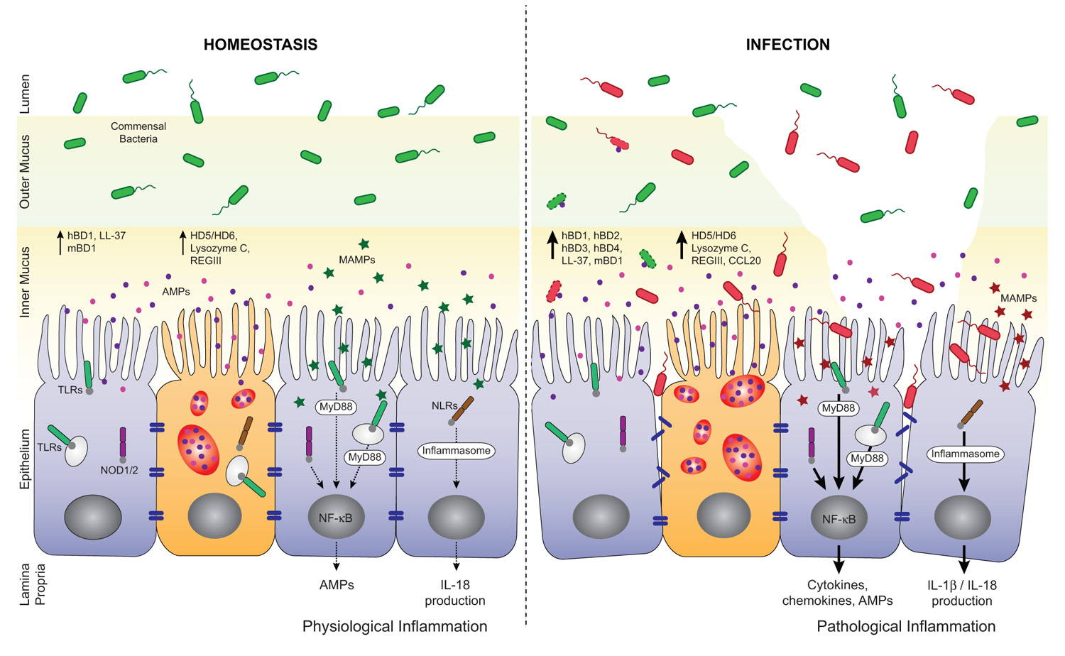
A depleted mucosal layer leads to inflammation and gut permeability.

It’s from a paper on the role of antimicrobial peptides in maintaining gut health. [1] The point of the left panel is that a healthy gut is characterized by a thick mucosal layer that shields our intestinal and immune cells from direct contact with bacteria. The inner mucus layer is infused with antimicrobial peptides to minimize its bacterial content. The outer mucus layer contains a population of friendly mucin-degrading bacteria – symbionts like Akkermansia who evolved to feed on our mucus. These friendly bacteria provide another layer of defense against infectious pathogens; bacteria tend to be quite good at keeping out competitors. Akkermansia has been found to prevent obesity.

In an unhealthy gut, on the other hand, the mucosal layer often gets stripped away. The image (right panel) attributes this to infection, which is one possibility, but nutritional factors also matter. For example:

  • Deficiencies of vitamins A or D will reduce production of antimicrobial peptides, making it easier for pathogens to reach our gut cells;
  • On very low-carb diets, production of mucin-2, the primary constituent of gut mucus, may be limited in order to preserve glucose for the brain (see “Dangers of Zero-Carb Diets, II: Mucus Deficiency and Gastrointestinal Cancers,” Nov 15, 2010);
  • Deficiencies of dietary fiber, vinegar, choline, and other nutrients may impair gut motility, leading to concentrations of partially digested food and bacteria at specific points in the intestine.

Regardless of why it happens, once bacteria come into direct contact with our gut and immune cells, they trigger inflammation and tend to loosen the gut barrier. This allows live bacteria and cell wall components from dead bacteria to enter the body from the gut.

Endotoxins – toxic compounds released when bacteria die, such as lipopolysaccharides from the cell walls of gram-negative bacteria – are immunogenic and inflammatory. A large influx of endotoxins into the body is “endotoxemia” – poisoning by endotoxins. As little as 2 nanograms LPS per kilogram body weight will induce fever, and 1 microgram LPS per kilogram of body weight will induce shock. [2]

Diseases caused by endotoxemia include:

  • Hepatitis [3]
  • Diabetes [4]
  • Heart disease [5]
  • Obesity [6]
  • Pulmonary hypertension [7]
  • Dyslipidemia [8]
  • Chronic kidney disease [9]

Many of today’s most prevalent diseases are caused by chronic endotoxemia.

So it behooves us to avoid it. If endotoxemia is fundamentally caused by the loss of a protective mucosal layer in the gut, how do we assure healthy production of mucus?

A recent paper sheds valuable light on that question.

Natural Whole Foods, High-Fat Diets, and Gut Health

It’s well known that in mice, “high-fat diets” induce endotoxemia. But these diets aren’t necessarily high in fat – any pelleted rodent food in which fat provides more than 20% of calories may be called “high-fat.” The critical difference of “high-fat diets” from chow is that they are composed of purified nutrients – starch, sugar, oil, vitamins, and minerals – whereas chow is composed of natural whole foods such as wheat, corn, and seeds.

A recent study tried to distinguish whether the cause of endotoxemia is the fat, or the purified starch, sugar, and oil. It made up three diets of varying fat content (8%, 48%, and 74% of energy respectively), but composed of natural whole foods. [10]

The result was remarkable:

[U]sing complex [i.e. natural whole foods] HFD, no associations were observed between dietary lipid amounts and the magnitude of endotoxemia, inflammation, and physiological alterations developed.

It turns out the endotoxemia that typically develops on high-fat diets is due to getting the calories from purified sources – starch, sugar, oil – rather than from whole foods. On a whole foods diet, the amount of endotoxin entering the body is more or less independent of the amount of fat consumed.

This is surprising in one respect. Lipopolysaccharide is fat-loving and enters the body along with dietary fat. So it stands to reason that a higher-fat diet would carry more endotoxin into the body.

But it turns out the body has mechanisms to regulate how much endotoxin enters the body. It wants a small amount so that the immune system can sample the gut microbiome, but not so much as to cause inflammation.

The primary mechanism for controlling endotoxin influx? Mucus production. The study noted that the mice eating higher-fat “had an increased number of goblet cells … [and] an increased MUC2 production.” MUC2 is mucin-2, the primary component of mucus in the gut.

Here is a picture with mucin-2 in the mucin-producing goblet cells stained red:

disease begins in mucus 02

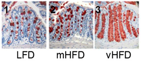
It’s obvious that mucin production goes up dramatically as the fat content of the diet increases.

The study concludes,

“We show that, in complex HFDs based on chow ingredients and milk fat, there was no association between dietary lipid amounts and the magnitude of metabolic endotoxemia or low-grade inflammation.”

If high-fat diets are healthy, we can thank our mucin-producing goblet cells.

One last note: the fact that mice can produce healthy amounts of mucus on a 74% fat diet does not necessarily mean that humans can do the same. Humans have much larger brains than mice, and as a result our carbohydrate needs are larger. It’s possible that mice can maintain mucus production on a low-carb diet better than humans can.

References

[1] Muniz LR, Knosp C, Yeretssian G. Intestinal antimicrobial peptides during homeostasis, infection, and disease. Front Immunol. 2012 Oct 9;3:310. http://pmid.us/23087688.

[2] Warren HS et al. Resilience to bacterial infection: difference between species could be due to proteins in serum. J Infect Dis. 2010 Jan 15;201(2):223-32. http://pmid.us/20001600.

[3] Parlesak A, Schäfer C, Schütz T, Bode JC, Bode C. Increased intestinal permeability to macromolecules and endotoxemia in patients with chronic alcohol abuse in different stages of alcohol-induced liver disease. J Hepatol. 2000 May;32(5):742-7. http://pmid.us/10845660.

[4] Moreno-Navarrete JM et al. Circulating lipopolysaccharide-binding protein (LBP) as a marker of obesity-related insulin resistance. Int J Obes (Lond). 2012 Nov;36(11):1442-9. http://pmid.us/22184060.

[5] Lepper PM et al. Association of lipopolysaccharide-binding protein and coronary artery disease in men. J Am Coll Cardiol. 2007 Jul 3;50(1):25-31. http://pmid.us/17601541.

[6] Cani PD et al. Metabolic endotoxemia initiates obesity and insulin resistance. Diabetes. 2007 Jul;56(7):1761-72. http://pmid.us/17456850.

[7] Dschietzig T, Alexiou K, Richter C, Bobzin M, Baumann G, Stangl K, Brunner F. Endotoxin causes pulmonary hypertension by upregulating smooth muscle endothelin type-B receptors: role of aldose reductase. Shock. 2008 Aug;30(2):189-96. http://pmid.us/18091567.

[8] Lassenius MI et al. Bacterial endotoxin activity in human serum is associated with dyslipidemia, insulin resistance, obesity, and chronic inflammation. Diabetes Care. 2011 Aug;34(8):1809-15. http://pmid.us/21636801.

[9] McIntyre CW et al. Circulating endotoxemia: a novel factor in systemic inflammation and cardiovascular disease in chronic kidney disease. Clin J Am Soc Nephrol. 2011 Jan;6(1):133-41. http://pmid.us/20876680.

[10] Benoit B et al. Increasing fat content from 20 to 45 wt% in a complex diet induces lower endotoxemia in parallel with an increased number of intestinal goblet cells in mice. Nutr Res. 2015 Apr;35(4):346-56. http://pmid.us/25687164.


Neu5Gc and Autoimmunity: Hashimoto’s Hypothyroidism

In Part I of this series, I reviewed the biology of Neu5gc (“Neu5gc, Red Meat, and Human Disease: Part I,” January 14, 2015). Now it’s time for Part II: a look at whether mammalian meats (beef, lamb, pork, dairy) may help provoke Hashimoto’s hypothyroidism.

Background on Autoimmunity

If you only care about health and what to eat, skip to the next section; but for those who want to understand mechanisms, here is the key background:

  • Neu5gc is abundant in nearly all mammals, but is absent in humans, ferrets, and new world monkeys.
  • Neu5gc is a sialic acid. It provides the terminal end of the carbohydrates which coat cells and glycoproteins. Cells need to be coated in these acids, because in water acids become ions and give the cells a charge, which repels other cells. When sialic acids are bound by antibodies, the charge is lost, and cells clump or aggregate. In fact, clumping of cells after pig serum was injected into humans was the first sign that humans form antibodies to pig cells. The main antigens in pig cells are “alpha-gal” and Neu5gc.
  • Although humans cannot manufacture Neu5gc due to a mutation that occurred 3 million years ago, we retain the ability to utilize it. So Neu5gc from food can appear on the surface of human cells.
  • To generate a broad-spectrum immune response, the DNA of B cells is re-arranged to create novel combinations of segments on the light- and heavy-chain portions of antibodies. This enables the body to generate more than 10^12 different antibodies. [1] To avoid generating antibodies to human antigens, any B cells that generate antibodies while still in bone marrow are destroyed. But Neu5gc from food doesn’t reach bone marrow, so there is nothing to stop the formation of white blood cells capable of generating antibodies to Neu5gc.

So Neu5gc has the potential to accumulate in human cells, especially intestinal cells which are directly exposed to food; and we can form antibodies to Neu5gc, which then may react to human cells which have incorporated Neu5gc into their carbohydrate coat.

One might think that this would be enough to generate autoimmunity, but more is needed. Although everyone has antibodies that can react to Neu5gc, the “preimmune repertoire” of antibodies binds to Neu5gc with very low affinity, and “low concentrations of anti-Neu5Gc antibodies do not seem to have any effect on Neu5Gc containing cells.” [4] In order to make high affinity antibodies, B cells must be repeatedly stimulated by Neu5gc-containing antigens.

The best stimulation is provided by bacterial cell wall components. As one paper states,

Bacteria are potent immunogens because they express a number of factors that can act as immune stimulants. Gram-negative bacteria universally produce endotoxins that have been shown to be powerful immune system modulators through the Toll-like receptor 4 (TLR4) on a variety of human immune cells. Lipoproteins on Gram-positive and Gram-negative cell surfaces can also interact with TLR2, resulting in release of cytokines involved in B cell and T cell proliferation. In addition, bacterial DNA has been known for many years to have the capability to stimulate the immune system. [2]

So to generate autoimmunity against Neu5gc incorporated in human cells, B cells must first be triggered to form high-affinity anti-Neu5gc antibodies by meeting bacterial pathogens who have incorporated Neu5gc into their cell walls.

This can happen because some bacteria do incorporate sialic acids from their local environment into their cell walls; and thus gut bacteria will incorporate Neu5Gc from food into cell walls.

A primary reason for doing this is that, by coating themselves in sialic acids acquired from their host, they look like a “host cell” and are shielded from immune attack. [5] Many pathogens have learned this trick:

Many pathogens secrete a sialidase that releases sialic acid from [nearby cells] … [O]ther sialic acid-utilizing bacteria, such as the respiratory pathogen H. influenzae, lack genes for a sialidase …. Presumably free sialic acid is made available to such pathogens by other, sialidase-expressing bacteria living in the same niche, or … by host sialidases that are activated in the course of inflammation. [3]

Among the pathogens known to use host sialic acids to shield themselves from human immunity is Neisseria gonorrhoeae, the pathogen that causes gonorrhea. It is possible that gonorrhea infection could lead to autoimmunity through this mechanism.

To date, the published research on this topic has focused on the possibility of pathogens incorporating Neu5Ac, the primary human sialic acid, from human cells into their cell walls, and subsequently triggering autoimmunity against Neu5Ac. There has been little study of the possibility that gut pathogens will incorporate Neu5Gc from food into their cell walls, potentially triggering autoimmunity against Neu5Gc incorporated in human cells.

Yet a recent study [4] comparing the levels of Neu5Gc antibodies in human blood against the prevalence of Hashimoto’s hypothyroidism suggests that this may be a significant pathway for autoimmunity.

Hashimoto’s Hypothyroidism and Neu5Gc Antibody Levels

This is one paper in which it’s almost enough just to present the data. Here are levels of anti-Neu5Gc antibodies in patients with hypothyroidism vs healthy controls:

Neu5gc hashis fig 1

This is Figure 1. [4] Patients with Hashimoto’s have, on average, 7-fold higher anti-Neu5Gc antibody levels than the general population. Patients with hypothyroidism, some of whom have Hashimoto’s and some don’t, have an intermediate level of anti-Neu5Gc antibodies.

Here are antibody levels in the healthy population (Figure 2a):

Neu5gc hashis fig 2a

Few healthy patients had more than 16 mcg/mL of anti-Neu5Gc antibodies, and none had more than 24 mcg/mL.

Here are antibody levels in Hashimoto’s patients (Figure 2c):

Neu5gc hashis fig 2c

Only 3% of Hashi’s patients had less than 12 mcg/mL of anti-Neu5Gc antibodies, and 57% had more than 24 mcg/mL.

This is a very good separation of the two groups. It looks like if you can generate large numbers of anti-Neu5Gc antibodies, then you are almost certain to get Hashimoto’s hypothyroidism.

About 50% (in this study, 47.9%) of hypothyroidism cases are autoimmune in origin. The 52.1% of hypothyroid patients who didn’t have Hashimoto’s generally had low levels of anti-Neu5Gc antibodies, similar to the healthy controls. This observation strengthens the association between anti-Neu5Gc antibodies and autoimmune hypothyroidism. It looks like anti-Neu5Gc antibodies are strongly linked to autoimmunity.

Adding plausibility, “both autoantigens related to Hashimoto disease [thyroid peroxidase and/or thyroglobulin] are glycoproteins and N-linked carbohydrates containing sialic acids have been detected in both molecules.” [4] So it’s possible Neu5Gc is incorporated directly into thyroid peroxidase and thyroglobulin.

The study authors state, “this is the first study investigating the association of anti-Neu5Gc antibodies with autoimmune diseases such as hypothyroidism.” [4]

They also tested for anti-Neu5Gc antibodies in rheumatoid arthritis patients, but found no connection there. Rheumatoid arthritis patients do not have elevated levels of anti-Neu5Gc antibodies.

Their paper has not yet been cited by any other papers. It looks like the investigation of Neu5Gc-mediated autoimmunity is at its very beginnings.

Jim Beecham’s Experience

Jim Beecham, MD, responded to my previous post with a personal story:

I read with interest your post about Neu5Gc. I am anxious to read Part 2 which I understand is coming. Meanwhile I have been doing a little research on the subject.

I suffered badly with childhood asthma, and I still get a sort of asthmatic tightness of my breathing once in a while. In the past few weeks have I realized this is on days after I eat cheese and/or beef. This has ceased upon my cutting out red meat and dairy this past week.

I also get hypothyroid symptoms of cold face and backs of hands from time to time. I wonder if this is also linked to Neu5Gc …

In a second comment Jim added:

Here’s another thought re: Neu5Gc…which I cannot prove but think is likely.

When an upsurge of titer of anti-Neu5Gc antibodies float in body fluids, they have opportunity cause inflammatory reaction.

One researcher postulated this mechanism for hemolytic uremic syndrome.

My own experience is I develop a groin ‘heat rash’ type reaction and irritable bladder a day or so after eating too much cheese and red meat.

Jim’s personal experiences add further evidence that Neu5Gc-driven inflammation and autoimmunity is a real phenomenon.

The paper linking Neu5Gc to hemolytic uremic syndrome is [6].

Conclusion

There’s an excellent chance that Hashimoto’s hypothyroidism is brought about by a complex of factors:

  1. An infection in the gut by bacterial pathogens that acquire Neu5Gc from food (primarily beef and pork) and incorporate it into their cell walls.
  2. A leaky gut that (a) allows Neu5Gc from food to enter the body for subsequent incorporation into human cells, such as thyroid cells, and (b) creates either a systemic invasion of Neu5Gc-bearing gut pathogens, or a “metabolic endotoxemia” in which Neu5Gc-bearing cell wall components of gut bacteria enter the body, triggering formation of high-affinity anti-Neu5Gc antibodies.
  3. Significant consumption of beef and pork, providing the Neu5Gc to drive the autoimmune process.

If this is the case, then the strategy for overcoming Hashi’s would involve:

  1. Improving gut barrier integrity and mucosal immunity,
  2. Normalizing or diversifying the gut flora, and
  3. Reducing dietary Neu5Gc by replacing beef, dairy, lamb, and pork with seafood and bird meats.

Neu5Gc-mediated autoimmunity does not play a role in rheumatoid arthritis, but it may play a role in other autoimmune diseases. The most likely organ to be affected is the gut, which is directly exposed to food; endothelial cells, which line blood vessel walls, and immune cells which circulate in blood, as blood is the next location after the gut exposed to food molecules entering the body; and lastly organs which interact closely with the blood, such as the thyroid. Hemolytic uremic syndrome is a condition of endothelial cell dysfunction.

So: it looks like reduction of mammalian meats, replacing them with seafood and bird meats, may be a prudent part of a “Hashimoto’s protocol.” In autoimmune disorders affecting the gut, blood vessels, or immune cells, it may be worth trying a 30-day elimination of mammalian meats.

Perfect Health Retreat

We have a few spots remaining for the May 2-9 Perfect Health Retreat, and will soon be taking reservations for the October retreat, either October 3-10 or 10-17 (or both). If you are interested, visit here for more info or email me at paul@perfecthealthretreat.com.

PHRetreat_img7_300x200px

References

[1] “Generation of Antibody Diversity,” in Alberts B, Johnson A, Lewis J, et al., Molecular Biology of the Cell, 4th edition, New York: Garland Science; 2002, http://www.ncbi.nlm.nih.gov/books/NBK26860/.

[2] Harvey HA, Swords WE, Apicella MA. The mimicry of human glycolipids and glycosphingolipids by the lipooligosaccharides of pathogenic neisseria and haemophilus. J Autoimmun. 2001 May;16(3):257-62. http://pmid.us/11334490.

[3] Severi E, Hood DW, Thomas GH. Sialic acid utilization by bacterial pathogens. Microbiology. 2007 Sep;153(Pt 9):2817-22. http://pmid.us/17768226. Full text: http://mic.sgmjournals.org/content/153/9/2817.long.

[4] Eleftheriou P et al. Prevalence of anti-Neu5Gc antibodies in patients with hypothyroidism. Biomed Res Int. 2014;2014:963230. http://pmid.us/25003133.

[5] Varki A, Gagneux P. Multifarious roles of sialic acids in immunity. Ann N Y Acad Sci. 2012 Apr;1253:16-36. http://pmid.us/22524423. Full text: http://www.ncbi.nlm.nih.gov/pmc/articles/PMC3357316/

[6] Löfling JC et al. A dietary non-human sialic acid may facilitate hemolytic-uremic syndrome. Kidney Int. 2009 Jul;76(2):140-4. http://pmid.us/19387473.


Neu5gc, Red Meat, and Human Disease: Part I

A number of people asked for comments on the most recent red meat scare, including Nicole, Ryan, and Mishkin on the blog, JT Olds on Twitter, and others on Facebook. You probably saw some of the headlines:

The article Nicole linked is a bit more scientifically inclined: “Possible Link Between Red Meat Consumption And Increased Cancer Risk Identified” (IFL Science). Here’s the press release version from UCSD: “Sugar Molecule Links Red Meat Consumption and Elevated Cancer Risk in Mice”. In the blogosphere, Stephan has summarized the issue in the context of a post on red meat and cancer.

The headlines are based on a paper [01] that reported that, in mice genetically altered to lack a sugar (Neu5gc) that humans also lack, feeding Neu5gc and injecting anti-Neu5gc antibodies generates inflammation which can promote the growth of cancers.

Significance of Neu5gc

The paper itself is a rather artificial scenario whose significance will be determined by future work. So analyzing this single paper would not be interesting. But I think it’s worthwhile to look into the broader idea that eating Neu5gc-bearing meats might be inflammatory or a source of autoimmunity.

In terms of PHD recommendations, this could affect the relative emphasis we place on different meats. If Neu5Gc is a true health risk, then we would want to emphasize seafood more and red meat less.

Another benefit to thinking about Neu5gc is that it may give us some insight into what a PHD “autoimmune protocol” should look like.

Background: Evolutionary History of Neu5gc

All cells in multicellular organisms are coated in carbohydrates, and the carbohydrates terminate in one of 43 sialic acids. In mammals, two forms of predominate: Neu5Ac and Neu5Gc. Each mammalian cell has tens or hundreds of millions of molecules of Neu5Ac and Neu5Gc on its surface. [02]

Neu5Gc is made from Neu5Ac, but the gene for making Neu5Gc was inactivated in the human lineage shortly before the emergence of Homo. The mutation occurred 3.2 million years ago and reached fixation – that is, all ancestral hominids had come to have the mutated gene – 2.9 million years ago. [03] This very rapid fixation indicates there was strong selection in support of the mutation.

In fact, this mutation by itself may have led to a speciation event, after which our ancestors could no longer mate with other apes. From that point on, Neu5gc-less females had difficulty producing children with males who retained the Neu5gc gene, because they would form antibodies against Neu5gc-coated sperm, making fertilization unlikely. [04]

Why was losing Neu5gc selected in our ancestors? Two possibilities are likely:

  • Loss of Neu5gc improved brain function.
  • Loss of Neu5gc (temporarily) reduced vulnerability to (ancestral) pathogens.

It should be noted that Neu5Gc has been lost independently in some other mammals as well – ferrets and new world monkeys. New world monkeys such as capuchins and spider monkeys also experienced a brain expansion, and ferrets are notably smart, so either explanation might be relevant to these cases of “convergent evolution.”

Neu5gc and Brain Function

Carbohydrates are extremely important for intercellular interactions. Indeed, the incorporation of carbohydrates into cell membranes and extracellular matrix is what made possible the rise of multicellular life.

In no organ are intercellular interactions as complex or consequential as in the brain. Not surprisingly, then, carbohydrates including the sialic acids are important to brain function.

The human brain is extraordinarly rich in sialic acids: neural membranes have 20 times more sialic acids than membranes of other human cell types. Animal brains are also enriched in sialic acids relative to their other tissues, but not as much as in humans; the human brain has 2-4 times more sialic acids the brains of other mammals. [05]

Curiously, though, Neu5gc is rare in the brains of all animals. Neu5gc is strongly suppressed, by about 98%, in the brains of all vertebrates, suggesting that its presence inhibits brain function. [06] It appears that Neu5gc is somehow toxic to brain function.

Loss of the gene for Neu5gc completely eliminated Neu5gc from the hominid brain. If Neu5gc does impair brain function, mutational inactivation of Neu5gc would have improved brain function. If so, the mutational inactivation of Neu5gc could have been driven by the same evolutionary forces that, soon after, selected for the tremendous expansion of the hominid brain.

Incidentally, dietary sialic acids — except for Neu5Gc – appear to be nutritious for humans, and especially for the developing infant brain. Breast milk is exceptionally rich in sialic acids, almost all of it Neu5Ac. Formula, by contrast, has much lower levels of sialic acids (0.21 mmol/L compared to 3.72 mmol/L in breast milk). Breast fed infants have nearly twice as many sialic acids in saliva than formula fed infants, confirming that milk sialic acids are taken up by the body and utilized.

Animal studies show that sialic acids in breast milk nourish the brain. Sialic acids facilitate neurotransmission between neurons. When piglet milk is supplemented with sialic acids, brain sialic acid levels are increased, and the piglets learn faster and make fewer mistakes in maze tests. [05] Rodents also perform better on tests of learning and memory after sialic acid supplementation. [07]

Not only does formula have fewer sialic acids than breast milk, cow milk based formulas have some Neu5Gc. [05] It has been observed that formula-fed infants have lower IQs than breast-fed infants. Sialic acids might help explain that. The lack of nourishing Neu5Ac and the presence of toxic Neu5Gc in formula might lastingly impair brain function in formula-fed infants.

Neu5Gc and Infection Risk

As the outermost molecules in the carbohydrate coat surrounding cells, sialic acids are the first contact point for pathogens seeking entry to the cell, and for immune cells seeking to detect whether the cell is native or foreign.

There has been a “Red Queen” evolutionary arms race in which pathogens evolved ways to utilize sialic acids for cell entry, or to hide from the immune system; and animals evolved changes to their sialic acids to frustrate the pathogens. [08]

Many pathogens interact with sialic acids in order to adhere to and gain entry into the cell. Pathogens generally rely on a single specific endocytic route for cell entry. This often requires binding to a specific sialic acid as the initial point of attachment.

Pathogens that specifically utilize Neu5Gc to enter cells include canine and feline parvoviruses [09]; pathogens that specifically utilize Neu5Ac include adeno-associated viruses and the minute virus of mice (MVM) [10].

A human pathogen that uses sialic acids to enter cells is the malaria protozoan. Plasmodium falciparum causes severe disease in humans and enters cells via Neu5Ac; Plasmodium reichenowi causes milder disease in chimpanzees and gorillas and enters cells via Neu5Gc. Plasmodium falciparum appears to have evolved recently – possibly reaching its current form only 10,000 years ago when the rise of agriculture and animal husbandry brought humans and mosquitos into closer proximity – while Plasmodium reichenowi is thought to resemble the ancestral form that would have afflicted hominids and apes 3.5 million years ago.

Possibly, the gene for Neu5Gc was inactivated to protect ancestral hominids from malaria. With the loss of Neu5Gc, hominids would have become immune to P. reichenowi. [11] [12]

Unfortunately, after P. falciparum’s adaptation to Neu5Ac which is overabundant in humans, we now suffer from more severe malaria than chimpanzees or gorillas (the “malignant malaria” mystery). [13]

In addition to entry points for microbes, sialic acids can be entry points for microbial toxins. For example, Shiga toxin from shigatoxigenic E. coli binds to Neu5Gc. [14]

Sialic Acid Concealment and the Gut Microbiome

The immune system is sensitive to the composition of the carbohydrate coat on a cell. White blood cells have a number of sialic acid detectors on their surfaces (called Siglecs, for sialic acid Ig-superfamily lectins). Some, which bind to human sialic acids, inhibit immune responses. Others, which bind to non-human sialic acids, activate immune responses.

Thus, when white blood cells contact a cell bearing human sialic acids, the immune system interprets it as “self” and tamps down immunity. When it detects foreign sialic acids, the immune system treats the cell as “foreign” and is more likely to attack it.

Some microbes – including commensal gut microbes – have been living in humans long enough that they have learned to take up sialic acids, chiefly Neu5Ac, and incorporate them into lipopolysaccharides on their cell membranes. This suppresses immunity toward them. [15]

A number of human pathogens have learned the same trick. Pathogens that incorporate sialic Neu5Ac into their cell membranes for the purpose of mimicking human cells and evading human immune defenses include Escherichia coli K1, Haemophilus influenzae, Pasteurella multocida, Neisseria spp., Campylobacter jejuni and Streptococcus agalactiae. [16]

Due to this “molecular mimickry” of human molecules, it has been suggested that these bacteria – especially Haemophilus influenzae and Neisseria spp. – may be sources of autoimmunity. [17]

While some bacteria can synthesize sialic acids themselves, most obtain it from their environment. These bacteria release enzymes called sialidases to cleave the sialic acids from food in the digestive tract, from surrounding cells, or from mucus. [15] Bacteria can obtain Neu5Ac from human tissue and mucus as well as food, but Neu5Gc only from food, chiefly beef and pork.

Neu5Gc in Human Tissue

Although humans can no longer synthesize Neu5Gc, we still have all the cellular machinery for utilizing it. When dietary Neu5Gc is absorbed into the body and enters cells, it can be incorporated into glycoproteins bound for the cell surface glycocalyx, just as Neu5Ac is.

As a result, Neu5Gc of dietary origin appears at low levels on the surface of human cells.

Neu5gc is found at high levels in all mammals except humans, ferrets, and new world monkeys; birds and reptiles do not produce Neu5Gc at all, and fish and shellfish produce only low levels. So, of the four major meat groups – beef, pork, chicken, and fish – Neu5gc is obtained predominantly from the red meats, beef and pork.

Among human cells, Neu5Gc appears at highest levels on tumor cells, especially metastatic cells. [21] This makes Neu5Gc a potential target for cancer therapy.

Neu5Gc as an Immunogen

Neu5gc expressed on cell walls is a potential immunogen. When pig organs are transplanted to humans, Neu5Gc is the second most important cause of rejection, after the α1,3-galactose (αGal) epitope. [20]

Anti-Neu5gc antibodies have been found in 85% of humans. [18] It is thought that antibodies form in early childhood after dietary Neu5Gc is incorporated by certain gut bacteria into lipooligosaccharides that can generate antibodies. Some of these antibodies may cross-react with compounds human cells form from dietary Neu5Gc; these human molecules are then known as “xeno-autoantigens.” [21]

Summary

Neu5Gc from mammalian meats, such as beef and pork, is incorporated into the cell surface coats and walls of gut microbes and some human cells, mainly in the gut and in tumors. Neu5gc in bacterial walls is immunogenic and 85% of people have detectable antibodies to Neu5Gc. Eating beef and pork supplies antigens for these antibodies, potentially triggering inflammation. There are concerns that this inflammation may have negative health effects.

Next up: Neu5Gc and autoimmunity.

Note: May Perfect Health Retreat

Interested in a luxury vacation combined with an education in how to be healthy for the rest of your life? Science classes, cooking classes, movement and relaxation classes, personal health coaching, gourmet food, all on a magnificent beach, with hot tubs and salt water pools? Come to the Perfect Health Retreat! Spaces are currently 2/3 filled and the retreat is expected to sell out early. Visit here for more info or email me at paul@perfecthealthretreat.com.

PHRetreat_img7_300x200px

References

[01] Samraj AN et al. A red meat-derived glycan promotes inflammation and cancer progression. Proc Natl Acad Sci U S A. 2014 Dec 29. pii: 201417508. [Epub ahead of print]. http://pmid.us/25548184.

[02] Kraemer PM. Sialic acid of mammalian cell lines. J Cell Physiol. 1966 Feb;67(1):23-34. http://pmid.us/5327858. Was 21

[03] Hayakawa T, Aki I, Varki A, Satta Y, Takahata N. Fixation of the human-specific CMP-N-acetylneuraminic acid hydroxylase pseudogene and implications of haplotype diversity for human evolution. Genetics. 2006 Feb;172(2):1139-46. http://pmid.us/16272417. Full text: http://www.ncbi.nlm.nih.gov/pmc/articles/PMC1456212/. Was 22

[04] Ghaderi D et al. Sexual selection by female immunity against paternal antigens can fix loss of function alleles. Proc Natl Acad Sci U S A. 2011 Oct 25;108(43):17743-8. http://pmid.us/21987817. was 2

[05] Wang B. Molecular mechanism underlying sialic acid as an essential nutrient for brain development and cognition. Adv Nutr. 2012 May 1;3(3):465S-72S. http://pmid.us/22585926. Full text: http://www.ncbi.nlm.nih.gov/pmc/articles/PMC3649484/. Was 31

[06] Davies LR, Varki A. Why Is N-Glycolylneuraminic Acid Rare in the Vertebrate Brain? Top Curr Chem. 2013 Mar 8. [Epub ahead of print] http://pmid.us/23471785. was 8

[07] Wang B. Sialic acid is an essential nutrient for brain development and cognition. Annu Rev Nutr. 2009;29:177-222. http://pmid.us/19575597. was 32

[08] Varki A. Colloquium paper: uniquely human evolution of sialic acid genetics and biology. Proc Natl Acad Sci U S A. 2010 May 11;107 Suppl 2:8939-46. http://pmid.us/20445087. was 51

[09] Löfling J et al. Canine and feline parvoviruses preferentially recognize the non-human cell surface sialic acid N-glycolylneuraminic acid. Virology. 2013 May 25;440(1):89-96. http://pmid.us/23497940. was 54

[10] Wu Z et al. Alpha2,3 and alpha2,6 N-linked sialic acids facilitate efficient binding and transduction by adeno-associated virus types 1 and 6. J Virol. 2006 Sep;80(18):9093-103. http://pmid.us/16940521. was 55

[11] Varki A, Gagneux P. Human-specific evolution of sialic acid targets: explaining the malignant malaria mystery? Proc Natl Acad Sci U S A. 2009 Sep 1;106(35):14739-40. http://pmid.us/19717444. was 57

[12] Martin MJ et al. Evolution of human-chimpanzee differences in malaria susceptibility: relationship to human genetic loss of N-glycolylneuraminic acid. Proc Natl Acad Sci U S A. 2005 Sep 6;102(36):12819-24. http://pmid.us/16126901. was 58

[13] Rich SM et al. The origin of malignant malaria. Proc Natl Acad Sci U S A. 2009 Sep 1;106(35):14902-7. http://pmid.us/19666593/.

[14] Byres E et al. Incorporation of a non-human glycan mediates human susceptibility to a bacterial toxin. Nature. 2008 Dec 4;456(7222):648-52. http://pmid.us/18971931.

[15] Varki A, Gagneux P. Multifarious roles of sialic acids in immunity. Ann N Y Acad Sci. 2012 Apr;1253:16-36. http://pmid.us/22524423. Full text: http://www.ncbi.nlm.nih.gov/pmc/articles/PMC3357316/

[16] Severi E, Hood DW, Thomas GH. Sialic acid utilization by bacterial pathogens. Microbiology. 2007 Sep;153(Pt 9):2817-22. http://pmid.us/17768226. Full text: http://mic.sgmjournals.org/content/153/9/2817.long.

[17] Harvey HA, Swords WE, Apicella MA. The mimicry of human glycolipids and glycosphingolipids by the lipooligosaccharides of pathogenic neisseria and haemophilus. J Autoimmun. 2001 May;16(3):257-62. http://pmid.us/11334490.

[18] Zhu A, Hurst R. Anti-N-glycolylneuraminic acid antibodies identified in healthy human serum. Xenotransplantation. 2002 Nov;9(6):376-81. http://pmid.us/12371933.

[19] Takahashi T et al. N-glycolylneuraminic acid on human epithelial cells prevents entry of influenza A viruses that possess N-glycolylneuraminic acid binding ability. J Virol. 2014 Aug;88(15):8445-56. http://pmid.us/24829344.

[20] Park JY et al. α1,3-galactosyltransferase deficiency in germ-free miniature pigs increases N-glycolylneuraminic acids as the xenoantigenic determinant in pig-human xenotransplantation. Cell Reprogram. 2012 Aug;14(4):353-63. http://pmid.us/22775484.

[21] Samraj AN, Läubli H, Varki N, Varki A. Involvement of a non-human sialic Acid in human cancer. Front Oncol. 2014 Feb 19;4:33. http://pmid.us/24600589.

Farewell Mathias

Longtime readers will remember Mathias and Zachary, the unfortunate children suffering from Neurodegeneration with Brain Iron Accumulation, a genetic disorder that leads to horrifically painful spasms beginning in early childhood, and death as a teenager. I wrote about their case in Ketogenic Diet for NBIA (Neurodegeneration with Brain Iron Accumulation), February 22, 2011.

The ketogenic version of PHD had remarkable effects for the NBIA kids. Mathias and another boy who tried the diet, Zachary, regained control their limbs, and the spasms and pain went away. Kindy, Mathias’s mom, wrote in 2011:

Both boys have begun smiling and laughing all the time.

Nothing inspires happiness more surely than the cessation of extreme pain!

My speculation is that a ketogenic (or high-fat) diet helps in NBIA by allowing Coenzyme A, the crucial enzyme which is under-generated in NBIA, to be redistributed from organs like the liver and muscle, where it is manufactured in abundance, to the brain where it is most needed. On a ketogenic or high-fat diet, more CoA is created and it is more often bound in water-soluble forms (such as acetyl-CoA, acetoacetyl-CoA, and HMG-CoA) that can cross cell membranes and enter the brain.

Mathias and Zachary continued to do well on our diet for over three years. Kindy recently wrote:

Zach is actually doing really well.  He is following your diet still (not into the ketogenic range but otherwise following it more or less precisely) …  He is off nearly all of his medicines and is able to do things that he never could in his life.   He is not well – but he is not in pain and has no spasms, and is doing school work etc.

An aside: I’ve been hearing recently from a number of people who experienced great benefits in neurological conditions – NBIA, epilepsy, migraines, and others – following the ketogenic version of our diet, and later transitioned to the regular version of PHD with more carbs and less fat, and continued to maintain all the neurological benefits they had first achieved on the ketogenic diet. Perhaps it was not the ketosis that was crucial, but some other aspect of PHD, such as reduction of inflammation or improved nutrition.

Mathias also was doing very well, until he developed pneumonia last summer. Possibly his genetic mutations disturbed immune function; in any case, the pneumonia led to fatal complications. Kindy wrote:

I want to let you know that on June 23, Mathias died of septic shock.  He went into the hospital 10 days prior with pneumonia and we were packing to go home on the following Friday when he got a sudden fever.  The doctors asked us to stay one more day – his lung x-rays were clear but they were concerned about the fever.

On Saturday, his fever went to 41 degrees Centigrade.  On Sunday, it went to 42 degrees.  Despite every available antibiotic and all other attempts to save him, Mathias died peacefully with a strong heart (153 beats per minute – and breathing on his own).

He was surrounded (even in Intensive Care) by his whole family, plus his aunt, and two of his long time helpers – plus two of his nurses and two doctors.  We thought it would be a few more days and we were all hugging him, and laughing with him and telling him stories.  From one second to the next, his heart stopped.

We choose to believe that Mathias decided – down to the last second – what and how he was going to leave his earthly body.  He had no cramps, no spasms, no pain. He just let go surrounded by love.

We are privileged and honored to have known such a brave, smiling, incredible person.  He did more and affected more people in his 9 years of life than most people do in their entire lives. He was always happy, always smiling – a gift to everyone around him.

Thank you for being part of the forces around his life who helped support him, love him, and provide him with the best life that was possible for him. Thank you from the bottom of our hearts!

Mathias RIP

Farewell Mathias. May you rejoice in God’s kingdom, where all love and all are lovable, and all tears are wiped away. Requiescat in pace.