Category Archives: Vitamin C

Are Bigger Muscles Better? Antioxidants and the Response to Exercise

We’re positive toward some dietary antioxidants:

  • Vitamin C is one of our supplement recommendations.
  • We also recommend higher-than-typical dietary intakes of zinc and copper, key ingredients of the antioxidant zinc-copper superoxide dismutase.
  • We recommend high intakes of extracellular matrix material in soups and stews. This is rich in glycine, a component of glutathione, a key antioxidant. N-acetylcysteine, which provides the other amino acid component of glutathione, is one of our therapeutic supplements.
  • Although we don’t normally recommend supplementing vitamin E, it is listed among our optional supplements, and we believe significant numbers of people might benefit from supplementing mixed tocotrienols.
  • Although we don’t recommend supplementing it, our book notes the importance of dietary intake of selenium, which is critical for an enzyme that recycles glutathione.

A fashionable contrary view has arisen: antioxidants not only don’t help, they can do harm by interfering with oxidative signaling pathways.

Adel Moussa, proprietor of Suppversity, has promulgated this view, especially the idea that antioxidants interfere with the response to exercise. In a post last week, “Bad News For Vitamin Fans – C + E Supplementation Blunts Increases in Total Lean Body and Leg Mass in Elderly Men After 12 Weeks of Std. Intense Strength Training,” he looked at a new study by Bjørnsen et al [1]. In the comments here, Spor asked me to address Adel’s post, and other readers expressed interest too.

The Bjørnsen Study: Muscle Size

In this new study, Bjørnsen and collaborators put a group of elderly men on a strength training regimen. Half the men were put on supplements – 1000 mg per day of vitamin C and 235 mg (350 IU) per day of the alpha-tocopherol form of vitamin E – and half on placebo. They then assessed the response to 12 weeks of exercise.

Both groups gained muscle mass, but the placebo group gained more. Total lean mass, and the thickness of the rectus femoris muscle (one of the quadriceps), increased more in the placebo than the antioxidant group. The lean mass increase averaged 3.9% in the placebo group, 1.4% in the antioxidant group. Here’s the plot of muscle thickness in the rectus femoris:

Bjornsen Fig 1

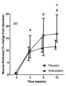
This muscle increased in thickness an average of 16.2% in the placebo group, 10.9% in the antioxidant group.

So far, so solid: it looks like muscle size will be larger if you don’t take antioxidants.

Adel concludes:

[Y]ou shouldn’t fall for … the bogus false promise that suffocating all the flames by using exorbitant amounts of antioxidants (and I am as this study shows talking about 10x the RDA not just 100x the RDA) would be good for you, let alone your training progress and muscle gains…. [Y]ou cannot recommend extra-vitamins for people who work out – specifically not the elderly.

I disagree.

The Trouble with Biomarkers

We have to use biomarkers to assess health, because the things we really care about – like how long we will live – cannot be determined on the time scales we need to make decisions in.

But no biomarker is a perfect assessment of health. There are always regimes in which a biomarker can look “better” but health can worsen.

Muscle size is no different. Other things being equal, more muscle is better than less muscle. But there are ways to increase muscle size that harm health. We have to look at the increase in muscle size in the placebo group and ask: was this good or bad?

The Bjørnsen Study: Strength

Fortunately, Bjørnsen and colleagues also reported another key biomarker: strength, as indicated by 1 rep maximum weight. Here is the data:

Bjornsen Fig 2

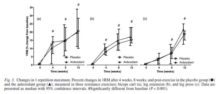
The exercises that utilize the rectus femoris muscle, the one that grew biggest in the placebo group, are the leg extension (b) and leg press (c). And here we see something interesting: in both cases, the antioxidant group increased their 1RM by more than the placebo group. Yes, the improvement was not statistically significant. But it was there. According to the text, on average, the antioxidant group increased their leg press 1RM by 18.7%, the placebo group by 15.8%.

So the antioxidant group gained more strength but less size than the placebo group. Which group was made healthier by the program?

I’ll put my money down on this: the smaller muscle that can exert more force is the healthier muscle. A gargantuan but weak muscle is an unhealthy muscle.

Large Muscle Size Can Be a Sign of Poor Health

To see that large muscles may be unhealthy, consider the health condition of cardiomegaly – an enlarged heart. When the heart tissue is dysfunctional and incapable of exerting as much strength as it should, the heart grows larger to compensate. People who have such an oversized but weak heart often die an early death.

We should consider whether something similar was going on in the placebo group of the Bjørnsen study. Their leg muscles grew larger to compensate for weakness. They needed more mass to accomplish less than the antioxidant group.

What causes cardiomegaly? One contributing factor is a deficiency of antioxidants. When antioxidants are deficient, oxidative stress generated during exertion leads to lipid peroxidation and tissue necrosis.

One of my favorite nutritional studies – I always show it at the Perfect Health Retreat to demonstrate the existence of multi-nutrient deficiency diseases, diseases that appear only when you are deficient in multiple nutrients at the same time – is a study by Kristina Hill and collaborators at Vanderbilt in 2001. [2] The running title is “Myopathy resulting from combined Se and E deficiency” and that summarizes it well. Guinea pigs were put on one of four diets – a control diet, the control diet but deficient in vitamin E, the control diet but deficient in selenium, or the control diet with both vitamin E and selenium removed.

Something interesting happened to the guinea pig muscles:

Bjornsen Fig 3

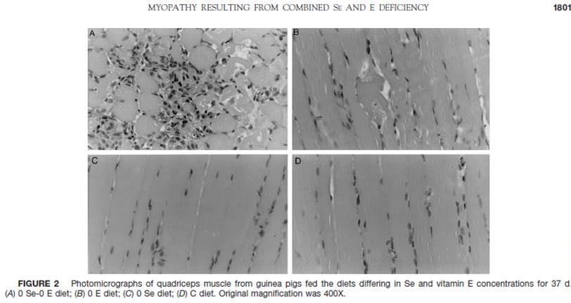
These are quadriceps muscles – the same muscle whose size was altered in the Bjørnsen study. Panel (D) shows a healthy muscle from the control group. The muscle fibers are long, straight, and parallel to one another. Panels (B) and (C), the low vitamin E and low selenium groups respectively, are mildly damaged but still functional. However, in panel (A), the group deprived of both selenium and vitamin E, the muscle fibers are severely damaged. This muscle cannot exert force.

In the group deprived of both selenium and vitamin E, the loss of strength continued until the guinea pigs could no longer stand or move. At that point they lost the ability to feed and began to die of starvation. This happened in as little as 30 days. Here was the survival curve:

Bjornsen Fig 4

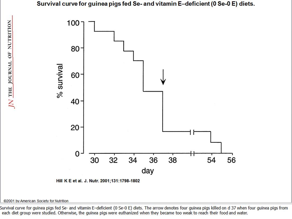
The last guinea pig died after 55 days.

Why did this happen? The guinea pig muscles were damaged by lipid peroxidation leading to cell death. They didn’t have enough antioxidants.

Hill et al didn’t measure muscle thickness, but it wouldn’t surprise me if at 20 days the guinea pigs on their way to an early death had the thickest and most massive quadriceps.

Can Muscle Size Be Used as an Indicator of Overtraining?

If growth in muscle size may indicate muscle damage from either overtraining or antioxidant deficiencies, we might be able to use the response to exercise to assess nutrition or exercise load.

Let’s look again at the Bjørnsen study. If the cross-sectional area of a muscle is proportional to the square of muscle thickness, then we can get a measure of strength per unit cross-section by taking the ratio of leg press 1RM to the square of rectus femoris thickness. That is 1.187/1.109^2 = 0.965 in the antioxidant group, 1.158/1.162^2 = 0.857 in the placebo group. Per unit cross-section, the antioxidant group lost 3.5% in strength, the placebo group lost 14.3%.

It looks like both groups may have damaged their muscles; the antioxidant group just did much less damage. It appears both groups were overtraining relative to their nutritional status. Perhaps if nutrition were better, the response to exercise would have been better, and strength per unit cross-section would have increased. Maybe the missing nutrients included antioxidants, and taking even more antioxidants may have enabled this rigorous training regimen (designed “to stimulate as much muscle growth as possible”) to take place without any impairment of health.

Is Bodybuilding Safe?

If large size can be an indication of damage in muscle, then many techniques which cause muscular hypertrophy will be health-damaging. The healthiest strength gains might come with only small size gains, as the muscle becomes more efficient. It is only unhealthy muscle that becomes super large.

If so, then bodybuilders, who are judged on the size of their muscles, not their strength, will be tempted to use health-damaging and muscle-damaging techniques, like antioxidant deficiencies, to expand their muscle mass. Presumably the winning bodybuilders will be those who use all effective techniques to grow muscle, including the health-damaging ones. So we should expect champion bodybuilders to die young.

I have not seen statistical evidence, but anecdotal lore suggests that champion bodybuilders do, indeed, die young, often of heart diseases (indicating muscle damage). Here are two Youtube videos memorializing bodybuilders who died young:

Conclusion

If you want me to believe that antioxidants are bad, the Bjørnsen study is not going to do it. It looks to me that the elderly men who were in the antioxidant group were the lucky ones. The 1000 mg of vitamin C and 350 IU of vitamin E they were taking daily improved their response to exercise. Indeed, for all we know their antioxidant intake may have been less than optimal!

The elderly men who didn’t get the antioxidants should worry about their hearts. If their leg muscles became large but weak, their hearts may have also.

Everyone who works out should be aware: when it comes to muscles, bigger is not the same as better. The healthiest muscles are those in a wiry physique – modest size, but able to exert a lot of force.

Finally, a pitch for our upcoming October 10-17 Perfect Health Retreat. Our advice is sensible, comprehensive, and increasingly well supported by guest experience. If you want to learn how to optimize health for the rest of your life, and have a great time doing it, please come join us.

PHRetreat_img3_600x400px

References

[1] Bjørnsen T et al. Vitamin C and E supplementation blunts increases in total lean body mass in elderly men after strength training. Scand J Med Sci Sports. 2015 Jul 1. http://pmid.us/26129928.

[2] Hill KE et al. Combined selenium and vitamin E deficiency causes fatal myopathy in guinea pigs. J Nutr. 2001 Jun;131(6):1798-802. http://pmid.us/11385070.


Danger of Zero-Carb Diets III: Scurvy

I started low-carb Paleo dieting in late 2005. I ate a lot of vegetables but no starches and hardly any fruit. In retrospect, I would call it a near zero-carb diet. At that time I was 12 years into a chronic illness that got a little worse each year and was quite mysterious to me. Adopting a low-carb diet brought immediate changes: it made what I would much later recognize as a chronic bacterial infection better (in parts of the body, not the brain) and made a chronic fungal infection worse.

Within about a year I had developed scurvy. It took me an embarrassingly long time to figure out what it was. By the time knew what it was, I had 3 cavities; had lost 25 pounds; had developed diverticulitis and an abdominal aorta that visibly swelled with every heartbeat; and had minor skin wounds – scrapes and scratches – that hadn’t healed in 6 months.

Developing scurvy was a surprise, because I was eating many vegetables plus taking a multivitamin containing 90 mg of vitamin C. I had never had any signs of vitamin C deficiency before adopting a low-carb diet.

Four grams a day of vitamin C for two months cured all the scurvy symptoms. It would be several more years before I figured out the infections, but this experience taught me the importance of micronutrition. The experience persuaded me that I needed to research diets and nutrition closely, and started us down the path of writing Perfect Health Diet.

Scurvy on a Ketogenic Diet

My experience is not unique. Here’s one case we mention in the book: the story of a young girl with epilepsy.

KM was a 9-year old girl … diagnosed with epilepsy at six months old. She started a ketogenic diet in October 2003, as her multiple antiepileptic drugs were proving to be less than effective; indeed she was having as many as 12 tonic seizures per day with prolonged periods of non-convulsive status epilepticus. After the diet was prescribed the seizure frequency reduced markedly and there were a number of long periods of time in which she had no seizures.

KM’s mother gave a history of her daughter having had bleeding gums since the beginning of September 2006; she described them as being very dark red, swollen and bleeding. In addition, she explained that her daughter had dry, crusted blood peri-orally. The family’s general dental practitioner had explained that this was probably caused by erupting teeth and instructed her to use 0.2% chlorhexidine gluconate gel and to continue her regular oral hygiene regimen; however this had no effect. About a month later the patient’s right arm became swollen. It was thought that she had sustained a fracture or a dislocation; however she was discharged from the local hospital’s fracture clinic because there was clinical improvement and radiographs showed no callus formation.

In early November KM inhaled a primary molar tooth while she was having her teeth cleaned (Fig. 1). This required an emergency bronchoscopy to retrieve it; at the same time the surgeons extracted her remaining primary teeth in order to avoid a recurrence of the problem….

At that time an appointment was made to attend a paediatric dentistry consultant clinic at the Leeds Dental Institute; however this was never kept as about three weeks after the extractions the patient was admitted to hospital with low grade fever, persistently bleeding gums, oedema of her hands and feet and a petechial rash on her legs. [1]

This girl was eating a typical amount of vitamin C: her dietary intake was calculated at 73 mg/day, well above the US RDA for 9-13 year olds of 45 mg/day. Yet her blood level was only 0.7 µmol/l. Scurvy is diagnosed at levels below 11 µmol/l.

The symptoms of scurvy are sufficiently insidious that it is easy to miss the diagnosis. In KM’s case, it happened that a “senior house officer” – a junior doctor in training – from India recognized the symptoms of scurvy. Otherwise, it might have never have occurred to the doctors to test her vitamin C level. [2]

What Is the Cause of Low-Carb Scurvy?

So what causes scurvy to develop on low-carb diets even with vitamin C intake well above the US RDA?

It seems to be a confluence of two factors:

  • An infection or some other stress (e.g. injury, cancer) leads to the oxidation of extracellular vitamin C; and
  • On a low-insulin or glutathione-deficiency-inducing diet, oxidized vitamin C is not recycled.

Infection and Vitamin C

The immune response to infections generates reactive oxygen species, which oxidize vitamin C. Oxidation removes a hydrogen atom from vitamin C, turning it into “dehydroascorbic acid,” or DHAA. If DHAA remains in the blood, it degrades with a half-life of 6 minutes. [3]

Infections can cause vitamin C deficiency on any diet. During the “acute phase response” to infection or injury, vitamin C often becomes deficient. Here is a nice paper in which French doctors surveyed their hospital patients for scurvy:

We determined serum ascorbic acid level (SAAL) and searched for clinical and biological signs of scurvy in 184 patients hospitalized during a 2-month period.

RESULTS: The prevalence of hypovitaminosis C (depletion: SAAL<5 mg/l or deficiency: SAAL<2 mg/l) was 47.3%. Some 16.9% of the patients had vitamin C deficiency. There was a strong association between hypovitaminosis C and the presence of an acute phase response (p=0.002). [4]

So half were at least depleted in vitamin C and 17% had outright deficiency, which if it persisted would produce scurvy.

We’ve previously written of how important it is to supplement with vitamin C during infections:

I might add here that in sepsis, an extremely dangerous inflammatory state brought on by bacterial infections, intravenous vitamin C reverses some of the worst symptoms. [5] If you have a loved one suffering from a dangerous infection, it might not be a bad idea to get them some vitamin C.

Insulin Dependence of Vitamin C Recycling

DHAA can be recycled back into vitamin C, but only inside cells.

In order to enter cells, DHAA needs to be transported by glucose transporters. GLUT1, GLUT3, and GLUT4 are the three human DHAA transporters; GLUT1 does most of the work. [6]

DHAA transport is crucial for brain vitamin C status. There is no direct transport of vitamin C into the brain, yet the brain is one of the most vitamin C-dependent tissues in the body. The brain relies entirely on GLUT1-mediated transport of DHAA from the blood for its vitamin C supply. Within the brain, DHAA is restored to vitamin C by glutathione.

Supplying DHAA to stroke victims (of the mouse persuasion) as late as 3 hours after the stroke can reduce the stroke-damaged volume by up to 95%:

DHA (250 mg/kg or 500 mg/kg) administered at 3 h postischemia reduced infarct volume by 6- to 9-fold, to only 5% with the highest DHA dose (P < 0.05). [7]

This is a fascinating reminder of the importance of vitamin C for wound repair and protection from injury.

Glucose transporters are activated by insulin. Thus, DHAA import into cells is increased by insulin, leading to more effective recycling of vitamin C [8]:

Insulin and IGF-1 promote recycling of DHAA into ascorbate. Source.

Confirming the role of insulin in promoting vitamin C recycling, Type I diabetics (who lack insulin) have lower blood levels of vitamin C, higher blood levels of DHAA, increased urinary loss of vitamin C metabolites, and greater need for dietary vitamin C. [9, 10]

Now we have a mechanism by which zero-carb diets reduce vitamin C recycling: by lowering insulin levels they inhibit the transport of DHAA into cells, preventing its recycling into vitamin C. Instead, DHAA is degraded and excreted. As a result, vitamin C is lost from the body.

Glutathione and Vitamin C Recycling

Once inside the cell, DHAA is recycled back to ascorbate, mainly by glutathione inside mitochondria:

Dehydroascorbate, the fully oxidized form of vitamin C, is reduced spontaneously by glutathione, as well as enzymatically in reactions using glutathione or NADPH. [11]

A GLUT1 transporter on the mitochondrial membrane is needed to bring DHAA into mitochondria, possibly squaring the effect of insulin on vitamin C recycling.

Since glutathione recycles vitamin C, glutathione deficiency is another possible cause of vitamin C deficiency.

Glutathione is recycled by the enzyme glutathione peroxidase, a selenium-containing enzyme whose abundance is sensitive to selenium status. One difficulty with zero-carb diets is that they seem to deplete selenium levels.

Selenium deficiency is a common side effect of ketogenic diets. Some epileptic children on ketogenic diets have died from selenium deficiency! [12]

So here we have a second mechanism contributing to the development of scurvy on a zero-carb diet. The diet produces a selenium deficiency, which produces a glutathione deficiency, which prevents DHAA from being recycled into vitamin C, which leads to DHAA degradation and permanent loss of vitamin C.

Conclusion

Zero-carb dieters are at high risk for vitamin C deficiency, glutathione deficiency, and selenium deficiency. Anyone on a zero-carb diet should remedy these by supplementation.

These deficiencies are exacerbated by chronically low insulin levels. Insulin helps recycle vitamin C, which supports glutathione status. Lack of insulin increases vitamin C degradation and loss.

The failure of the body to efficiently recycle vitamin C and maintain antioxidant stores on a zero-carb diet is evidence of an evolutionary maladaption to the zero-carb diet.

There was no reason why our ancestors should have become adapted to a zero-carb diet; after, all they’ve been eating starches for at least 2 million years. It seems a risky step to try to live this way.

Related Posts

Other posts in this series:

  1. Dangers of Zero-Carb Diets, I: Can There Be a Carbohydrate Deficiency? Nov 10, 2010.
  2. Dangers of Zero-Carb Diets, II: Mucus Deficiency and Gastrointestinal Cancers A Nov 15, 2010.
  3. Dangers of Zero-Carb Diets, IV: Kidney Stones Nov 23, 2010.

References

[1] Willmott NS, Bryan RA. Case report: Scurvy in an epileptic child on a ketogenic diet with oral complications.  Eur Arch Paediatr Dent. 2008 Sep;9(3):148-52. http://pmid.us/18793598.

[2] Willmott NS, personal communication.

[3] “Dehydroascorbate,” Wikipedia, http://en.wikipedia.org/wiki/Dehydroascorbate.

[4] Fain O et al. Hypovitaminosis C in hospitalized patients. Eur J Intern Med. 2003 Nov;14(7):419-425. http://pmid.us/14614974.

[5] Tyml K et al. Delayed ascorbate bolus protects against maldistribution of microvascular blood flow in septic rat skeletal muscle. Crit Care Med. 2005 Aug;33(8):1823-8. http://pmid.us/16096461.

[6] Rivas CI et al. Vitamin C transporters. J Physiol Biochem. 2008 Dec;64(4):357-75. http://pmid.us/19391462.

[7] Huang J et al. Dehydroascorbic acid, a blood-brain barrier transportable form of vitamin C, mediates potent cerebroprotection in experimental stroke. Proc Natl Acad Sci U S A. 2001 Sep 25;98(20):11720-4. http://pmid.us/11573006.

[8] Qutob S et al. Insulin stimulates vitamin C recycling and ascorbate accumulation in osteoblastic cells. Endocrinology. 1998 Jan;139(1):51-6. http://pmid.us/9421397.

[9] Will JC, Byers T. Does diabetes mellitus increase the requirement for vitamin C? Nutr Rev. 1996 Jul;54(7):193-202. http://pmid.us/8918139.

[10] Seghieri G et al. Renal excretion of ascorbic acid in insulin dependent diabetes mellitus. Int J Vitam Nutr Res. 1994;64(2):119-24. http://pmid.us/7960490.

[11] Linster CL, Van Schaftingen E. Vitamin C. Biosynthesis, recycling and degradation in mammals. FEBS J. 2007 Jan;274(1):1-22. http://pmid.us/17222174.

[12] Bank IM et al. Sudden cardiac death in association with the ketogenic diet. Pediatr Neurol. 2008 Dec;39(6):429-31. http://pmid.us/19027591. (Hat tip Dr. Deans.)

Nutrients Are Needed to Heal Wounds and Injuries

Abby asked for suggestions to accelerate healing of her injuries. What should be done when a wound won’t heal?

More often than not, I think, slow healing wounds reflect nutritional deficiencies. Tissue regeneration is a nutrient-intensive process, and a lack of nutrients can radically slow it down.

Osteoporosis Epidemic Indicates a Widespread Deficiency of Bone Nutrients

Tissues are not static:  they are constantly broken down and regenerated. So just maintaining tissues requires a steady supply of nutrients.

Bone is particularly in need of certain nutrients: vitamins C, D, and K2; magnesium; and others. Unfortunately, the nutrients needed by bone are precisely the ones in which Americans are most deficient.

I believe that deficiencies in these nutrients are the main cause of the osteoporosis epidemic. Take vitamin K2. Most Americans are deficient in vitamin K2, which is needed for bone calcification. Non-vertebral fractures are five-fold more common in people with vitamin K2 deficiency. [1] The rise in fracture rate in women after menopause may be due to the fact that estrogen improves vitamin K2 status. [2]

Vitamin D is another nutrient critical for bone health. Bone mineral density peaks in the range 32 to 45 ng/ml. [3]

Vitamin C is a third nutrient necessary for bone health. Vitamin C is needed for collagen to form a meshwork that can then be mineralized by calcium, magnesium and other minerals. In the absence of vitamin C, bone is malformed.

Interestingly, cow’s milk has only one-fifth the vitamin C of human breast milk, and vitamin C is destroyed during pasteurization, so formula-fed babies before the days of vitamin C supplementation were prone to scurvy. Some believe that vitamin C and vitamin D deficiencies, not malicious parents, are responsible for “Shaken Baby Syndrome.” [4,5]

Vitamin Levels Determine the Success of Orthopedic Surgery

Today I read a press release about a study that found that 40% of all patients arriving for orthopedic surgery, and 52% of those coming in for trauma service, were deficient in vitamin D. Deficiency was defined as 25(OH)D levels below 20 ng/ml.

(We recommend keeping 25(OH)D between 35 and 50 ng/ml.)

What happened?  Those who had surgery with vitamin D deficiency failed to heal properly, while those who were vitamin D sufficient generally did well. Concluded the doctors:

“In the perfect world, test levels, fix and then operate,” said Joseph Lane, M.D., professor of Orthopedic Surgery and chief of the Metabolic Bone Disease Service at HSS, who led the study. “If you put people on 2,000-4,000 [milligrams] of vitamin D based on what their deficient value was, you can usually get them corrected in four to six weeks, which is when you are really going to need the vitamin D. If you are really aggressive right before surgery, you can correct deficient levels quickly, but you have to correct it, measure it, and then act on it.”

According to Dr. Lane, bone remodeling or bone tissue formation, a part of the healing process, occurs about two to four weeks after surgery. This is the critical stage when your body needs vitamin D….

“With arthroplasty, there is a certain number of patients that when you put in the prothesis, it breaks the bone adjacent to the protheses, which can really debilitate patients.” This could be prevented or minimized by rectifying vitamin D levels. Dr. Lane also explained that they now perform procedures where they grow a bone into a prosthesis without using cement. “In those people, it would be an advantage to have adequate vitamin D, because it matures the bone as it grows in, it is really healing into the prosthesis,” he said.

“The take home message is that low vitamin D has an implication in terms of muscle and fracture healing, it occurs in about 50 percent of people coming in for orthopedic surgery, and it is eminently correctable,” Dr. Lane said. “We recommend that people undergoing a procedure that involves the bone or the muscle should correct their vitamin D if they want to have an earlier faster, better, result. What we are saying is ‘wake up guys, smell the coffee; half of your patients have a problem, measure it, and if they are low, then fix it.'” [6]

Conclusion

If you have any sort of injury, make sure you are well nourished.

If an injury refuses to heal, consider it a red flag:  you are probably missing one or more crucial micronutrients. Take steps to identify the deficiencies and remedy them as quickly as possible.

References

[1] Cockayne S et al. Vitamin K and the prevention of fractures: systematic review and meta-analysis of randomized controlled trials. Arch Intern Med. 2006 Jun 26;166(12):1256-61. http://pmid.us/16801507

[2] Shea MK et al. Genetic and non-genetic correlates of vitamins K and D. Eur J Clin Nutr. 2009 Apr;63(4):458-64. http://pmid.us/18030310.

[3] Bischoff-Ferrari HA et al. Positive association between 25-hydroxy vitamin D levels and bone mineral density: a population-based study of younger and older adults. Am J Med. 2004 May 1;116(9):634-9. http://pmid.us/15093761.

[4] http://legaljustice4john.com/scurvySigns.htm.

[5] http://www.vitamindcouncil.org/newsletter/vitamin-d-deficiency-rickets-another-shattered-family.shtml.

[6] Hospital for Special Surgery (2010, October 7). Vitamin D deficiency rampant in patients undergoing orthopedic surgery, damaging patient recovery. ScienceDaily. October 7, 2010, http://www.sciencedaily.com/releases/2010/10/101006141556.htm.  Journal citation: L. Bogunovic, A. D. Kim, B. S. Beamer, J. Nguyen, J. M. Lane. Hypovitaminosis D in Patients Scheduled to Undergo Orthopaedic Surgery: A Single-Center Analysis. The Journal of Bone and Joint Surgery, 2010; 92 (13): 2300 DOI: 10.2106/JBJS.I.01231.

Fighting Viral Infections by Vitamin C at Bowel Tolerance

Alan Smith’s remarkable recovery from a seemingly fatal infection, discussed here and here, thanks to administration of 100 g/day vitamin C over vigorous opposition from his doctors, highlights both the slow pace of medical progress and the potential benefits of natural healing methods that cooperate with human biology.

The doctors’ adamant opposition to vitamin C treatment is hard to fathom. There is ample clinical experience demonstrating that high doses of vitamin C can help the body defeat viral infections. Moreover, it is among the safest of known interventions.

Vitamin C in Animals

Vitamin C apparently first developed in plants and land animals about 500 million years ago. An ability to synthesize vitamin C has been retained by most land animals and birds. [1]

Over 4,000 species of mammals can synthesize vitamin C from glucose. In good health, mammals synthesize the equivalent in humans of 2 to 13 g/day; under disease or stress, vitamin C synthesis increases up to 100-fold. [2] Under such conditions, vitamin C synthesis may constitute over 90% of glucose utilization – placing a heavy burden on the liver and arguing that vitamin C must have exceedingly important functions in diseased and stressed states.

Three times in mammals – in bats, certain rodents (guinea pigs and capybaras), and the Haplorrhini branch of the primates (containing tarsiers, monkeys, apes, and humans) – the ability to synthesize vitamin C was lost. [1] The loss in the Haplorrhini line was triggered by a retroviral infection about 60 million years ago.

The loss of vitamin C must have had a selective advantage at the time in order to triumph. Whether that selective advantage would still hold today is unknown. Since most Haplorrhini today obtain the human equivalent of 1 to 4 g/day of vitamin C from diet, it’s plausibly the case that any advantage from loss of C synthesis would hold only for those obtaining gram doses of vitamin C through diet or supplements.

Clinical Experience With High-Dose Vitamin C Therapy

A few unconventional doctors have generated most of the clinical experience with high-dose vitamin C therapy.

Fred R. Klenner, a general practitioner from Reidsville, North Carolina, was the pioneer. In the 1940s and 1950s, he found that viral diseases, notably pneumonia and polio, could be cured or greatly improved by intravenous sodium ascorbate of up to 200 g/day. (He favored the pH-neutral sodium ascorbate over conventional acidic versions.) Vitamin C was first isolated in 1932 and first synthesized in 1934, so Klenner was a very early adopter. [3]

Klenner’s maxim was that the patient should “get large doses of vitamin C in all pathological conditions while the physician ponders the diagnosis.” [4]

Irwin Stone and Linus Pauling popularized vitamin C therapies in the late 1960s and early 1970s.

Persuaded by Stone and Pauling, a doctor named Robert Cathcart, who had previously invented an improved artificial hip, began using high-dose vitamin C when he took up general practice in Incline Village, Nevada.

He soon made an interesting discovery:

In 1970, I discovered that the sicker a patient was, the more ascorbic acid he would tolerate by mouth before diarrhea was produced. At least 80% of adult patients will tolerate 10 to 15 grams of ascorbic acid … The astonishing finding was that all patients … can take greater amounts of the substance orally without having diarrhea when ill or under stress. [5]

Cathcart presented this figure showing how various diseases affected vitamin C tolerance:

Over the next two decades, Dr. Cathcart treated over 12,000 patients with high-dose vitamin C. He found that vitamin C “markedly alters the course of many diseases.” [3] In his experience, Dr. the limit of bowel tolerance was a “convenient and clinically useful measure of ascorbate need”:

The maximum relief of symptoms which can be expected with oral doses of ascorbic acid is obtained at a point just short of the amount which produces diarrhea. The amount and timing of the doses are usually sensed by the patient. The physician should not try to regulate exactly the amount and timing of these doses because the optimally effective dose will often change from dose to dose… The patient tries to TITRATE between that amount which begins to make him feel better and that amount which almost but not quite causes diarrhea. [5]

Recall that in animals, vitamin C synthesis rises as much as 100-fold under disease. In humans, the limit of bowel tolerance rises up to 20-fold during illness. This suggests that bowel tolerance limit is an indicator of need.

Dr. Cathcart found that vitamin C was helpful in combination with antibiotics against bacterial infections:

The effect of ascorbic acid is synergistic with antibiotics and would appear to broaden the spectrum of antibiotics considerably….

A most important point is that patients with the bacterial infections would usually respond rapidly to ascorbic acid plus a basic antibiotic determined by initial clinical impressions. [5]

Unfortunately, vitamin C didn’t help much against fungal infections:

Although ascorbic acid should be given in some form to all sick patients to help meet the stress of disease, it is my experience that ascorbate has little effect on the primary fungal infections. Systemic toxicity and complications can be reduced in incidence. [5]

Overall, Dr. Cathcart’s experience was that benefits of vitamin C therapy could be huge.

The method produces such spectacular effects … as to be undeniable. [5]

It’s Not Easy to Test Vitamin C In Clinical Trials

Cathcart notes:

Either this titration method or large intravenous doses are absolutely necessary to obtain excellent results. Studies of lesser amounts are almost useless. The oral method cannot by its very nature be investigated by double blind studies because no placebo will mimic this bowel tolerance phenomenon. [5]

The Danger of Induced Scurvy

I developed scurvy at the nadir of my own chronic infection, so I pass this from Dr. Cathcart along as a warning:

Well-nourished humans usually contain not much more than 5 grams of vitamin C in their bodies….

If a disease is toxic enough to allow for the person’s potential consumption of 100 grams of vitamin C, imagine what that disease must be doing to that possible 5 grams of ascorbate stored in the body. A condition of ACUTE INDUCED SCURVY is rapidly induced. [5]

Anyone with a chronic infection, certain or suspected, should be careful to supplement gram doses of vitamin C.

How Does Vitamin C Work?

Dr. Cathcart’s explanation for the benefits of high-dose vitamin C is that exogenous vitamin C comes with high-energy electrons suitable for transfer to the molecules involved in respiratory bursts:

Conventional wisdom is correct in that only small amounts of vitamin C are necessary for this [antioxidant] function because of its [recycling and] repeated use. The point missed is that the limiting part in nonenzymatic free radical scavenging is the rate at which extra high-energy electrons are provided through NADH to re-reduce the vitamin C and other free radical scavengers. When ill, free radicals are formed at a rate faster than the high-energy electrons are made available. Doses of vitamin C as large as 1-10 g per 24 h do only limited good. However, when ascorbate is used in massive amounts, such as 30-200+ g per 24 h, these amounts directly provide the electrons necessary to quench the free radicals of almost any inflammation. Additionally, in high concentrations ascorbate reduces NAD(P)H and therefore can provide the high-energy electrons necessary to reduce the molecular oxygen used in the respiratory burst of phagocytes. In these functions, the ascorbate part is mostly wasted but the necessary high-energy electrons are provided in large amounts. [6]

This explanation would be consistent with cell biology studies showing that vitamin C is consumed rapidly during phagocytosis:

The high concentration of ascorbate in leucocytes and its rapid expenditure during infection and phagocytosis suggests a role for the vitamin in the immune process. Evidence published to date shows an involvement in the migration and phagocytosis by macrophages and leucocytes … [7]

However, other mechanisms have been proposed by which vitamin C may support immunity:

  • Another early view was that vitamin C helps by destroying histamine, which may be produced in excess under conditions of stress. [8]
  • A 2008 paper from Japan points to a third mechanism: that the oxidized form of vitamin C, dehydroascorbate, has strong antiviral activity in vitro. It may be that the benefit from ingesting high-dose vitamin C comes not from the C itself, but from higher levels of its oxidized waste product, dehydroascorbate [9].

The Japanese finding may supply the best explanation for why, in clinical experience, vitamin C is most effective against viral infections. Phagocytic respiratory bursts are more important for bacterial than viral defense.

Since animals upregulate ascorbate production under all kinds of stress, not just viral infections, it seems probable that vitamin C aids health by multiple pathways, not only by antiviral activity.

Safety

Vitamin C is extremely safe. Intravenous doses of 120 g/day given to cancer patients have been well tolerated. [10]

One of the reasons doctors give for avoiding vitamin C is fear that it might cause kidney stones. However, Cathcart believed vitamin C was beneficial for kidney stones:

It is my experience that ascorbic acid probably prevents most kidney stones. I have had a few patients who had had kidney stones before starting bowel tolerance doses who have subsequently had no more difficulty with them. Acute and chronic urinary tract infections are often eliminated; this fact may remove one of the causes of kidney stones. [5]

Veterinarians Aren’t Afraid to Supplement Vitamin C

In animals such as chickens that lack the ability to synthesize vitamin C, vitamin C is recognized as a means of supporting bacterial and viral immunity. [11]

Conclusion

Based on Cathcart’s testimony, thousands of patients with infectious diseases have benefited from high-dose vitamin C. Although mechanisms are not well understood, vitamin C probably helps along multiple pathways.

A well-tested therapeutic strategy would be to take 4 g vitamin C every hour with water until bowel intolerance is reached. The therapy is extremely safe, and its effectiveness is usually apparent within days.

Given its safety and the ease of testing its effectiveness, why shouldn’t every seriously ill person try this therapeutic strategy? Is there any good reason NOT to try it for at least a few days to see if it has an effect?

References

[1] “Vitamin C,” http://en.wikipedia.org/wiki/Vitamin_C.

[2] Lewin S. Vitamin C: its molecular biology and medical potential. New York: Academic Press, 1976, p 109. Cited in Ely JT. Ascorbic acid role in containment of the world avian flu pandemic. Exp Biol Med (Maywood). 2007 Jul;232(7):847-51. http://pmid.us/17609500. Full text: http://ebm.rsmjournals.com/cgi/content/full/232/7/847.

[3] Klenner FR. Massive doses of vitamin C and the virus diseases. South Med Surg. 1951 Apr;113(4):101-7. http://pmid.us/14855098.

[4] “Fred R. Klenner,” http://en.wikipedia.org/wiki/Fred_R._Klenner

[5] Cathcart RF. Vitamin C, titrating to bowel tolerance, anascorbemia, and acute induced scurvy. Med Hypotheses. 1981 Nov;7(11):1359-76. http://pmid.us/7321921.

[6] Cathcart RF. A unique function for ascorbate. Med Hypotheses. 1991 May;35(1):32-7. http://pmid.us/1921774.

[7] Thomas WR, Holt PG.  Vitamin C and immunity: an assessment of the evidence. Clin Exp Immunol. 1978 May;32(2):370-9. http://pmid.us/352590.

[8] Nandi BK et al. Effect of ascorbic acid on detoxification of histamine under stress conditions. Biochem Pharmacol. 1974 Feb 1;23(3):643-7. http://pmid.us/4132605.

[9] Furuya A et al. Antiviral effects of ascorbic and dehydroascorbic acids in vitro. Int J Mol Med. 2008 Oct;22(4):541-5. http://pmid.us/18813862.

[10] Riordan HD et al. A pilot clinical study of continuous intravenous ascorbate in terminal cancer patients. P R Health Sci J. 2005 Dec;24(4):269-76. http://pmid.us/16570523. Hoffer LJ et al. Phase I clinical trial of i.v. ascorbic acid in advanced malignancy. Ann Oncol. 2008 Nov;19(11):1969-74. http://pmid.us/18544557.

[11] Andreasen CB, Frank DE. The effects of ascorbic acid on in vitro heterophil function. Avian Dis. 1999 Oct-Dec;43(4):656-63. http://pmid.us/10611981.