Monthly Archives: August 2010

The Amazing Curative Powers of High-Dose Vitamin D in Aging and Autism

In a comment to my post “Vitamin D Dysregulation in Chronic Infectious Diseases,” Charles Colenaty, who is in his 80s, reports that high doses of vitamin D, assisted by curcumin, have cured his high blood pressure, age-related macular degeneration, bone and tooth decay, enlarged prostate, and graying hair:

Stumbled upon your site while searching for information abouit vitamin d dysreguiation and was so impressed that I had to tell you so. You gave me a much more comprehensive insight into some of vitamin D’s ecosystem that I had never imagined might be the case.

All of which prompts me to mention my vitamin D enigma that has my doctor stumped. When I retired 15 years ago as a consulting psychologist I moved from the San Francisco Bay area to the Seattle area to be close to my son. Then I got so caught up in using the computer to follow a range of interests that I seldom got out of doors — and the latitude here limits the D I could get from sunlight anyway — and I get virtually no vitamin D from my diet since I am allergic to seafood. The upshot was that as I moved into my 80’s I was confronted with a variety of physical changes that that I now think were due to severe vitamin D deficiency. Three or four teeth just crumbled over a period of a month or so, I developed adult scoliosis, and my blood pressure (always a bit high) went out of control, hitting the 190’s and low 200’s. I refused blood pressure pills since I had previously been damaged by them, and instead began taking increasing amounts of vitamin D. When I hit 15,000 a day it began to drop, and settled at the 150 to 175 range. Three months ago my vitamin D level was measured as part of a yearly physical exam, and when my doctor found that my NgL level was 92 he said that he had never seen one that high and asked me to cut my intake to 10,000 units for starters. I had tried to do that three times previously, and my blood pressure went back up the first two times and the third time my face began to swell. This fourth time didn’t work either, with my blood pressure going up after a few days of starting. I stuck it out for two weeks and then went back to the 15,000 IUs. But, as opposed to my three earlier tries, when blood pressure was back to my normal in a week, this time it took a six weeks before the blood pressure came down again. So the enigma that I have has to do with this weird relationship between my vitamin D “requirement” and my blood pressure.

Otherwise I feel better than fine. My Google research led me to a curcumin program a while back and that has brought back my original dark brown hair color, and recently I found that I now longer had to get up to go to the bathroom every night(as has been the case for years). And as of a week ago I found that my prostate is shrinking. More importantly, the AMD I have in both eyes is gradually reversing to the point where I no longer am a member of the enlarged print gang. So as far as I know everything is working fine and I don’t have a chronic anything!

These conditions rarely regress on conventional medical treatments, so to achieve this degree of success is a medical miracle.

Like Charles’s doctor, I was stumped by this at first, but then I thought to look up some other case reports of patients who benefited from super-normal 25OHD. Autism reports from Dr. John Cannell of the Vitamin D Council gave me an idea that might solve Charles’s enigma.

Background on Vitamin D

For most people, health is optimized by obtaining about 4,000 IU/day of vitamin D3 from sun or supplements, leading to a serum 25-hydroxyvitamin D (25OHD) level of 35 to 50 ng/ml in people of Eurasian ancestry or 30 to 40 ng/ml in people of African ancestry.

Not long ago I did a post on the characteristic pattern of vitamin D dysregulation in chronic infections. In chronic infectious diseases, low 25OHD is often found with elevated levels of the more active metabolite 1,25-dihydroxyvitamin D (1,25D). Possible mechanisms for this include:

  • Infections making cell membranes leaky to 1,25D, causing it to spill out of cells into the blood, thus reducing activation of the nuclear membrane’s vitamin D receptor (VDR).
  • Infections obstructing or downregulating the VDR, causing the body to attempt to upregulate VDR activation by increasing conversion of 25OHD to 1,25D. Both forms of vitamin D are active ligands for the VDR, but 1,25D is far more active, so converting 25OHD to 1,25D means more activation of the VDR.

Inventing ways to block the VDR or move 1,25D out of the cell would be fitness-enhancing mutations for bacteria or viruses, since activation of the VDR triggers production of antimicrobial peptides that are central to intracellular immunity. Since bacteria evolve a lot faster than humans, it should be no surprise that pathogens have been able to evolve these capabilities.

But Some Diseases Have The Opposite Pattern

But some people have diseases that produce the opposite pattern. In their diseases, “normal” 25OHD levels are associated with impaired health, while unnaturally high 25OHD levels normalize health.

Charles is a great example:

  • He is taking super-normal amounts of vitamin D: Sunshine alone will generally not produce sustained creation of more than 4,000 IU/day. (Yes, I know that 10,000 IU can be produced in half an hour in D-deprived individuals, but if that person went out in the sun every day vitamin D production would soon decrease.) So 15,000 IU/day is roughly four times the normal dose.
  • He is achieving super-normal levels of 25OHD that would probably be toxic for most adults. The maximum 25OHD levels achievable through sunshine vary among persons, but are generally between 48 and 80 ng/ml. [1] Moreover, human cells turn on the gene CYP24A1, which codes for the main vitamin D-degrading enzyme, at 25(OH)D levels below 100 ng/ml. [2] It seems that evolution has designed us to keep 25OHD levels around 50 ng/ml or lower – certainly below 80 ng/ml. So Charles’s 92 ng/ml is well above the levels achievable by natural methods.

Since both 25(OH)D production and degradation have been strongly selected for by evolution, we can be confident that in healthy people of reproductive age it’s not a good idea to supplement at 15,000 IU/day or drive serum 25(OH)D to 92 ng/ml.

And what limited clinical evidence we have supports that conclusion. Those tropical lifeguards who get their serum 25(OH)D levels up to 80 ng/ml? They have three times the rate of heart attacks of those with normal 25(OH)D. [3]

Aside:  Their high rate of heart disease may be due to vitamin K2 deficiency. Charles, please be sure to supplement vitamin K2, preferably a mix of MK-4 and MK-7 forms, along with your D.

Yet whereas healthy younger people would experience toxicity at Charles’s vitamin D dose or 25OHD level, Charles’s health improves.

Autism and Vitamin D

Let’s consider a few other cases where super-physiological 25OHD levels have cured diseases. Dr. John Cannell of the Vitamin D Council is the most prolific writer on the subject of vitamin D, and in his newsletter has collected a number of reports of diseases being cured by pharmacologic doses of vitamin D.

Here’s a sample case report of the recovery of an autistic child, from the January 2010 newsletter. My comments are italicized within brackets:

At age 2.5 years, between December 2007 and January 2008, my son experienced a fairly dramatic onset of symptoms that led to his diagnosis of autism….

Neither the DAN Doctor nor our pediatrician would write a prescription for a therapy light, so we purchased one on our own and found it made no discernible impact on his symptoms. [PJ: No matter how much sunlight or UV light the child is exposed to, it is not possible to raise 25OHD levels enough to impact the disease.]…

I … decided we would try a vitamin D supplement. Our pediatrician did not encourage any dose higher than 400 i.u. (that found in a typical multivitamin) but did write a script to have his 25-hydroxy level tested. In August his level was 37, so we started him on 5,000 iu daily [PJ: Since vitamin D needs scale by body weight and this is a young child, this is a very high dose – comparable to Charles’s 15,000 IU] and had his level retested on October 21st. By October his level was 96 ng/ml [PJ: A super-normal level, close to Charles’s 92 ng/ml] The pediatrician was concerned that this was too high and told us he should not have more than 400 iu per day.

Knowing that Nov–March are typically his worst months, we reduced the dosage down only to 3,000 iu from October through mid-December. At an appointment in December our son was doing wonderfully (none of his usual fall/winter symptoms yet evident) and the pediatrician told us 3,000 iu was too much and that we should be giving no more than 400 iu. In mid-December we reduced the dose to 1,500 iu. [PJ: This would still be a high dose for a normal 4 year old]  By the beginning of January we noted a marked loss of eye contact. [PJ:  But this “high” dose is insufficient] We also noted that our son was again interchanging his right hand for writing and eating (after using his left hand exclusively for 8+ months). We increased his vitamin D level to 4,000 iu daily in early January. On January 11 we had his 25-Hydroxy level checked on January 11 and found that it was 89. [PJ: Again, the disease is present at a “normal” 25OHD of 37 ng/ml but absent at a super-normal level around 90 ng/ml.] By the end of January, we and his grandparents noted improvement in his eye contact.

In January 2010 we attended his preschool conferences. The teacher had marked cards with the following code (1=age appropriate, 2=developing, 3=area of concern). Our son received 1s in all areas with the exception of hopping on one foot and balance beam where he received 2s. We were told that he is on par with or ahead of his peers in all areas (academic, fine motor, etc.), and that his teacher had noted no unusual symptoms or concerns.

So the child’s autism is essentially cured on super-normal doses of vitamin D that raise serum 25OHD to around 90 ng/ml.

Is it just a coincidence that Charles and the autistic child experienced a normalization of health at the same 25OHD level? And that in both cases, the normalization occurs after a few weeks of high-dose vitamin D supplementation?

Hypothesis:  Impaired Production of 1,25D from 25OHD

Let’s step back for a moment and think about what would cause health to normalize with super-normal 25OHD.

Suppose that for some reason, cells were unable to convert 25OHD to 1,25D. What would happen?

First, cells would have unusually low levels of 1,25D for any given level of 25OHD. Since 1,25D is more than a hundred-fold more active as a VDR ligand than 25OHD, this means that their level of VDR activation would be reduced. 

By how much?  In many cells, there seems to be a nearly equal balance between 25OHD and 1,25D activation of the VDR. As one paper notes:

the high serum concentration of 25(OH)D3 [500–1000 times higher than 1,25(OH)2D3] overcomes its low affinity for the receptor [500 times lower than 1,25(OH)2D3]. [4]

If the higher activity of 1,25D is almost precisely balanced by its lower abundance, then a cell’s loss of ability to make 1,25D will cut VDR activation in half.

So to restore VDR activation to normal levels, you would need to raise 25OHD to double normal levels: 70 to 100 ng/ml.

This would fit the cases of the autistic child and of Charles, both of whom reached normal health at around 90 ng/ml.

Genetic Defects: Pseudo-Vitamin D Deficiency Rickets

Mutations in the gene CYP27B1, which codes for the enzyme that turns 25(OH)D into 1,25D, create a disease called pseudo-vitamin D deficiency rickets (PDDR) or vitamin D-dependent rickets type I (VDDR I). [5]

PDDR is characterized by muscle weakness and rickets.

One nice thing about diseases caused by a single genetic defect is that they are easily reproduced in animals. PDDR can be reproduced in mice by knocking out the CYP27B1 gene.

CYP27B1 knockout mice are growth retarded, hypocalcemic, and have poor bone mineralization. The negative effects are all apparent at normal 25OHD levels of 36 ng/ml. But when the mice were given high doses of vitamin D, raising 25OHD levels to 144 ng/ml, their health was normalized. [4]

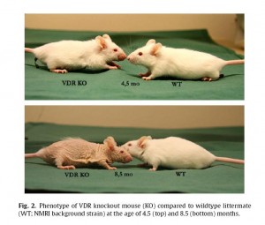
Other insights into inadequate 1,25D production have been obtained through mice deficient in vitamin D receptors. Their characteristics:

VDR mutant mice have growth retardation, osteoporosis, kyphosis, skin thickening and wrinkling, alopecia, ectopic calcification, progressive loss of hearing and balance as well as short lifespan. [6]

“Alopecia” is hair loss. “Kyphosis” is the familiar hunchback that many elderly develop. Osteoporosis is a familiar symptom of aging, as is loss of muscle, wrinkled skin, hardening of the arteries and stiffening of joints (“ectopic calcification”), loss of hearing and balance, and – approaching death.

These are all symptoms of a syndrome that is commonly called “aging.”

Here is what VDR knockout mice look like [7] (click to enlarge):

Note what happens when you can’t activate the VDR: hair loss, wrinkled skin. You get old before your time. VDR knockout mice die at an age of 10.6 months, compared to 20.5 months in wild-type mice. [7]

Our Cases Resemble PDDR

In his essay “Vitamin D Theory of Autism,” (http://www.vitamindcouncil.org/health/autism/vit-D-connection.shtml) Dr. Cannell notes similarities between PDDR and autism:

While no one has assessed afflicted [with PDDR] children for signs of autism, these children clearly display autistic markers such as hypotonia (flabby muscles), decreased activity, developmental motor delay, listlessness, and failure to thrive.

It is quite possible that autism results, as Dr. Cannell argues, from insufficient activation of the VDR during developmental ages. [8]

Similarly, what about the conditions Charles suffered from?  Tooth loss, bone mineral deficiencies, and scoliosis are all classic manifestations of rickets, and vitamin D deficiency is a known risk factor for high blood pressure and for arterial hardening. Finally, his recovery of hair color might be a result of restored vitamin D function:  the VDR promotes hair cycling. [9]

What Mechanisms Might Produce a CYP27B1 Deficiency in the Elderly?

It’s a safe bet that Charles does not have a genetic defect in CYP27B1. If he has a CYP27B1 dysfunction, it must have been acquired in old age.

What could have created the problem?  I don’t know, but speculation is permitted at PerfectHealthDiet.com. Two possibilities are:

  • Infection with a pathogen that interferes with CYP27B1. Pathogens have evolved ways to interfere with other human proteins in order to suppress the immune response. Since CYP27B1 creates 1,25D which enhances immunity, it would not be a surprise if some pathogen had evolved a way to interfere with CYP27B1.
  • Mitochondrial dysfunction. The enzyme coded by CYP27B1 operates in the inner mitochondrial membrane. Only in mitochondria can 1,25D be created. The “mitochondrial theory of aging” holds that mitochondrial decay is the primary cause of aging. Perhaps in elderly people suffering from mitochondrial dysfunction, CYP27B1 does not operate properly.

Conclusion

Whatever the mechanism of CYP27B1 loss-of-function may be, it appears that doubling 25OHD levels remedies much of the loss-of-function within a few weeks.

It might not be amiss for elderly patients and autistic children with symptoms of vitamin D deficiency to experiment with raising 25OHD to twice normal levels. In those with a CYP27B1 defect, this may produce an amazing recovery.

Further recovery might be possible. If the cause is infectious, appropriate antibiotics could help. If the cause is mitochondrial decay, then mitochondrial supplements might help.

The centrality of vitamin D function to optimal aging raises another thought. What if the main cause of aging is not the decay of mitochondria in general, but a specific decay in their support for 1,25D formation in the mitochondrial inner membrane? What if this loss of intracellular 1,25D is widespread among the elderly?

In that case, following Charles’s protocol and raising 25OHD in the elderly might significantly extend lifespans. And improve hair and skin at the same time!

Related Posts

“Vitamin D Dysregulation in Chronic Infectious Diseases,” https://perfecthealthdiet.com/?p=421, August 21, 2010.

References

[1] Heaney RP. Vitamin D in health and disease. Clin J Am Soc Nephrol. 2008 Sep;3(5):1535-41. http://pmid.us/18525006.

[2] Lou YR et al. 25-Hydroxyvitamin D(3) is an agonistic vitamin D receptor ligand. J Steroid Biochem Mol Biol. 2010 Feb 15;118(3):162-70. http://pmid.us/19944755.

[3] Rajasree S et al. Serum 25-hydroxyvitamin D3 levels are elevated in South Indian patients with ischemic heart disease. Eur J Epidemiol. 2001;17(6):567-71. http://pmid.us/11949730.

[4] Rowling MJ et al. High dietary vitamin D prevents hypocalcemia and osteomalacia in CYP27B1 knockout mice. J Nutr. 2007 Dec;137(12):2608-15. http://pmid.us/18029472.

[5] Takeda E et al. Vitamin D-dependent rickets type I and type II. Acta Paediatr Jpn. 1997 Aug;39(4):508-13. http://pmid.us/9316302.

[6] Tuohimaa P. Vitamin D and aging. J Steroid Biochem Mol Biol. 2009 Mar;114(1-2):78-84. http://pmid.us/19444937.

[7] Keisala et al. Premature aging in vitamin D receptor mutant mice. J Steroid Biochem Mol Biol. 2009 Jul;115(3-5):91-7. http://pmid.us/19500727.

[8] Cannell JJ. On the aetiology of autism. Acta Paediatr. 2010 Aug;99(8):1128-30. http://pmid.us/20491697. Cannell JJ. Autism and vitamin D. Med Hypotheses. 2008;70(4):750-9. http://pmid.us/17920208.

[9] Haussler MR et al. The nuclear vitamin D receptor controls the expression of genes encoding factors which feed the “Fountain of Youth” to mediate healthful aging. J Steroid Biochem Mol Biol. 2010 Jul;121(1-2):88-97. http://pmid.us/20227497.

NZ Man Left for Dead by Doctors, Cured by Vitamin C

Modern doctors are often deeply over-invested in the use of drugs, and amazingly ignorant of the power of the human immune system, when supported by a healthy diet and optimal nutrition, to defeat disease.

They sometimes exhaust their repertoire of drugs without ever considering using nutritional supplements to support the patient’s immune defense.

An extraordinary illustration comes from New Zealand. It began when Alan Smith, a New Zealand farmer, contracted swine flu:

He caught the Swine Flu (probably while on a fishing trip in Fiji), so badly that his lungs had “white out”, which is to say they were so full of fluid that they didn’t show up on an x-ray. The doctors also said he had got leukemia and he ended up being put on a life support machine.

The doctors told the family the machine should be turned off … [1]

The diagnosis of leukemia is suspicious. Both infections and leukemia lead to “leukocytosis” or a very high white blood cell count. In one case the white blood cells are multiplying to fight the infection, in the other a malignant population is multiplying. The difference is that in leukemia the population is monoclonal, i.e. all the new white blood cells are genetically identical, while in normal people with infections the white blood cells are created with genetic diversity. (Keywords for those who wish to investigate: T-cell antigen repertoire and B-cell immunoglobulin repertoire.)

As subsequent events showed, the leukemia “diagnosis” was mistaken. I wonder if it was made just for “family management” – in order to help persuade the family his case was hopeless and support the recommendation to end life support.

… but the family asked that he be given high dosages of Vitamin C. After a fight (one of many), one of the doctors agreed. Alan began getting better; his lungs showed pockets of air. Then he began to get worse and the family found out the doctors had stopped the Vit C.

Many more fights ensued, the patient getting better while having the Vit C, and getting worse when he was taken off. Alan’s wife describes one of the doctors sitting back in his chair, arms folded, rolling his eyes, looking at the ceiling, telling her that no way could the vitamin C be helping. The family hired a lawyer, forcing the doctors to continue the vit C treatment (albeit in slow dosage, until he got better enough to eat and his wife brought along sachets of large dosage herself for him to take).

Eventually Alan fully recovered, no trace of leukemia even. [1]

He should have been given high doses of vitamin D and iodine as well. Iodine supports leukocyte respiratory bursts of reactive oxygen species which destroy pathogens; vitamin C supports respiratory bursts by recycling glutathione and providing antioxidant protection for leukocytes against their own respiratory bursts, and also supports anti-viral immunity; vitamin D creates antimicrobial peptides that kill many pathogens.

Other possibly beneficial supplements in cases of elevated leukocyte counts due to infection: selenium, to support both glutathione and iodine/thyroid function; iron, for myeloperoxidase (respiratory burst enzyme) and catalase (antioxidant defense); N-acetyl cysteine (for glutathione production) and glutathione; zinc and copper (for the anti-oxidant zinc-copper superoxide dismutase).

Thank goodness the family had the sense to try vitamin C, and that that was enough for him to recover. It would have been a shame if he died for lack of vitamin D and iodine.

New Zealand was a pioneer of socialized medicine in the English-speaking world. Economists say that people respond to incentives; one wonders if the doctors were more motivated to tend to the interests of the bureaucrats who controlled their budgets, than to the health of the family and patient who weren’t paying them. Perhaps “free” medical care has unexpected costs.

References

[1] “Indictment of Our Medical Profession,” New Zealand Conservative, http://nzconservative.blogspot.com/2010/08/indictment-of-our-medical-profession.html; hat tip to Jewel at http://health.groups.yahoo.com/group/infection-cortisol/message/1760.

Retroviruses and Chronic Fatigue Syndrome

One of the themes of this blog is that chronic infections, exacerbated by bad diets and malnutrition, are at the root of nearly all health problems.

With the invention of new tools for microbiology over the last 20 years, scientists are for the first time able to study chronic parasitic infections, albeit with difficulty. I mentioned a few weeks ago that this should be the dawn of a “golden era of antimicrobial medicine.” And maybe it is: careful studies are now linking specific pathogens to chronic diseases and discovering the mechanisms by which they cause disease.

A good example of emerging science is the progress made since 2002 in understanding a retrovirus family that is now firmly linked to cancer and chronic fatigue syndrome and may soon be linked to other diseases.

Beginning of the Story:  Human Anti-Viral Immunity and Chronic Fatigue Syndrome

Our story begins back in the 1970s with studies of the role of interferons in defending human cells against viruses. Interferons are a key part of the immune defense against intracellular pathogens – the ones that cause most human chronic diseases.

Following the effects of interferons, researchers discovered an enzyme known as ribonuclease L (RNase L). RNase L is upregulated by interferons and its function is to degrade RNA, both viral and human, to stop viral replication. [1]

Aside: High levels of RNase L destroy so much human RNA that the cell dies. This is probably adaptive for the host, since cell apoptosis also kills many pathogens within. However, it shortens lifespan. RNase L knockout mice have extended lifespans. [1]

In 1997, RNase L was found to be strongly upregulated in chronic fatigue syndrome patients. [2] This showed that chronic fatigue patients usually have viral infections. Whether the viruses were causing chronic fatigue, or just “hitchhiking” with a disease that suppressed the immune system (perhaps via a bacterial infection?), remained an open question.

A Link Between RNase L and Prostate Cancer

By the early 2000s it was established that a common (allele frequency 35%) gene mutation, the “R462Q” mutation which substitutes a glutamine for an arginine in the “hereditary prostate cancer 1” locus, raised the risk of prostate cancer. A man with two copies of this mutation has twice the risk of prostate cancer; one copy raises the risk by 50%. About 13% of prostate cancer cases were attributable to this mutation. [1, 3, 4]

It was important, therefore, to determine which protein this locus coded for. A breakthrough finding, made in 2002, was that the “hereditary prostate cancer 1” locus was the gene for RNase L. [5]

It was soon shown that the R462Q mutation decreased the effectiveness of RNase L at cleaving viral RNA. This placed prostate cancer in a new light: it implied that an unknown virus against which RNase L defends was a probable cause of prostate cancer. When RNase L function was impaired by the R462Q mutation, the infection became more virulent, and prostate cancer rates were higher. [1]

The search for this unknown virus was on.

The discovery of “xenotropic murine leukemia virus-related virus” (XMRV)

The strategy was basically to take prostate tumors and search for viral RNA, looking for viruses that were most common in patients who had the double R462Q mutation.

In 2006 one of these searches yielded fruit.  A new gamma retrovirus was found in 8 of 20 prostate cancer patients with double R462Q mutations, but only 1 of 66 patients without the double mutation. [6]

This gamma retrovirus shared a lot of RNA with a family known as the xenotropic murine leukemia viruses (MuLVs). It was dubbed “xenotropic murine leukemia virus-related virus” (XMRV). Despite the sound, it is not a murine (mouse) leukemia virus; it merely shares a lot of nucleic acids with those viruses.

Back to chronic fatigue syndrome

In 2009 a paper was published in Science reporting that XMRV was found in peripheral blood cells of 67% of chronic fatigue patients but only 3.7% of healthy controls. [7] This study was done by a group at the Whittemore Peterson Institute in Reno, Nevada.

Aside:  The Whittemore Peterson Institute has a nice Q&A about this virus and its role in chronic fatigue syndrome here.

A number of researchers tried and failed to reproduce these results. For instance, a group from the Centers for Disease Control failed to detect XMRV proteins in 51 chronic fatigue and 53 healthy patients. [8]

Perhaps proteins are just not the right molecules for detecting this virus. A new paper has just appeared that links XMRV more strongly than ever to chronic fatigue. It looked at DNA for viral genes inserted into the human genome and found XMRV sequences in 86.5% of chronic fatigue patients but only 6.8% of controls. [9] This paper was held back from publication since June because of its conflict with the CDC paper (see “Why I Delayed XMRV Paper”), but has now been released.

These percentages are impressive and, if they hold up, would seem to make it unlikely that XMRV is merely a “passenger” virus hitchhiking on a suppressed immune system. It may be causal for chronic fatigue.

Will anti-retroviral therapies be effective?

Clinical trials are extremely expensive and the drug companies seem to be waiting for XMRV to be proven as the cause of chronic fatigue before undertaking trials. From the Wall Street Journal:

Norbert Bischofberger, chief scientific officer at Gilead Sciences Inc., the leading maker of HIV drugs, said the company might consider a small pilot trial but would like to see stronger evidence that the viruses cause CFS before launching a large trial. Still, “I’m very open, and this would be a great opportunity,” he said.

A spokesman for Merck & Co., another major manufacturer of HIV drugs, said: “A clinical trial program would be possible to develop only after further substantial evidence of an association with CFS.” [10]

But some aren’t waiting for trials. Anti-retroviral drugs developed for AIDS are being prescribed off-label:

Jamie Deckoff-Jones, 56 years old, a doctor and CFS patient in New Mexico, has been blogging about her experiences and those of her 20-year-old daughter. Both tested positive for XMRV and are taking a combination of three anti-retrovirals.

Dr. Deckoff-Jones said a year ago she could only get up for short periods during the day. After five months on the drugs, she flew last week to Reno for an XMRV conference. Her daughter was able to go to a party and is enrolling in community college. “This is all very new, and there is no way to know if improvement will continue,” Dr. Deckoff-Jones wrote in an email, “but we appear to be on an uphill course.” [10]

Chronic fatigue patients are celebrating the progress:

Many [CFS patients] were ecstatic at news that the second study was being published.

“We’re really hoping this will blow the lid off,” said Mary Schweitzer, a historian who has written and spoken about having the illness. “Patients are hopeful that now the disease itself might be treated seriously, that they’ll be treated seriously, and that there might be some solution.” [11]

It’s sad that for decades many haven’t taken chronic diseases seriously. The absence of a known cause reflected only the lack, until recently, of microbiological tools capable of detecting and characterizing intracellular pathogens.

Had doctors taken these diseases seriously, the accumulating evidence that these were chronic infectious diseases caused by intracellular parasites might have encouraged them to look for the sort of dietary and nutritional therapies for chronic disease that we advocate on this blog. Though diet and nutrition by themselves will probably not cure these diseases, they can greatly slow disease progression and improve the odds of a cure.

A new name for XMRV: Human Gamma Retrovirus

The Whittemore Peterson Institute recently hosted the first official scientific symposium on XMRV. Dr. Joseph J. Burrascano reported from the symposium:

We formed a working group to be in constant touch and we plan to meet regularly because advances are coming so rapidly.

Big news that everyone should know and adopt is that we have proposed a name change for the virus.

This virus is a human, not mouse virus, and it is the first and so far only gamma-retrovirus known to infect people. Also, it is clearly not an “endogenous” retrovirus (one that is present in all genomes due to ancient infection).

Because of all of this, and because of the desire to begin on the right track, the new name of the virus is HGRV- Human Gamma Retro Virus. The illness caused by this infection is named HGRAD- Human Gamma Retrovirus Associated Disease.

We plan to announce this at the upcoming NIH retroviral conference this September.

Definitely stay tuned- the volume of new and important information about this virus and its disease associations is increasing rapidly and in my opinion should be a concern to every patient with chronic neuro-immune diseases, including those with chronic Lyme. [12]

It sounds like some exciting findings may be on the way.

Conclusion

This case is a fascinating illustration of the twisting turns that scientific research can take. The early discovery of a link between anti-viral immunity and prostate cancer may now lead to a cure for chronic fatigue syndrome. At least, we can hope so.

As one of the pioneers, Dr. Robert Silverman, describes it,

One of the remarkable aspects of being a scientist, is that you never know where your scientific journey will lead. [1]

Science takes a lot of patience, diligence, and persistence. It’s gratifying when all that work is rewarded by discovery.

References

[1] Silverman RH. A scientific journey through the 2-5A/RNase L system. Cytokine Growth Factor Rev. 2007 Oct-Dec;18(5-6):381-8. http://pmid.us/17681844.

[2] Suhadolnik RJ et al. Biochemical evidence for a novel low molecular weight 2-5A-dependent RNase L in chronic fatigue syndrome. J Interferon Cytokine Res. 1997 Jul;17(7):377-85. http://pmid.us/9243369.

[3] Silverman RH. Implications for RNase L in prostate cancer biology. Biochemistry. 2003 Feb 25;42(7):1805-12. http://pmid.us/12590567.

[4] Casey G et al. RNASEL Arg462Gln variant is implicated in up to 13% of prostate cancer cases. Nat Genet. 2002 Dec;32(4):581-3. http://pmid.us/12415269.

[5] Carpten J et al. Germline mutations in the ribonuclease L gene in families showing linkage with HPC1. Nat Genet. 2002 Feb;30(2):181-4. http://pmid.us/11799394.

[6] Urisman A et al. Identification of a novel Gammaretrovirus in prostate tumors of patients homozygous for R462Q RNASEL variant. PLoS Pathog. 2006 Mar;2(3):e25. http://pmid.us/16609730.

[7] Lombardi VC et al. Detection of an infectious retrovirus, XMRV, in blood cells of patients with chronic fatigue syndrome. Science. 2009 Oct 23;326(5952):585-9. http://pmid.us/19815723.

[8] Switzer WM et al. Absence of evidence of xenotropic murine leukemia virus-related virus infection in persons with chronic fatigue syndrome and healthy controls in the United States. Retrovirology. 2010 Jul 1;7:57. http://pmid.us/20594299.

[9] Lo S et al. Detection of MLV-related virus gene sequences in blood of patients with chronic fatigue syndrome and healthy blood donors. PNAS Epub before print August 23, 2010. http://www.pnas.org/content/early/2010/08/16/1006901107.abstract.

[10] Amy Dockser Marcus, “New Hope in Chronic Fatigue Fight,” Wall Street Journal, Aug 23, 2010, http://online.wsj.com/article/SB10001424052748703846604575447744076968322.html.

[11] David Tuller, “Study Links Chronic Fatigue to Virus Class,” New York Times, Aug 23, 2010, http://www.nytimes.com/2010/08/24/health/research/24fatigue.html.

 [12] http://www.forums.aboutmecfs.org/showthread.php?7001-News-from-WPI-symposium-Name-change-to-be-proposed-for-XMRV (hat tip http://health.groups.yahoo.com/group/infection-cortisol/message/1753).

Vitamin D Dysregulation in Chronic Infectious Diseases

Commenter qualia recently got his serum 25-hydroxy vitamin D levels tested and found a surprising result: He had doubled his vitamin D intake from 5,000 IU to 10,000 IU per day, but his 25(OH)D levels didn’t budge – they were at 61 and 62 nmol/l, equivalent to 24.4 ng/ml in American units.

24 ng/ml is well below the optimal level for healthy people of 40 ng/ml. When a healthy person supplements vitamin D, the serum 25(OH)D level usually rises linearly with dose up to about 40 ng/ml, then it rises very slowly thereafter as the body tries to keep 25(OH)D from rising by putting vitamin D into storage.

So it was natural for qualia to expect his serum 25(OH)D level to rise when he doubled his dose.

But it didn’t. The stability of his 25(OH)D levels suggests that his body has reached an equilibrium at 24 ng/ml. Instead of plateauing at 40 ng/ml with abundant vitamin D3 as a healthy person would, he is plateauing at a lower level.

Why does that happen?

Well, I don’t know. But I would like to provide qualia with a little bit of background, because this could be a clue that helps diagnose his condition and optimize treatments.

Normal vitamin D regulation strictly controls 1,25D levels

I suggested in the comment thread that qualia get his 1,25D levels measured as well as 25(OH)D.

Levels of 1,25D are not routinely measured, even in scientific studies, because they rarely vary. Blood levels of 1,25D control calcium homeostasis and are tightly regulated. In healthy people, as vitamin D intake rises from zero, serum 1,25D levels shoot up to normal levels before 25(OH)D levels reach 12 ng/ml. They then stay in a normal range no matter how high 25(OH)D levels rise. The kidney is the primary controller of blood 1,25D levels. The normal range is about 16 to 42 pg/ml (42 to 110 pmol/L).

While all human cells can convert 25(OH)D to 1,25D, most cannot release 1,25D into the blood. 25(OH)D freely crosses cell membranes and maintains the same level throughout the body; but 1,25D does not cross membranes. This allows every cell in the body to “personalize” its 1,25D levels to its own needs.

Both 25(OH)D and 1,25D are active ligands for the Vitamin D Receptor (VDR), a nuclear receptor.  [1] When either 25(OH)D or 1,25D binds to the VDR, the compound is imported into the nucleus, where it combines with a vitamin A-activated Retinoid X-Receptor (RXR) to form a transcription factor which, among other roles, upregulates production of antimicrobial peptides (AMPs) that are crucial for cellular defense against intracellular pathogens.

The difference between 25(OH)D and 1,25D is that 1,25D is about fifty-fold more likely to bind to the VDR than 25(OH)D. So by converting more 25(OH)D to 1,25D, cells can upregulate their VDR activation and upregulate their immune defense against pathogens. Meanwhile, uninfected cells can keep their 1,25D levels low. Across human cells, there is a thousand-fold variation in the rate of conversion of 25(OH)D to 1,25D. [1]

In chronic infectious diseases, blood 1,25D is dysregulated

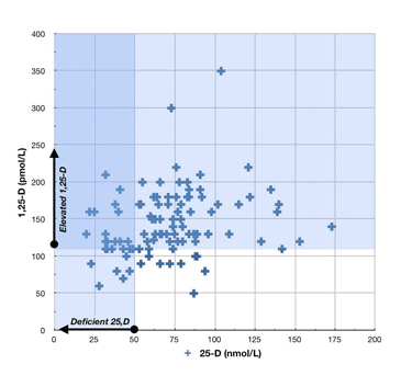
However, in people with chronic infectious diseases, 1,25D levels range all over the map, and are largely uncorrelated with 25(OH)D levels.  Here is a scatter plot from a paper by Dr. Greg Blaney [2]:

The patients in this sample were 100 chronic disease patients: 29 with fibromyalgia, 27 with chronic fatigue syndrome, 12 with post-treatment Lyme Disease, 9 with metabolic disease, 6 with osteoarthritis, 4 with irritable bowel syndrome, 4 with psoriatic arthritis, 3 with multiple sclerosis, 3 with seronegative arthritis, and 27 with other diseases.

Probably all of these diseases are caused by chronic parasitic infections.

A few things to note from this plot: (1) 25OHD levels in a lot of chronic disease patients cluster around the 61 nmol/L level that qualia has; and (2) most chronic disease patients have 1,25D levels well above the normal range, even though their 25OHD levels are mostly below the optimal level in healthy people.

What Causes 1,25D Dysregulation?

Here’s where we get into speculation. There just hasn’t been research exploring this question. Researchers are only just realizing that these diseases are infectious in origin and that vitamin-D-mediated innate immunity is critical to the intracellular immune defense.

Rather than speculate, I’m just going to mention a couple of possibilities.

First, in granulomatous diseases like sarcoidosis, it’s common to have low 25(OH)D and very high 1,25D. Granulomas are nodules where immune cells have been unable to eliminate some foreign matter and instead have built a barrier around it that walls it off from the body.  Granulomas often release 1,25D to the body. Some other granulomatous diseases: 

  • Tuberculosis
  • Leprosy
  • Schistosomiasis
  • Histoplasmosis
  • Cryptococcosis
  • Crohn’s disease

These are all infectious diseases, some of them protozoal in origin.

Second, nearly all human pathogens manufacture proteins or RNA that interfere with the innate immune response. Some are known to interfere with the VDR or with other aspects of vitamin D biology. (The HIV virus blocks the VDR entirely, one reason why it predisposes AIDS patients to infections.) It’s possible that vitamin D dysregulation is brought about by direct pathogen actions to disrupt cellular vitamin D pathways.

Conclusion

The only thing we can conclude with confidence from qualia’s vitamin D tests is that he must have a chronic infectious disease … but he knew that already.

Qualia would be best served by getting advice from an infectious disease specialist with experience in chronic diseases. Such a doctor might be able to narrow down the diagnosis. A diagnosis would help determine which antibiotics might be appropriate to help fight the infection.

Until a doctor’s diagnosis or qualia’s personal experience indicates otherwise, it’s probably prudent to continue with a reasonable intake of vitamin D and to increase iodine as quickly as possible. (Even this is not certain: the standard advice is to minimize vitamin D in granulomatous diseases.) Other infection-fighting supplements, like vitamin C, N-acetylcysteine, and glutathione are likely to be helpful also.

Finally, I always recommend that anyone with a chronic disease find a good discussion forum, like the one at http://cpnhelp.org, and try to find people with similar disease histories and learn from their experiences.

Best of luck, qualia, and please keep us posted.

References

[1] Lou YR et al. 25-Hydroxyvitamin D(3) is an agonistic vitamin D receptor ligand. J Steroid Biochem Mol Biol. 2010 Feb 15;118(3):162-70. http://pmid.us/19944755.

[2] Blaney GP et al. Vitamin D metabolites as clinical markers in autoimmune and chronic disease. Ann N Y Acad Sci. 2009 Sep;1173:384-90. http://pmid.us/19758177.