Category Archives: Cancer - Page 2

An Anti-Cancer Diet

Our cancer series resumes today with some tentative advice for cancer patients. (Note: This post is designed for solid tumor cancers, not blood cancers. However, most of the advice would also be applicable to blood cancers.)

This series began with Toward an Anti-Cancer Diet (Sep 15, 2011). There we advocated trying to shift cells away from the cancer phenotype via 8 anti-cancer strategies.

Future posts will explore in detail how to implement those strategies via diet and lifestyle. Today, I’m just going to give a general overview of what I would do if I had cancer.

Eat the Perfect Health Diet

This may sound self-serving, but it’s my best advice. Our diet is designed to optimize health generally, and that’s exactly what you want to do against cancer.

I said in the introduction that cancer is a disease in which cells lose their “humanness” – their proclivity to collaborate with other human cells to create a human organism. Instead, they lose recently evolved features and “remember” an identity similar to that of our distant evolutionary ancestors from the early days of multicellular life. This regression is possible because we retain the genes of our primitive evolutionary ancestors, and silencing of only a few hundred genes may cause a human cell to resemble, genetically, bacteria or fungi.

Many gut bacteria can take on two modes of behavior – a commensal or harmless phenotype, or a virulent harmful phenotype – depending on whether their environment is benign. In beneficial environments, bacteria tend to be cooperative with their host; in harsh environments, bacteria begin to look out for their own interests “selfishly,” and begin to display virulence traits which harm their host but help them move to a better environment.

Something similar may happen with “proto-cancer” cells. In a healthy environment, they are pleased to cooperate with their host – to retain their “humanness.” But in a harsh environment, they are more likely to withdraw from their neighbors and go their own way. An abused cell is more likely to become a cancer cell.

This may sound like anthropomorphization, but the metaphor is probably sound. Bruce Ames has remarked upon the fact that almost every compound is a carcinogen in large enough doses. Why? Because any unbalanced environment is harsh, and any harsh environment makes the cell more likely to develop the cancer phenotype.

It’s not only by discouraging “cancer virulence” that a good diet helps. A healthy diet also optimizes immune function.

Immune function is highly variable. Under stress, we suppress immunity so that all the body’s resources are available to meet “fight or flight” needs. Contrariwise, peaceable happiness is stimulating to immune function. A nutrient-rich diet, savory meals, happiness, calm, restful time spent in conversation – all of these things tell the body it has no pressing concerns and that available resources can be devoted to immunity and healing.

After cancer diagnosis, from a similar medical condition, those who are under stress tend to succumb to cancer, while those who are happy, cheerful, and sociable tend to recover from it. It is believed that this difference is primarily due to improved immune function in those under less stress.

I believe that a healthy, tasty diet is also a stimulant for immune function. Make your food nourishing and enjoyable.

Specific Dietary Aspects

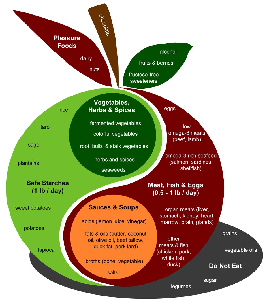
A few aspects of an anti-cancer diet deserve special mention. Let’s look at the PHD Food Plate:

Some aspects I would emphasize for cancer patients:

  • Safe starches. I recommend obtaining 400 to 600 glucose calories a day, mainly from safe starches. I believe it is important to avoid a glucose deficiency, since glycosylated proteins are the means of intercellular coordination, and defects in glycosylation are characteristic of the cancer phenotype. (See, eg, this paper.) You don’t want to aggravate this with a self-induced glucose deficiency.
  • Low omega-6 meats. Omega-6 fats can be very damaging to mitochondria and can promote metastasis. Our needs for them are minimal, and they are everywhere. It’s important to choose foods that minimize omega-6 levels. Among meats, prefer seafood, shellfish, and red meats; obtain eggs, milk, and organ meats from pastured and naturally raised animals. Eat tropical plant oils like coconut and palm.
  • Omega-3 and omega-6 balance. The diet should include some marine sources of omega-3 fats, like salmon or sardines.
  • Bone broth soups and gelatin (cooked collagen). Collagen is 30% of our body’s protein and forms much of the extracellular matrix scaffolding which is crucial to maintainance of tissue health. The extracellular matrix is broken down in cancer. An anti-cancer diet should be rich in cooked joint tissue, such as can be found in Ox Feet Broth soups. Vitamin C and sulfur, discussed below, are also required for collagen formation; be sure you’re not deficient in these.
  • Fermented vegetables, yogurt, and acids. A diverse portfolio of gut bacteria can be helpful to the fight against cancer by several mechanisms. Probiotic flora from fermented  foods help shield against the entry of cancer-promoting pathogens to the body through the gut; they generate by-products, like short-chain fats and vitamin K2, which have anti-cancer effects; and they can modulate immunity in a favorable direction. Acids such as vinegar and lemon juice can also favorably modify gut bacteria.
  • Vegetables, herbs, and spices.Fiber is probably beneficial against cancer. Butyrate, which is produced by gut bacteria from the digestion of many types of fiber including “resistant starch” from safe starches, has anti-cancer properties. Moreover, many vegetables and traditional herbs and spices have been shown to have anti-angiogenic effects. Foods with anti-angiogenic properties include:
    • Garlic.
    • Tomato.
    • Green tea.
    • Dark chocolate / cocoa.
    • Maitake mushroom.
    • Bok choy.
    • Kale.
    • Many berries.
    • Cherries.
    • Ginseng.
    • Turmeric.
    • Oregano.
    • Parsley.
    • Polyphenol-rich extra virgin olive oils.
  • Organ meats and egg yolks. It’s important to be well nourished, and organ meats like liver and egg yolks tend to be rich in micronutrients. They are much better than plant foods for compounds like phospholipids. In particular, choline (and its phospholipid form phosphatidylcholine) is important for methylation status and epigenetic functioning – an important element in cancer prevention.
  • Sea vegetables, sea salt, and seafoods. These are good sources of trace minerals such as iodine, which is a critical anti-cancer nutrient.

In general cancer patients should focus on the foods in the apple of the PHD Food Plate more than the “pleasure foods.” However, there’s nothing wrong with some berries, dark chocolate, pistachios, and whipped cream for dessert, and some red wine with dinner. Above all, it’s important to enjoy your food. Try to obtain from every meal a sense of pleasure and well being!

Supplements

Much more could be said on this topic than I’m going to say today. One could make a very long list of supplements that might help against cancer (also a long list of those that hurt). However, the crucial five from my point of view are in our recommended supplement list:

  • Vitamin D
  • Vitamin K2
  • Iodine
  • Selenium
  • Magnesium

The tricky one here is the iodine. Iodine dosage should be built up very slowly from a low level, so as not to disrupt thyroid function. (Hyperthyroidism can strongly promote cancer, and hypothyroidism can inhibit immune function and healing, so any thyroid dysfunction is a serious risk.) Start at 500 mcg or less, and increase the dose no faster than a doubling per month. If you get either hypothyroid or hyperthyroid symptoms from an increase in dose, back off a bit (eg instead of going directly from 500 mcg to 1 mg per day, go to 500 mcg and 1 mg on alternate days). Be patient, but try to build up to 12 mg/day over a 6 month period. Then stay there. Be sure to get 200 mcg/day selenium along with the iodine.

I also recommend a multivitamin, for general nourishment; and make sure there is no deficiency of vitamin C, zinc, copper, or chromium. Also, when it comes to antioxidants, more is not better. Avoid most antioxidant supplements other than glutathione, vitamin C, selenium, zinc, copper, and manganese.

For magnesium, I recommend taking a 200 mg oral supplement of magnesium citrate or a magnesium chelate. Epsom salt baths might not provide magnesium, but they can be a useful source of sulfur (in the form of sulfate) which assists collagen formation.

Vitamin C is an unusual case. It supports collagen formation, and for this purpose and to avoid a deficiency I strongly suggest taking 1 g per day. In higher doses, vitamin C may be helpful because it has anti-viral properties (see Fighting Viral Infections by Vitamin C at Bowel Tolerance, Sep 26, 2010), and most cancers are probably viral in origin. Linus Pauling, of course, advocated high doses of vitamin C – either taken orally to bowel tolerance, or intravenously. However, there are arguments on the other side. Vitamin C can protect cancer cells from immune attack, and also makes them resistant to chemotherapies. Clinical trials have not yet proven high-dose vitamin C therapy, but it may help against a subset of cancers caused by viruses sensitive to vitamin C therapy.

If sufficient amounts are not obtained from diet, then choline should be supplemented.

Intermittent Fasting, Intermittent Ketosis, Intermittent Protein Restriction

This is an extremely important cluster of strategies that are probably highly effective against cancer.

Their common trait is that all three promote autophagy, or “self-eating,” which is both a means for cells to cope with resource scarcity and a central part of the intracellular immune response.

When resources are abundant, cells allow aged organelles and junk proteins to accumulate. When resources are scarce, they turn on autophagy and digest unnecessary components, recycling the resources.

Autophagy is the dominant innate immune mechanism inside cells – the primary way cells kill bacteria and viruses.

Autophagy also recycles damaged mitochondria, which can be digested, enabling remaining healthy mitochondria to multiply. The result is a healthier mitochondrial population.

Since viruses and damaged mitochondria promote cancer, autophagy helps transform cells from the cancer phenotype back to the normal human phenotype.

Fasting, by inducing resource scarcity, promotes autophagy. Scarcity of amino acids, which can be achieved by a protein restricted diet, also promotes autophagy. And ketosis, which is part of the metabolic profile of starvation, also promotes autophagy.

Note in my section heading the shared word: “intermittent.” We don’t want to sustain fasts or protein scarcity too long; that could create malnourishment and cause more harm than good. Permanent ketosis may promote fungal infections. The most helpful course is probably to follow these strategies intermittently:

  • Engage in daily intermittent fasting: eat only within a 6 to 8 hour window each day. Within the fasting period, eat some coconut oil or MCT oil to promote ketosis.
  • Eat high protein for a few weeks while engaging in resistance exercise to build muscle; then low protein for a few weeks.

A Note on Ketogenic Diets

Since we wrote our book, we’ve become a bit less excited about the therapeutic potential of ketogenic diets.

Ketogenic diets have demonstrated effectiveness in brain cancers, and several considerations suggest that they would be helpful against all cancers:

  • Cancer cells are dependent on glucose metabolism, a phenomenon called the Warburg effect. In ketosis, blood glucose levels can be decreased – a fall from 90 to 65 mg/dl is achievable – and reduced glucose availability should retard cancer growth.
  • Mitochondria do well on ketones, and some studies had shown that provision of ketones can restore the ability of mitochondria to trigger apoptosis, or the programmed cell death of cancer cells.

It’s too early to judge, but a few scraps of data published recently have made ketogenic diets seem a bit less exciting then hoped.

First, the group of Michael Lisanti has published work suggesting that tumors can evade the metabolic restrictions of a ketogenic diet by manipulating neighboring normal cells. The idea (here is an overview) is that cancer cells release hydrogen peroxide, which causes a stress response in neighboring cells, stimulating them to release lactic acid, which the cancer cells can metabolize. This process can happen nearly as well on a ketogenic as on a normal diet, so the effectiveness of a ketogenic diet in starving the cancer cells is reduced.

The Lisanti group results are hardly conclusive – indeed so far as I know no other group has supported their claims – and there are plenty of skeptics. Jimmy Moore gathered responses from a panel of low-carb experts.

Second, clinical experience with ketogenic diets has not yet shown them to be highly effective. The sort of data we have is well represented by a recent report in Nutrition and Metabolism. Sixteen patients with advanced metastatic cancer were put on ketogenic diets. The results:

One patient did not tolerate the diet and dropped out within 3 days. Among those who tolerated the diet, two patients died early, one stopped after 2 weeks due to personal reasons, one felt unable to stick to the diet after 4 weeks, one stopped after 6 and two stopped after 7 and 8 weeks due to progress of the disease, one had to discontinue after 6 weeks to resume chemotherapy and five completed the 3 month intervention period.

The conclusion: a ketogenic diet “has no severe side effects and might improve aspects of quality of life and blood parameters in some patients.”

Clinical trials with control groups and more statistical power are needed to evaluate whether ketogenic diets have therapeutic effect. For now, I think the most prudent course is intermittent ketosis and intermittent ketogenic fasting, rather than a continuously ketogenic diet.

UPDATE: Mario makes a great point in the comments: fasting prior to chemotherapy reduces toxicity to normal cells but increases toxicity to cancer cells. It is quite likely that a ketogenic diet might have the same effect during chemotherapy. So the combination of intermittent ketogenic dieting with chemotherapy should be given consideration.

Circadian Rhythm Enhancement

Many diseases become more likely, or more severe, if circadian rhythms are disrupted. Enhancement of circadian rhythms may be therapeutic for these diseases.

I’ve blogged about circadian rhythm therapies for hypothyroidism (“Intermittent Fasting as a Therapy for Hypothyroidism,” Dec 1, 2010) and for sleep disorders, psychiatric disorders, neurodegenerative disorders, and obesity (“Seth Roberts and Circadian Therapy,” Mar 22, 2011).

Well, cancer is another disease for which circadian disruption may be damaging. The International Agency on Research on Cancer (IARC) has recently classified “shiftwork that involves circadian disruption” as “probably carcinogenic to humans.”

It’s plausible that circadian enhancement may be therapeutic for cancer. Tactics that enhance circadian rhythms include:

  • Exposure to mid-day sunlight.
  • Sleeping in total darkness during hours of darkness.
  • Confining eating to daylight hours.
  • Socializing – especially, looking at faces and talking – during daylight hours. Seth Roberts found that looking at images of human faces can substitute for actual socializing.
  • Exercising during daylight hours. Even low-level activity – like standing instead of sitting – helps.
  • In people who are melatonin deficient due to a brain immune response, supplementation of melatonin just before bedtime.

Curiously, circadian rhythm disruption seems to make chemotherapy more effective. Also, timing treatments to match circadian rhythms may double their effectiveness.

Exercise and Other Lifestyle Factors

A number of lifestyle factors are important for cancer recovery. David Servan-Schreiber’s Anti-Cancer has an excellent overview of the evidence.

A recent study in the Lancet found that every additional 15 min of daily exercise beyond 15 min a day reduced all-cancer mortality by 1%. Exercise appears to be therapeutic even for late stage cancers. A meta-review found that two and a half hours of exercise a week could lower a breast cancer patient’s risk of dying or cancer recurrence by 40 percent, and could reduce a prostate cancer patient’s risk of dying from the disease by about 30 percent.

However, exercise should not be exhausting. Rather, it should be restful and relaxing; or build muscle. Resistance exercise on the “Body by Science” model of one intense workout per week, with more time spent in restful recovery than in stress, is probably a good strategy. Long walks outdoors in nature, and relaxing exercises like yoga or tai chi, are also great approaches to cancer therapy.

Being sociable, happy, calm, and optimistic are all important factors for cancer recovery. Those who have companions they love, and a purpose for living that makes them happy, have the best prognosis. Be grateful for what you have, and make your body understand that life is worth living.

Dealing with Anorexia and Nausea

Anorexia and nausea can seriously impair the ability of cancer patients to eat a nourishing diet and maintain their strength.

I haven’t had time to research this aspect of the disease yet, but there do seem to be some dietary and lifestyle interventions that help.

For instance, exercise can correct anorexia.

Among dietary interventions, ginger has been reported to reduce chemotherapy-induced nausea, reducing incidence in one study from 93% to 55%. (Hat tip: Healthy Fellow.)

Ginger teas are a traditional Asian folk remedy. Slice some ginger root in water, boil it on the stove, add some rice syrup for sweetness, and drink up!

Under-Utilized Therapies

There are a few therapies which are rarely prescribed, but might be more helpful than chemotherapies in treating cancer:

  • Low-dose naltrexone.
  • Anti-viral drugs.
  • Anti-fungal therapies.

Low-dose naltrexone is taken at night before bed. It temporarily blocks opioid receptors, which leads the body to increase production of endorphins and enkephalins – immune compounds which interact with opioid receptors. The following day, the naltrexone is gone and the opioid receptors are working again, but the endorphins are still around. Taking LDN thus increases endorphin levels. Endorphins inhibit cancer proliferation, and may enhance anti-cancer immunity. Here is a recent paper on anti-proliferative effects of LDN against ovarian cancer: http://pmid.us/21685240. Here is a recent paper on LDN plus alpha lipoic acid as a therapy against pancreatic cancer: http://pmid.us/20042414. For a general overview, see http://lowdosenaltrexone.org/.

Viruses cause or contribute to most cancers, and thus anti-viral drugs have great potential. A few cancer-causing viruses are famous, such as the Human Papilloma Virus for which there is a vaccine; however, most of the viruses that cause cancer remain unknown, though we know they exist because genetic mutations that impair viral immunity greatly increase cancer incidence.

Mario Renato Iwakura recently sent me a link to a paper that nicely illustrates the potential of antiviral therapies against cancer. Cytomegalovirus, also known as human herpes virus 5, is a common virus that infects 40% of adults worldwide and 50% to 80% of Americans. However, it is found in almost 100% of human tumors. It seems to be difficult to get cancer if you haven’t been infected by cytomegalovirus.

From the paper abstract:

Medulloblastomas are the most common malignant brain tumors in children…. Human cytomegalovirus (HCMV) is prevalent in the human population and encodes proteins that provide immune evasion strategies and promote oncogenic transformation and oncomodulation…. Remarkably, all of the human medulloblastoma cell lines that we analyzed contained HCMV DNA and RNA and expressed HCMV proteins at various levels in vitro. When engrafted into immunocompromised mice, human medulloblastoma cells induced expression of HCMV proteins. HCMV and COX-2 expression correlated in primary tumors, cell lines, and medulloblastoma xenografts. The antiviral drug valganciclovir and the specific COX-2 inhibitor celecoxib prevented HCMV replication in vitro and inhibited PGE2 production and reduced medulloblastoma tumor cell growth both in vitro and in vivo.

Tumor growth declined by 72% when treated with Valcyte (valganciclovir) and an NSAID drug. A press release notes that these drugs have “relatively good adverse effect profiles” and that “antiviral drugs are selective and largely affect infected cells.”

Yet another antimicrobial approach that may be helpful against cancer is antifungal therapy. Most cancer patients develop systemic fungal infections, and fungal infections such as Candida promote metastasis and tumor growth, and may also suppress anti-cancer immunity. An effective antifungal therapy may significantly retard cancer progression.

Conclusion

Much more remains to be said, and it’s certain that we’ll refine these suggestions after more thoroughly studying the literature. But I think this basic approach to an anti-cancer diet can’t be too far wrong.

Our prayers and best wishes go out to all those who are battling cancer.

Toward an Anti-Cancer Diet

Since starting the blog, I’ve gotten a number of emails from cancer patients or their family members. When the Q&A page started last week, the second question was from Lindsay, asking for a cancer diet:

In the past 3 weeks my partner has been diagnosed with stage 3 breast cancer. She is 28 and there is no family history….

Do you have suggestions aside from vit d and green tea that might be therapeutic?…

In my mind she simply needs to be extremely well nourished, but that is proving tricky due to nausea. I made a batch of chicken broth the other day and the smell alone sidelined her. Any thoughts on a way to sneak in dense nutrients without a strong odor?

I’ve delayed blogging about cancer and diet because of the complexity of the issue. Research has not yet determined the optimal diet for cancer patients, and there is reason to believe that the optimal diet may differ for different cancer patients. It is not easy to balance the many factors that should influence a cancer patient’s diet.

Today I’ll lay out my general perspective on cancer. The goal is to identify aspects of the disease that we can influence through diet. In subsequent posts, I’ll discuss foods, nutrients, and eating strategies. I hope the manner in which I’m addressing the issue will help cancer patients to understand the issues better and to design an effective personal eating strategy.

Cancer as a Progression of Diseases

Cancers often develop over long periods of time – typically decades. They usually cannot be detected or diagnosed at early stages. This is just as well, because most early cancers resolve spontaneously; they disappear or return to a normal state. Perhaps we should have a distinct name for these early and usually harmless cancers – “proto-cancers” perhaps.

Proto-cancers develop through a series of stages into life-threatening cancers. At each stage, the character of the disease changes. The purpose, and perhaps the nature, of dietary interventions may change with it.

Origins of Cancer

What characterizes these proto-cancers is that one or more cells develop an abnormal state of gene expression that I’ll call the cancer phenotype.

What causes a cell to develop the cancer phenotype? I believe the most common are:

  1. Infections, especially viral infections (since viruses are good at modifying gene expression).
  2. Toxins, especially DNA-modifying toxins such as those generated by peroxidation of polyunsaturated fats.
  3. Malnutrition, especially nutrient deficiencies that impair the ability to maintain epigenetic regulation of DNA.

The Wikipedia page “Infectious Causes of Cancer” says that 18% of human cancers are known to have infectious causes, but I suspect the fraction will get much larger. Read through our story of XMRV and chronic fatigue syndrome (“Retroviruses and Chronic Fatigue Syndrome, Aug 24, 2010) for an example of how difficult it is to identify the viruses that cause cancers. In this instance, a new human retrovirus may (it is still disputed) have been discovered because men with genetic impairments to anti-viral immunity have much higher rates of prostate cancer, and scientists searched prostate tumors of men with this genetic impairment for viruses. If finding the cancer-causing virus is so difficult when we know it is present in the tested tissue, you can easily imagine how many other viruses may have escaped scientific notice.

Interventions to prevent the original causes of cancer are great for cancer prevention, but they may also be therapeutic. Removing cancer-causing viruses may enable the body to defeat a cancer it otherwise could not.

Proto-Cancers and the Evolution of Cancer Cells

The cancer phenotype has various characteristics, but at early stages important characteristics may include:

  • Suppression of mitochondrial activity, especially apoptosis (programmed cell death).
  • Metabolic changes toward metabolism of glucose and away from metabolism of fatty acids or ketones.
  • Isolation of the cell from the rest of the body. Normal human cells closely coordinate their activities with the rest of the body, especially with neighboring cells, and respond to hormonal and other signals. Cancer cells tend to be more “individualistic,” less responsive to the body and to their neighbors.

Every once in a while one of these cells with a cancer phenotype will divide, creating two daughter cells. Perhaps in part because after metabolic impairment the cell has difficulty providing sufficient ATP to handle the complex motor tasks involved in cell division, these cancer cells often fail to divide properly, resulting in daughter cells with altered genetic state. Common changes include:

  • Aneuploidy. Most genes have two copies, one inherited from the mother and one from the father. In cell division, genes are first duplicated, and then divided among two daughter cells, so that each cell gets two copies. In aneuploidy, chromosomes are improperly separated so that one cell gets three copies of a chromosomal strand, the other one. Genes on that strand then become expressed 1.5-fold in the cell with three copies, half-fold in the cell with one copy.
  • Translocations. Chromosomes have a certain gene order. However, they can be re-assembled in an improper order, with one segment translocated to another place. This changes gene expression.
  • Epigenetic modifications. DNA comes with a protein scaffold that packages and organizes it, and can be modified so that gene expression is silenced (via methylation) or enhanced (via acetylation). Epigenetic modifications are usually inherited by daughter cells – but dividing cancer cells may experience less stability in the epigenome.

These changes mean that cell division causes cells with a cancer phenotype to evolve. Let’s say aneuploidy occurs affecting a gene that stimulates cell division. One daughter cell gets 3 copies and becomes more likely to proliferate; the other cell gets only 1 copy and becomes less likely to proliferate. Of course, every time a cell divides it creates two daughters, so a decade later the first cell may have thousands of descendants while the first cell has few. A proliferative phenotype has become more common in the “tumor.”

The Middle Stages of Cancer Development

As a result of cellular evolution, the early cancer phenotype becomes a later cancer phenotype with new traits, such as:

  • A tendency to proliferate. This is the trait people most commonly associate with cancer cells.
  • A tendency to stay alive indefinitely. Some cancer cells become “immortal.” For instance, HeLa cells are immortal cells taken from the cervical cancer of Henrietta Lacks, who died in 1951. So resistant to death were these cells, they were the first human cells ever to survive in vitro.

Both traits are promoted by infections. Viruses and other germs want to stay alive, and to do that they need to keep their host cell alive, since cell death typically kills any germs in the cell (thus programmed cell death is a major part of human immune defense). Viruses also want to replicate, and to do that they often piggyback on human DNA replication. So viruses have evolved ways to promote proliferation and immortality of host cells. Cancers caused by viruses, therefore, often have a head start on acquiring these traits. Cancers that appear at young ages are probably almost always viral in origin.

Once these traits are acquired, the cancer cells proliferate and form a tiny tumor. These micro-tumors can reach a size of about 0.5 mm in diameter. At that point, growth stalls for lack of oxygen and nutrients. Cells inside the tumor cannot get enough resources to continue their growth.

Often this is the end of the cancer; it never develops beyond this point. It’s been estimated that most adults have thousands of these microtumors, and most never go on to develop clinical cancer. It is generally impossible to diagnose the presence of these microtumors.

However, cells in the microtumor are still evolving. Cells, when nutrients are available, divide, and one daughter cell survives to divide again while another cell in the tumor dies to make room. There is a “survival of the fittest” contest in which cells become more adapted to the environment of the tumor.

Progression to Diagnosable Disease

At some point, one or more cells may gain the ability to manipulate neighboring cells to their own benefit. This is a crucial stage in the development of cancer: when the cancer phenotype extends to give the tumor new abilities to exploit its human host. A key capability is:

  • the ability to induce the formation of blood vessels. This process is called angiogenesis.

This process involves manipulation of the immune system, which is responsible for wound healing as well as defense against foreign bodies. Angiogenesis is part of the normal wound healing process, and when it becomes angiogenic, a tumor (in the famous phrase of Hal Dvorak) becomes a “wound that never heals.” That is, it acts like a wound to call forth the blood vessel generation process, but it never allows the wound healing process to terminate.

Once cancer cells can call forth new blood vessels from surrounding tissue, they have access to all the body’s nutrient and oxygen resources. There is no longer any limit to the tumor’s growth. This evolution of an angiogenic capability could be said to create the disease of cancer.

One of the interesting findings of recent research is that foods can significantly influence the likelihood that tumors will develop an angiogenic capability. A number of plant compounds from foods such as garlic, tomato, green tea, and turmeric have been shown to inhibit angiogenesis. If you read the excellent book Anti-Cancer by David Servan-Schreiber, you’ll find that these anti-angiogenic foods form the essence of his dietary advice.

Once tumors can induce angiogenesis, they can grow quite large. But even large single tumors are usually not deadly.

Progression to Deadly Disease

Cancers become deadly when another change evolves:

  • Some cancer cells become metastatic.

Metastatic cells migrate away from the tumor, interact with non-cancer cells, and may travel through the blood to distant sites where they establish new tumors. When cancer metastasizes, many tumors can develop and the cancer can become a devastating drain on the host.

Immune Suppression and Co-Infections

Other new cancer capabilities may also evolve. For instance:

  • Suppression of anti-cancer immunity.

When the immune system successfully attacks and destroys cancers, it is usually through an innate immune response involving natural killer cells and macrophages. Interestingly, this is also the same immune response which defends against fungal infections.

One of the interesting aspects of the evolution of cancer cells is that they often end up with many genes silenced, such that they lose many distinctively “human” genes and probably come to resemble our primitive evolutionary ancestors. In other words, cancer cells evolve to look more like fungal cells, so that a tumor may biologically resemble a mold colony.

Suppose cancer cells evolve a capability to suppress the NK cell and macrophage immune response. Then the tumor will flourish more readily – but so also will fungal infections.

It happens that late-stage cancer patients commonly develop systemic fungal infections.

It also works the other way: fungi that have evolved into obligate parasites of human hosts, like Candida, are good at suppressing human anti-fungal immunity. In doing so they also suppress human anti-cancer immunity. Thus, fungal infections are a risk factor for cancer. I saw a study recently in which a large fraction of people treated for systemic fungal infections were diagnosed with cancer in the following five years.

There is evidence that fungal infections of cancer cells increase the rate of metastasis. So the combination of cancer and fungal infections may be particularly deadly. This suggests that cancer patients might benefit from anti-fungal therapies.

There may be great variability in human immunity against cancer. Biologist Zheng Cui has found such variability in mice, and estimates that 10-15% of humans may be highly resistant to cancer. It is likely that diet can modulate this resistance, which suggests looking for dietary tactics that promote anti-cancer immunity.

Cachexia and Anorexia

Cachexia is the wasting syndrome that afflicts late-stage cancer patients. The tumors become a large drain on resources, their nutrient consumption is not met by diet, and the rest of the body is cannibalized in order to provide resources to the tumors. Muscle and other tissue wastes away until they can no longer sustain life.

Cachexia is often what kills cancer patients.

Now, if the cancer patient could eat sufficient food, even very large tumor burdens might be tolerable. Olympic swimmers eat 12,000 calories a day; pregnant women support 8 pound growths in their abdomen without risk.

Unfortunately, cancer also tends to diminish appetite. One of the consequences of cancer’s interactions with the immune system is that late-stage cancer generates a lot of inflammatory cytokines which can be imported into the brain where they affect the food regulatory systems that Stephan Guyenet has written about, causing anorexia.

Cancer-related anorexia makes food distasteful and causes cancer patients to cease eating. Lindsay mentioned her partner’s anorexia as one of the problems she hoped I could help her address.

Exercise and dietary strategies that promote muscle and tissue growth (“anabolic” strategies) such as those employed by bodybuilders and strength athletes might increase appetite, protect tissue, and delay the negative effects of cachexia. They might also have an anti-cancer effect by depriving the cancer of resources.

Interaction with Chemotherapies

Yet another complexity is that the standard therapies for cancer involve poisoning the body with chemotoxins.

This raises a conundrum. A healthy diet makes the body, and all its cells including cancer cells, more resilient to toxins. So a healthy diet may undermine the effectiveness of chemotherapies.

Some diet-chemotherapy interactions are well documented. Supplementation of vitamin C, glutathione, and omega-3 fats are all known to protect cancer cells against chemotherapies.

If beneficial foods reduce the effectiveness of chemotherapy, it might also be the case that toxic foods could increase their effectiveness. Thus, the optimal diet during chemotherapy might be quite different from the optimal diet when off chemotherapy.

I will not say much about these interactions, other than to advise that before undergoing chemotherapy cancer patients discuss their diet and supplement regimen with the oncologist.

Summary: Our Path to an Anti-Cancer Diet

So, we’ve identified a number of possible levers for attacking cancer. We can look for dietary steps to:

1)      Defeat viral or other infections that originally caused the cancer.

2)      Remove toxins and improve nutrition in order to promote DNA and epigenome stability.

3)      Deprive cancer cells of their favored glycolytic metabolic pathways, slowing their growth.

4)      Restore mitochondrial function, promoting apoptosis (programmed cell death) of cancer cells.

5)      Inhibit angiogenesis.

6)      Inhibit metastasis.

7)      Promote anti-cancer and anti-fungal immunity.

8)      Mitigate anorexia and cachexia.

Those who are trying to prevent cancer will want to focus on (1)-(5); those with early stage cancers on (1)-(7); those with late stage cancers on (1)-(8).

Our mission: understand how diet and nutrition can affect each of these; and then try to integrate various dietary tactics into an optimal anti-cancer strategy.

Conclusion

I think this gives us plenty to work on. Next week, I’ll provide short provisional answers. Over the next year, I’ll examine each type of cancer-diet interaction in detail and see if we can refine and improve our anti-cancer strategy.

Can Endurance Exercise Promote Cancer?

I got into a bit of trouble in the comments a few weeks back when I joked that Grete Waitz may have died from marathoning. Steve replied:

Paul, you said “… marathoning (from which Grete Waitz just died at 57)”

Gee. The news said cancer. How confident are you that she died “from” running marathons?

Of course, not confident at all. Maybe if she’d been a sprinter she would have died at 54. Maybe if Lance Armstrong had been a couch potato he would still have had testicular cancer metastasized to his brain and lung at age 25.

A few days ago I got an email. Two highly fit endurance athletes, both of whom have always tended to their health and been careful to eat “healthy” (i.e. vegetable and whole grain rich, meat and fat poor) diets, have contracted cancers in the prime of life and been given less than a year to live. My correspondent asked, “Why?”

Let’s look into this. Is it possible that endurance exercise, especially if combined with a high-carb diet, may promote cancer?

Oxidative Damage to DNA and Cancer

Human DNA is constantly being damaged and repaired. It’s been estimated that over the course of a cell cycle – that is, from the time a cell is formed to the time it divides into two daughter cells – a human cell develops 5,000 single-stranded DNA breaks due to oxidative damage from reactive oxygen species (ROS). The vast majority are repaired by the body’s DNA repair machinery. [1]

However, in typical human cells 0.1% or 5 are not successfully repaired; instead a corresponding break is created in the complementary DNA strand, resulting in a double-strand break. In people with Bloom syndrome, an inherited condition which creates a strong predisposition to cancer, fully 1% or 50 are not successfully repaired. [1]

The double-strand break leads to a re-arrangement or “translocation” of parts of the chromosome. Usually, this does not break the coding region for a protein, but it does break non-coding regions resulting in changes to gene expression.

These sorts of genetic changes are observed both in cancer and in aging. [1] In short, oxidative damage to DNA is considered a risk factor for cancer development.

Oxidative Damage to DNA Has Been Specifically Linked to Endurance Exercise

Diets and activities that increase oxidative stress – for instance, diets deficient in antioxidant minerals – can therefore increase cancer risk. And diets and activities that minimize oxidative stress can minimize cancer risk and facilitate recovery.

Endurance exercise generates oxidative stress. Marathon running “caused a large increase in the tissue content of oxidized glutathione (189%) at the expense of reduced glutathione (-18%).” [2]

Moreover, endurance exercise damages DNA:

Both a systemic inflammatory response as well as DNA damage has been observed following exhaustive endurance exercise….

Extremely demanding endurance exercise has been shown to induce both a systemic inflammatory response [15, 42, 53, 71] as well as DNA damage [21, 36, 58, 62, 80]….

Exercise-induced DNA damage in peripheral blood cells appear to be mainly a consequence of an increased production of reactive oxygen and nitrogen species (RONS) during and after vigorous aerobic exercise [58]. Besides oxidative stress, other factors such as metabolic, hormonal and thermal stress in addition to the ultra-structural damage of muscle tissue are characteristic responses to prolonged strenuous exercise, that can lead to the release of cytokines, acute phase proteins and to the activation or inhibition of certain lines of the cellular immune system [15, 29]. [3]

There seems to be a big difference between moderate exercise and exercise to exhaustion. Moderate exercise actually protects DNA by upregulating DNA repair:

Sato et al. showed that acute mild exercise as well as chronic moderate training does not result in DNA damage, but rather leads to an elevation in the sanitization system of DNA damage [66]. [3]

However, endurance exercise leads to increased DNA damage:

Increased levels of DNA strand breaks were observed after exhaustive treadmill running in subjects of different training status [22, 45]….

In conclusion, there is growing evidence that strenuous exercise can lead to DNA damage that with few exceptions [36] is predominantly observed not before 24 h after the resolution of exercise [21, 44, 45, 80]. [3]

In addition, strenuous endurance exercise induces hormonal and other changes which might promote cancer. An Ironman triathlon has significant effects on hormones and inflammatory markers, some of which persist for more than 19 days post-race:

Briefly, as described in details elsewhere [42], there were significant (P<0.001) increases in total leukocyte counts, MPO, PMN elastase, cortisol, CK activity, myoglobin, IL-6, IL-10 and hs-CRP, whereas testosterone significantly (P<0.001) decreased compared to pre-race. Except for cortisol, which decreased below pre-race values (P<0.001), these alterations persisted 1 d post-race (P<0.001, P<0.01 for IL-10). Five days post-race CK activity, myoglobin, IL-6 and hs-CRP had decreased, but were still significantly (P<0.001) elevated. Nineteen days post-race most parameters had returned to pre-race values, with the exception of MPO and PMN elastase, which had both significantly (P<0.001) decreased below pre-race concentrations, and myoglobin and hs-CRP, which were slightly, but significantly higher than pre-race [42]. [3]

In the opinion of the authors of this review, the biggest problem is production of reactive oxygen and nitrogen species (RONS) by damaged immune cells:

The most conclusive picture that emerges from the available data is that oxidative stress seems to be the main link between exercise-induced inflammation and DNA damage…. DNA damage in peripheral immuno-competent cells, indeed, most likely resulted from an increased generation of RONS due to initial systemic inflammatory responses or the delayed inflammatory processes in response to muscle damage (Fig. 1). [3]

What About High-Carb Diets?

Do high-carb diets contribute?

During strenuous exercise mitochondria produce oxidation products:

The mitochondrial electron transport system can trigger the formation of superoxide leading to increased production of H2O2 by superoxide dismutase [49], [50]. [4]

In a normal person at rest, about 1-2% of the oxygen utilized by mitochondria ends up in superoxide. [4]

Before we go further let’s take a brief detour into mitochondrial chemistry: specifically, something called the electron transport chain.

Here’s a stylized view:

Source: Wikipedia.

The main point for our purposes is that there are two points of entry into the chain, one that goes through complex I and one that bypasses it.

Glucose metabolism favors entry via complex I, while fatty acid metabolism is relatively more favorable to entry via complex II. Quantitatively, glucose metabolism produces 5 NADH molecules (entering at complex I) for every one succinate molecule (entering at complex II), while fatty acid metabolism produces only 2 NADH for every one succinate.

High-carb dieting tends to habituate the body to metabolism of glucose. Therefore, it increases utilization of complex I.

This is significant because complex I is vulnerable to production of excess oxidative stress under some circumstances.

In principle, every mitochondrial complex has the potential to operate cleanly with minimal production of superoxide. However, if mitochondrial function is in any way impaired, so that operation of a complex is inhibited, then ROS production can rise substantially.

If for some reason electrons cannot flow properly through the electron transport chain, then they leave as superoxide:

One factor which may sensitise cells to increased DNA damage is impaired mitochondrial function [74]…. Reduced electron flow through the mitochondrial respiratory chain, particularly through the inhibition of complex I or complex III, favours the enhanced production of superoxide and H2O2 [75]. Together, with the age-dependent increase in oxidative stress and decline in NAD+ and ATP content, we found a tendency to the reduction in the activity of the respiratory complexes with age in all organs. Sipos et al. (2003) showed that mitochondrial formation of H2O2 due to complex I inhibition is more clinically relevant than ROS production due to inhibition of complex III and IV in situ [76]. [4]

What exactly did Sipos et al. find?  They state:

ROS formation was not detected until complex III was inhibited by up to 71 +/- 4%, above that threshold inhibition, decrease in aconitase activity indicated an enhanced ROS generation. Similarly, threshold inhibition of complex IV caused an accelerated ROS production. By contrast, inactivation of complex I to a small extent (16 +/- 2%) resulted in a significant increase in ROS formation, and no clear threshold inhibition could be determined. [5]

Basically, superoxide can be generated in complexes I, III, and IV. However, in complexes III and IV, there is a high threshold of inhibition of electron transport before any superoxide is produced. In complex I, there is no threshold:  even very slight inhibition will generate ROS. This means that during practical living, the great majority of excess ROS is produced from complex I.

This means that high-carb dieting, which increases utilization of complex I, will tend to generate oxidative stress if there is any inhibition of complex I.

But in endurance exercise, there is inhibition of complex I. To name just one pathway, exercise increases levels of the hormone DHEA, and DHEA inhibits complex I. [6]

It looks like high-carb diets and endurance exercise may be a bad combination.

Are Whole Grains Especially Bad?

There may be specific problems with grain toxins. For instance, wheat germ agglutinin, a wheat toxin that is very effective at distributing itself through the body through transcytosis, is able to damage mitochondria:

WGA induced a loss of transmembrane potential, disruption of the inner mitochondria membrane, and release of cytochrome c and caspase-9 activation after 30 min of cell interaction. [7]

At high doses in test tubes this can lead to cell death. It’s conceivable that at physiological levels WGA damage to mitochondria might mildly inhibit complex I and increase oxidative stress.

Of course, any deficiency in antioxidant minerals zinc and copper, which dismutate superoxide to hydrogen peroxide which is then disposed of by glutathione peroxidase (a selenium containing enzyme), would increase oxidative stress. Wheat contains phytic acid which chelates minerals and reliance on wheat as a calorie source may impair antioxidant status.

Conclusion

I don’t want to exaggerate the risks of endurance sports. With the exception of melanoma [8], there isn’t a clear increase in cancer incidence among marathon runners. And if this post seemed a bit tortuous, it’s because there’s no simple “smoking gun” pathway connecting endurance exercise to cancer.

On the other hand, endurance exercise is probably not as healthy, in terms of cancer risk, as shorter-duration activities. Also, the risk may rise substantially on high-carb or wheat-based diets. There are at least a few plausible mechanisms, not all of which I’ve discussed here, that might connect endurance exercise on grain-based high-carb low-fat diets to cancer.

References

[1] Vilenchik MM, Knudson AG. Endogenous DNA double-strand breaks: production, fidelity of repair, and induction of cancer. Proc Natl Acad Sci U S A. 2003 Oct 28;100(22):12871-6. http://pmid.us/14566050.

[2] Cooper MB et al. The effect of marathon running on carnitine metabolism and on some aspects of muscle mitochondrial activities and antioxidant mechanisms. J Sports Sci. 1986 Autumn;4(2):79-87. http://pmid.us/3586108.

[3] Neubauer O et al. Exercise-induced DNA damage: is there a relationship with inflammatory responses? Exerc Immunol Rev. 2008;14:51-72. http://pmid.us/19203084.

[4] Braidy N et al. Age related changes in NAD+ metabolism oxidative stress and sirt1 activity in wistar rats. PLoS One. 2011 Apr 26;6(4):e19194. http://pmid.us/21541336.

[5] Sipos I et al. Quantitative relationship between inhibition of respiratory complexes and formation of reactive oxygen species in isolated nerve terminals. J Neurochem. 2003 Jan;84(1):112-8. http://pmid.us/12485407.

[6] Safiulina D et al. Dehydroepiandrosterone inhibits complex I of the mitochondrial respiratory chain and is neurotoxic in vitro and in vivo at high concentrations. Toxicol Sci. 2006 Oct;93(2):348-56. http://pmid.us/16849397

[7] Gastman B et al. A novel apoptotic pathway as defined by lectin cellular initiation. Biochem Biophys Res Commun. 2004 Mar 26;316(1):263-71. http://pmid.us/15003540.

[8] Ambros-Rudolph CM et al. Malignant melanoma in marathon runners. Arch Dermatol. 2006 Nov;142(11):1471-4. http://pmid.us/17116838.

DHA and Angiogenesis: The Bottom Line

So I thought I’d finish up the series on DHA and angiogenesis by discussing 2 issues:

1.      First, an assertion: The pathway by which oxidized DHA drives angiogenesis may be really important for human health.

2.      Second, the $64,000 question: Is there evidence that high levels of dietary DHA promotes diseases of pathological angiogenesis? What about other dietary factors bearing on DHA oxidation?

Significance of the Oxidized DHA Link to Angiogenesis

The papers discussed in Friday’s post about a major angiogenesis pathway stimulated by oxidized DHA (Omega-3s, Angiogenesis and Cancer: Part II, April 29, 2011) may not seem important to many readers. But to cancer researchers and pharmaceutical companies, this is blockbuster work.

A tumor is, in the words of Hal Dvorak, “a wound that never heals.” [1] To support growth, cancers invoke the wound healing process – especially, creation of new blood vessels, or angiogenesis. But the tumor prevents the wound healing process from completing. If it ever did complete, then the tumor itself would be healed. It would cease to grow and become benign.

It’s been recognized for decades that an ability to block angiogenesis would effectively constitute a cure for cancer. The William Li video explains why: nearly everyone gets microscopic tumors that never develop the ability to induce angiogenesis. Life-threatening cancer is the result of tumors that can induce angiogenesis. No angiogenesis, and no one would die of cancer.

But existing anti-angiogenic cancer therapies have produced disappointing results. Avastin, an anti-angiogenic drug targeting VEGF (vascular endothelial growth factor), has been estimated to extend colon cancer patient lifespan by only 6 weeks.  (Nevertheless, Avastin generated $7.3 billion in revenue last year. Imagine how much money there would be in an anti-angiogenic therapy that worked!)

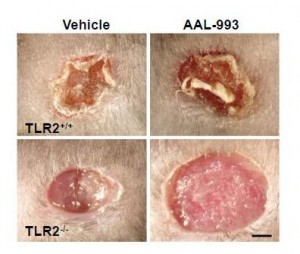
The work I discussed last Friday suggests a reason for that failure. Recall these pictures:

If only the VEGF pathway is blocked (upper right), there is almost as much angiogenesis and wound healing as in a normal wound (upper left). But when both the VEGF and TLR-2 angiogenic pathways are blocked (lower right), there is no wound healing.

If these are the operative pathways in cancer also, then blocking the TLR-2 angiogenesis pathway might be the key to cancer therapy.

But cancer is not the only disease of pathological angiogenesis. Others include:

  • Age-related macular degeneration, diabetic retinopathy, and retinopathy of prematurity – three common causes of blindness.
  • Atherosclerosis, which often features angiogenic vessels in thickened arterial walls.
  • Vascular malformations and tumors.
  • Obesity. Adipose tissue utilizes angiogenic pathways, and angiogenesis inhibition prevents the deposition of fat.
  • Rosacea, psoriasis, and some other skin conditions.
  • Endometriosis, uterine fibroids, and some other causes of female infertility.
  • Rheumatoid arthritis.
  • Crohn’s disease.
  • Preeclampsia.

It may be that the TLR-2 pathway is key to these diseases as well, and that a treatment that inhibits this pathway can cure or improve all of these diseases.

Add up the size of these markets and a pharmaceutical company executive would swoon.

Luckily, we’re not pharmaceutical company executives. But we can still get excited over possibilities to improve these diseases through diet and anti-microbial medicine.

Infections as Contributing Causes of These Diseases

TLR-2 is stimulated by other things besides oxidized DHA. In particular, TLR-2 is an immune molecule which is stimulated by pathogen proteins. As Wikipedia notes:

TLR-2 recognizes many bacterial, fungal, viral, and certain endogenous substances.

This tells us that many pathogens may stimulate angiogenesis through the TLR-2 pathway. As a result, anti-microbial medicines might help treat some diseases of pathological angiogenesis.

Some antibiotics, including doxycycline and minocycline, are known to exercise anti-angiogenic effects independent of the antibiotic effects. [2]

Diet-Induced Angiogenesis

Many foods affect angiogenesis. In fact, cancer studies have identified dozens of plant foods, from garlic to tomatoes to leeks, that possess anti-angiogenic properties.

However, foods can also promote angiogenesis. Let’s stick to the oxidized DHA pathway and see if there’s evidence that foods drive it.

You’ll recall the recipe was:

DHA + oxidative stress + retinyl protein = TLR-2 driven angiogenesis

If this pathway is important in human disease, then we should expect diseases of angiogenesis to be worsened by adding the ingredients on the left.

Specifically, cancer, AMD, rosacea, and so forth should be worsened by high doses of DHA, high doses of vitamin A, and low doses of antioxidant minerals like zinc or selenium.

Is there any evidence for that pattern?

Cancer Studies

First, let me give my bottom line on the Brasky study that kicked off this series. High tissue levels of DHA were associated with increased risk of high-grade prostate cancer, and the oxidized DHA angiogenesis pathway provides a mechanism for this association. What’s not clear is why tissue DHA levels were high. EPA levels were also elevated in the high-grade prostate cancers, but not by nearly as much as DHA levels. EPA and DHA appear together in fish and fish oil, so this suggests that fish consumption contributed to but was not the primary cause of the elevated tissue DHA. The drug finasteride greatly raised risk of high-grade prostate cancer, but the paper did not break down the DHA-cancer association between the finasteride and placebo arms. The most likely explanation, in my view, is that finasteride increases conversion of EPA to DHA and creates artificially high tissue DHA levels. The high DHA levels combined with oxidative stress drive cancer through the TLR-2 angiogenesis pathway.

A clever but unlikely alternative explanation was suggested by Peter at Hyperlipid: perhaps extra dietary fish oil raises testosterone levels. Prostate cancer is a hormone-dependent cancer and can be promoted by testosterone, just as breast cancer is promoted by estrogen. Possible supporting evidence comes from a paper showing an inverse association between metabolic syndrome / diabetes and prostate cancer. The trouble with this idea is that (a) this effect should have been strongest in the low-grade cancers, since diabetes reduced the incidence of low-grade cancers, but in the Brasky study DHA had no association with low-grade cancers, (b) fish oil lowers testosterone levels in rats, (c) in the Brasky study high-grade prostate cancers were strongly associated with obesity and the obese generally have low testosterone levels, and (d) surprisingly, high-grade prostate cancers are associated with low testosterone, not high. So one could argue that fish oil might promote high-grade prostate cancer by lowering testosterone!

A unified explanation along this line would be: Finasteride raises DHA levels, and DHA lowers testosterone. Low testosterone reduces incidence of low-grade prostate cancers but makes it much more likely they will progress to high-grade. Thus, finasteride reduces prostate cancer incidence but increases high-grade prostate cancer incidence and overall prostate cancer mortality. Fits all the facts. Could be.

My bottom line: the Brasky study is weak evidence for anything, but it does raise a whiff of evidence that high dietary fish oil intake might encourage a transition from low-grade to high-grade cancer.

What about other ingredients in the recipe? Does increasing retinyl levels raise cancer risk?

Retinyl palmitate (vitamin A) has been tested in clinical trials for its effect on cancer risk. The trials had to be cut short when it was found that vitamin A increased cancer mortality:

The Carotene and Retinol Efficacy Trial (CARET) was a multicenter randomized, double-blind placebo-controlled chemoprevention trial testing whether daily supplementation with 30 mg β-carotene + 25,000 IU retinyl palmitate would reduce lung cancer risk among 18,314 heavy smokers, former heavy smokers and asbestos-exposed workers. The intervention ended 21 months early in January, 1996 when interim analysis found evidence that the supplements increased the risk of lung cancer and total mortality in this high-risk population by 28% and 17%, respectively (10). [3]

After the study ended participants were tracked for years afterward. Those who had received vitamin A during the trial, but especially those in the vitamin A arm who took additional supplements (mainly multivitamins which are rich in A, but possibly also fish oil), had more high-grade prostate cancers:

As a proportion of the total prostate cancer cases, more men who were randomized to the active arm developed high-grade prostate cancer (Gleason 7-10) than in the placebo arm (44.6% vs. 40.1%, respectively)….

For aggressive prostate cancer, men in the CARET intervention arm who used additional supplements had a relative risk for aggressive prostate cancer (Gleason >or=7 or stage III/IV) of 1.52 (95% CI, 1.03-2.24; P < 0.05), relative to all others. [3]

Interestingly, in the placebo arm taking multivitamins and other supplements reduced cancer risk.

Other studies have found similar results.

Men with higher retinol concentrations at baseline were more likely to develop prostate cancer (quintile 5 vs. quintile 1 hazard ratio = 1.19, 95% confidence interval: 1.03, 1.36; P(trend) = 0.009). The results were similar for aggressive disease. Joint categorization based on baseline and 3-year retinol levels showed that men who were in the highest quintile at both time points had the greatest increased risk (baseline/3-year quintile 5/quintile 5 vs. quintile 1/quintile 1 hazard ratio = 1.31, 95% confidence interval: 1.08, 1.59). In this largest study to date of vitamin A status and subsequent risk of prostate cancer, higher serum retinol was associated with elevated risk, with sustained high exposure conferring the greatest risk. [4]

Carotenoids, which can generally be converted to vitamin A, are also associated with higher cancer risk. There is one exception – lycopene:

Lycopene was inversely associated with prostate cancer risk (comparing highest with lowest quartiles, odds ratio (OR) = 0.65, 95% confidence interval (CI): 0.36, 1.15; test for trend, p = 0.09), particularly for aggressive disease (comparing extreme quartiles, OR = 0.37, 95% CI: 0.15, 0.94; test for trend, p = 0.04). Other carotenoids were positively associated with risk. [5]

What’s special about lycopene? Wikipedia explains:

Lycopene may be the most powerful carotenoid quencher of singlet oxygen,[18] being 100 times more efficient in test tube studies of singlet-oxygen quenching action than vitamin E … The absence of the beta-ionone ring structure for lycopene increases its antioxidant action….

Lycopene is not modified to vitamin A in the body

So lycopene does not increase retinyl levels, but does act as an extraordinarily powerful antioxidant, thus reducing oxidative stress! If you wanted a good food for stopping the DHA – angiogenesis pathway, you’ve found it: tomatoes.

Hmmm, tomatoes go well with salmon …

That gets us to the third part of the recipe, oxidative stress. If oxidized DHA drives angiogenesis, then antioxidants should be preventative for these diseases.

The evidence here is rather mixed, because with the exception of the negative effects of vitamin A, most antioxidants seem to have little effect on cancer. Nevertheless, I’ll give some studies. Selenium is a antioxidant mineral due to its role in glutathione peroxidase:

Serum selenium was inversely associated with risk of prostate cancer (comparing highest to lowest quartiles, OR = 0.71, 95% CI 0.39-1.28; p for trend = 0.11), with similar patterns seen in both blacks and whites. [6]

Zinc is an antioxidant due to its role in zinc-copper superoxide dismutase. Prostate cancer is associated with low tissue levels of zinc. [7, 8] High dietary intake of zinc is associated with lower rates of prostate cancer. [9]

N-acetylcysteine is an antioxidant supplement that is a precursor to glutathione. N-acetylcysteine has been shown to prevent angiogenesis and has been proposed as a likely cancer preventative, but this is as yet untested. [10]

Other Diseases of Angiogenesis

I’ll skip those for now, other than to note that fish oil is a well-known trigger of rosacea. Is it possible that the mechanism is via TLR-2 activation by oxidized DHA?

Conclusion

At the moment there’s some puffs of smoke but no fire. Observational studies weakly link high DHA, high vitamin A, and low antioxidant status to diseases of angiogenesis such as cancer.

This pattern would be consistent with the idea that the natural pathway used in wound healing to trigger angiogenesis – DHA oxidation and combination with retinyl protein to trigger TLR-2 pathways – is also important for cancer progression.

It suggests a strategy of reduced fish oil and vitamin A consumption and increased intake of certain antioxidants (such as lycopene, zinc, selenium, or NAC) may be helpful against cancer.

However, this idea needs testing. No study in animal cancer models has tested this dietary combination.

Given the many proven benefits of moderate amounts of fish oil, I don’t see a reason yet to alter our recommendation that healthy people should eat a pound of fish per week. That said, I do think very high intakes of fish or fish oil are ill advised. And I’m intrigued by the idea that dietary changes may have the potential to play a powerful role in recovery from diseases of angiogenesis such as cancer.

References

[1] Dvorak HF. Tumors: wounds that do not heal. Similarities between tumor stroma generation and wound healing. N Engl J Med. 1986 Dec 25;315(26):1650-9. http://pmid.us/3537791.

[2] Yao JS et al. Comparison of doxycycline and minocycline in the inhibition of VEGF-induced smooth muscle cell migration. Neurochem Int. 2007 Feb;50(3):524-30. http://pmid.us/17145119.

[3] Neuhouser ML et al. Dietary supplement use and prostate cancer risk in the Carotene and Retinol Efficacy Trial. Cancer Epidemiol Biomarkers Prev. 2009 Aug;18(8):2202-6. http://pmid.us/19661078.

[4] Mondul AM et al. Serum retinol and risk of prostate cancer. Am J Epidemiol. 2011 Apr 1;173(7):813-21. http://pmid.us/21389041.

[5] Vogt TM et al. Serum lycopene, other serum carotenoids, and risk of prostate cancer in US Blacks and Whites. Am J Epidemiol. 2002 Jun 1;155(11):1023-32. http://pmid.us/12034581.

[6] Vogt TM et al. Serum selenium and risk of prostate cancer in U.S. blacks and whites. Int J Cancer. 2003 Feb 20;103(5):664-70. http://pmid.us/12494476.

[7] Sarafanov AG et al. Prostate cancer outcome and tissue levels of metal ions. Prostate. 2011 Jan 26. doi: 10.1002/pros.21339. [Epub ahead of print] http://pmid.us/21271612.

[8] Costello LC, Franklin RB. Zinc is decreased in prostate cancer: an established relationship of prostate cancer! J Biol Inorg Chem. 2011 Jan;16(1):3-8. http://pmid.us/21140181.

[9] Epstein MM et al. Dietary zinc and prostate cancer survival in a Swedish cohort. Am J Clin Nutr. 2011 Mar;93(3):586-93. http://pmid.us/21228268.

[10] Noonan DM et al. Angiogenesis and cancer prevention: a vision. Recent Results Cancer Res. 2007;174:219-24. http://pmid.us/17302199.