Category Archives: Omega-3 and Omega-6 Fats

Omega-3 Fats and Cancer

On Wednesday a new paper reported that higher levels of long-chain omega-3 fats (EPA, DPA, and DHA) in blood are associated with a 43% increased risk of prostate cancer and a 71% increased risk of aggressive prostate cancer. [1] This built on earlier work by the same group. [2] In a press release, the authors stated:

“We’ve shown once again that use of nutritional supplements may be harmful,” said Alan Kristal, Dr.P.H., the paper’s senior author …

“[W]e have confirmed that marine omega-3 fatty acids play a role in prostate cancer occurrence,” said corresponding author Theodore Brasky, Ph.D.

They sound confident! Is there anything to it, and should it affect our dietary advice?

Mechanisms Linking Omega-3 Fats to Cancer

In our book and on this blog, we’ve already discussed two mechanisms linking excessive omega-3 intake to cancer risk.

First, there is the issue of lipid peroxidation. Of all fatty acids, long-chain omega-3 fats are the most readily peroxidized:

PUFA relative peroxidizability

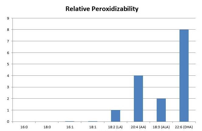
Peroxidation of PUFA generates highly toxic compounds, such as aldehydes, which mutate DNA and turn proteins into advanced lipoxidation end products (ALEs). [3, 4] These lipid peroxidation products have been implicated as causal factors in cancer. [5]

Second, oxidation products of DHA promote angiogenesis – the creation of new blood vessels to feed tumors. These products make cancers grow rapidly. I’ve blogged about this (DHA and Angiogenesis: The Bottom Line, May 4, 2011; Omega-3s, Angiogenesis and Cancer: Part II, April 29, 2011; Omega-3 Fats, Angiogenesis, and Cancer: Part I, April 26, 2011).

So there are known mechanisms by which the long omega-3s in fish oil may promote cancer.

The Brasky et al Papers

The new study by Brasky et al measured omega-3 fat levels in plasma phospholipids. Thus, it doesn’t measure any omega-3s in cells, only omega-3s in serum particles like LDL, HDL, and VLDL; and even in those particles it excludes omega-3 fats found in triglycerides.

This is a very different biomarker than the Omega-3 Index of William Harris, which looks at the omega-3 phospholipids in red blood cell membranes. [6] This biomarker might behave quite differently than the Omega-3 Index.

The study measured plasma phospholipid omega-3s in a group of people, then followed them for 6 years or so to see who developed cancer. Here are the group averages [1]:

Brasky 2013 Table 2

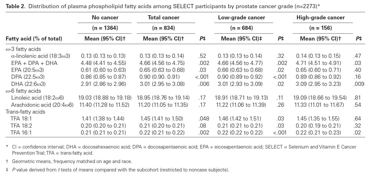
Statistically the most reliable data is in the no cancer vs total cancer comparison. There we find that subjects who went on to develop prostate cancer averaged 3% more DHA and 4% more EPA+DPA+DHA in plasma phospholipids than those who didn’t develop cancer.

Does This Variation Reflect Dietary Intake?

Chris Kresser kindly sent a link to an analysis of the study published at LecturePad by William Harris: “Omega-3 Fatty Acids and Risk for Prostate Cancer.” Harris tells us how to translate the plasma numbers to the corresponding Omega-3 Index numbers:

Based on experiments in our lab, the lowest quartile would correspond to an HS-Omega-3 Index of <3.16% and the highest to an Index of >4.77%).

Even the top quartile of the Brasky et al subjects had quite low omega-3 levels:

In Framingham, the mean Omega-3 Index of participants who were not taking fish oil supplements was 5.2% and for those taking supplements, it was 7.5% [7]. Both of these numbers are considerably higher than the values reported by Braskey et al., even in their highest quartile.

The trial asked its participants not to take supplements, and it looks like they drew a study population whose fish intake was much lower than that of Framingham, Massachusetts, residents.

If dietary omega-3 intake was low in all subjects and varied only slightly among participants, how do we know that this biomarker is related in any way to dietary intake? There could be other factors – genetics, oxidative environment, omega-6 fat intake, antioxidant intake, changes in the proportions of VLDL, LDL, and HDL, to name a few – that affect this biomarker.

Does High Dietary Intake Lead to More Cancer?

If high dietary intake of omega-3s caused more cancer, we would expect cultures that consume lots of fish oil to have higher prostate cancer rates. But epidemiological studies have found that high omega-3 intakes seem to be associated with low cancer rates. For instance, the Japanese eat eight times more omega-3 fatty acids than Americans and their blood levels are twice as high, yet the prostate cancer rates are only one-sixth the American rate.

Of course, there are many confounders in epidemiological studies. Harris helpfully provides a summary of clinical trials in which fish oil was provided as part of the study and cancer outcomes measured:

Harris reply to Brasky 2013

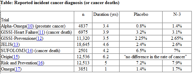
Although none of these studies produced a statistically significant link between omega-3 intake and cancer, incidence of cancer diagnosis or death was increased in every one of the clinical trials except the GISSI-Heart Failure study and perhaps the Origin study. A meta-analysis might find a small cancer promoting effect of omega-3s.

UPDATE: Vladimir Heiskanen points me to an interesting paper in which the effect of dietary fatty acids on cancer metastasis was examined. Colon carcinoma cells were injected into the portal vein (which leads from intestine to liver) of rats and 3 weeks later rats were sacrificed and their livers were examined for metastases. The rats were on three diets — low-fat, high omega-6 (safflower oil), high omega-3 (fish oil). The results:

At 3 weeks after tumor transplantation, the fish oil diet and the safflower oil diet had induced, respectively, 10- and 4-fold more metastases (number) and over 1000- and 500-fold more metastases (size) than were found in the livers of rats on the low-fat diet. [7]

I wish they’d used a saturated fat or monounsaturated fat diet, rather than a low-fat diet, as the control, as this would have clarified that polyunsaturates specifically promote metastasis; in the study the rats’ food was mixed with fish oil or safflower oil, greatly increasing the fat fraction and decreasing the carbohydrate, protein, and micronutrient fractions, so the control diet deviates in many respects from the high-PUFA diets. However, the results are consistent with the idea that fish oil is more cancer-promoting than the less peroxidizable safflower oil, perhaps because of the unique pro-angiogenic effects of DHA products.

Conclusion

There might be biological contexts in which omega-3 fats promote cancer.

This doesn’t mean we should refrain from eating omega-3 fats. Cardiovascular disease causes more deaths than cancer, and omega-3 fats are protective against CVD.

However, I think these studies support the PHD advice:

  • Eat enough oily marine fish to achieve omega-6 and omega-3 balance;
  • Minimize omega-6 intake so that omega-6 and omega-3 balance is achieved at the lowest possible intake of polyunsaturated fats.

All nutrients can be eaten in excess, and omega-3 fats surely fall into this category. The right amount of oily fish is probably about one to two meals per week.

References

[1] Brasky TM et al. Plasma Phospholipid Fatty Acids and Prostate Cancer Risk in the SELECT Trial. J Natl Cancer Inst. 2013 Jul 10. [Epub ahead of print] http://pmid.us/23843441.

[2] Brasky TM et al. Serum phospholipid fatty acids and prostate cancer risk: results from the prostate cancer prevention trial. Am J Epidemiol. 2011 Jun 15;173(12):1429-39. http://pmid.us/21518693.

[3] Hulbert AJ et al. Life and death: metabolic rate, membrane composition, and life span of animals. Physiological Reviews 2007 Oct;87(4):1175–213, http://pmid.us/17928583.

[4] Hulbert AJ. Metabolism and longevity: is there a role for membrane fatty acids? Integretive and Comparative Biology 2010 Nov;50(5):808–17, http://pmid.us/21558243.

[5] Nair U, Bartsch H, Nair J. Lipid peroxidation-induced DNA damage in cancer-prone inflammatory diseases: a review of published adduct types and levels in humans. Free Radic Biol Med. 2007 Oct 15;43(8):1109-20. http://pmid.us/17854706.

[6] Harris WS, Von Schacky C. The Omega-3 Index: a new risk factor for death from coronary heart disease? Prev Med. 2004 Jul;39(1):212-20. http://pmid.us/15208005.

[7] Griffini P et al. Dietary omega-3 polyunsaturated fatty acids promote colon carcinoma metastasis in rat liver. Cancer Res. 1998 Aug 1;58(15):3312-9. http://pmid.us/9699661.

Bengali Fish Curry (Machher Jhal), I: Health Benefits

Dr. Shilpi Bhadra Mehta is a Doctor of Optometry, a Board Member of the Archaeological Institute of America, and leader of the Boston Paleo group, Living Paleo in Boston. I asked her to tell us about Indian cuisine, and she offered a discussion of Bengali Fish Curry. We’ll do it in two parts: first, a discussion of its health benefits; second, a recipe with pictures. — Paul

My husband, Amit, grew up in a vegetarian family and never cared much for fish. But when he went Paleo for health reasons he fell in love with this traditional Bengali recipe, so I make it almost weekly! Bengal is part of India and Bangladesh, it is the home of Bengal tigers, but it is most famous for eating and cooking fish.

When Amit and I first tried Paleo we had some minor setbacks, but our experience improved wonderfully on the Perfect Health Diet version of Paleo so we are grateful for the Jaminets’ wonderful book and website. Amit and I are organizers of the Boston Paleo Meetup Group, and hosted a great potluck and lecture by the Jaminets in October 2011. It’s a pleasure to give back by providing a Perfect Health Diet recipe for you!

Health Benefits of Fish Oil, Ginger, and Turmeric

I’m a practicing optometrist and recommend that all my patients regardless of age or health (except those on blood thinners or a week before surgery) eat about a pound of oily fish per week (about 4-5 servings) like wild salmon, sardines, and sablefish (black cod). For those resistant to eating fish, I recommend fish oils (about 1-3 grams total/day depending on age and health). Fish is great for the whole body – especially the heart, brain, and eye!

There are many health benefits to the omega-3 fatty acids in oily fish. They:

  • Exercise anti-inflammatory effects throughout the body.
  • Help with brain and mental issues such as ADHD, autism spectrum disorders, anxiety, depression (including prenatal and postpartum), mood, cognition, Huntington’s disease, bipolar, schizophrenia, etc.
  • Help prevent and reduce age related macular degeneration (AMD/ARMD – a blinding eye disease) even in those with a strong genetic history.
  • Improve cardiovascular health in aspects such as blood pressure, circulation, triglycerides, VLDL, heart attacks, and stroke.
  • May improve immune function, rheumatoid arthritis, and insulin sensitivity.

The long-chain omega 3s EPA (Eicosapentaenoic acid) and DHA (Docasahexaenoic acid) are especially important. DHA is the most common Omega 3 in the brain and the retina – the neural part of the eye that senses light, and part of the central nervous system (CNS)! About 60% of the polyunsaturated fatty acids (PUFA) in the retina are from DHA, and 40% of the brain’s PUFA is DHA. Low levels of DHA are associated with senility, depression, and suicide risk.

DHA is also important for sperm and erythrocytes (red blood cells). In the often blinding eye disease Retinitis Pigmentosa (RP), RP patients compared to normals had far lower DHA in blood and sperm. The RP patients had fewer and lower quality sperm. Since DHA is found in many tissues, the abnormalities in one tissue may share a similar biochemical cause as in other tissues.

The best and most easily absorbed source of omega-3 fatty acids is wild fish. EPA and DHA are most abundant in oily fish and in the breast milk of women who consume fish. Algae, pastured/grass-fed animals, and pastured dairy and eggs from grass-fed animals are other possible sources.

The body can convert some ALA (alpha linolenic acid) found in plants such as flax and chia into EPA and DHA, but this conversion is often poor – as low as 0.05-15% in healthy humans and worse in older people and those with some medical conditions! ALA has not shown the cardiovascular improvements of fish oil.

Although I typically recommend 1-3 grams of fish oil/per person a day for dry eye and other health issues, I cannot recommend the amounts of flaxseed that would be needed to deliver equivalent amounts of omega-3s. In flaxseed this would require 6-60 grams/per person a day which might cause diarrhea, intestinal blockage, nausea, constipation and other GI side effects.

I cannot recommend flaxseed also for women due to estrogenic effects that could negatively affect hormonal conditions like PCOS, endometriosis, fibroids, or any reproductive cancers. I believe flaxseed should be avoided in women of childbearing age – especially pregnant and breastfeeding women – since in animal studies and some human epidemiological studies it has been associated with preterm birth. In rodent studies flaxseed affected menstrual cycle, lowered birth weight, and altered reproduction in offspring including infertility.

In addition to oily fish, Bengali Fish Curry provides healthy plant foods such as onion, ginger, turmeric, and lemon/lime.

Ginger may help improve mood since it affects serotonin receptors. It helps with nausea from morning sickness, chemotherapy, and seasickness. Ginger also may have some antimicrobial properties. In animal studies it prevents skin cancer, kills ovarian cancer cells, and reduces diabetic complications such as cataracts.

Turmeric has anti-oxidant, anti-inflammatory, antimicrobial, and antitumor properties. A component known as curcumin has been shown to help cancer, osteoarthritis, Alzheimer’s disease, pancreatitis, psoriasis, and some infections.

There are so many benefits to Bengali Fish Curry, and probably more will be discovered in time – that is why I recommend it!

Selected References

Wikipedia also has a good introduction to each.

Ginger

Ernst and Pittler. Efficacy of Ginger for Nausea and Vomiting: A Systemic Review of Randomized Clinical Trials. British Journal of Anaethesia. 2000. 84 (3) 367-371. http://bja.oxfordjournals.org/content/84/3/367.full.pdf+html

Kato et al. Inhibitory Effects of Zingiber officinale Roscoe Derived Components on Aldose Reductase Activity in Vitro and in Vivo. Journal of Agricultural and Food Chemistry. 2006. 54 (18), 6640-6644. http://pubs.acs.org/doi/pdfplus/10.1021/jf061599a

Turmeric/Curcumin

Frautschy et. al. A Potential Role of the Curry Spice Curcumin in Alzheimer’s Disease.” Current Alzheimer Research. 2005. Apr; 2(2): 131-6. http://www.ncbi.nlm.nih.gov/pmc/articles/PMC1702408/pdf/nihms3585.pdf

Rajasekaran, Sigrid. Therapeutic Potential of Curcumin in Gastrointestinal Diseases. World Journal Gastrointestinal Pathophysiology. 2011 February 15; 2(1): 1–14. http://www.ncbi.nlm.nih.gov/pmc/articles/PMC3097964/?tool=pubmed

Omega 3 Fatty Acids/DHA/EPA/Flaxseed

Amminger et al. Long-chain omega-3 fatty acids for indicated prevention of psychotic disorders: a randomized, placebo-controlled trial. Archives General Psychiatry. 2010 Feb;67(2):146-54. http://www.eiyh.org.uk/silo/files/fatty-acids-for-indicated-prevention.pdf

Tou et al. “Flaxseed and Its Lignan Precursor, Secoisolariciresinol Diglycoside, Affect Pregnancy Outcome and Reproductive Development in Rats.” Journal of Nutrition. 1998 Nov;128(11):1861-8. http://jn.nutrition.org/content/128/11/1861.long

Ho et al. Reducing the genetic risk of age-related macular degeneration with dietary antioxidants, zinc, and ?-3 fatty acids: the Rotterdam study. Archives Ophthalmology. 2011 Jun;129(6):758-66. http://www.ncbi.nlm.nih.gov/pubmed/21670343

Barker et al. Nutritional manipulation of primate retinas, V: effects of lutein, zeaxanthin, and n-3 fatty acids on retinal sensitivity to blue-light-induced damage. Investigative Opthalmology & Visual Science. 2011 Jun 6;52(7):3934-42. Print 2011 Jun. http://www.ncbi.nlm.nih.gov/pmc/articles/PMC3175953/pdf/z7g3934.pdf

Wong et al. Prevention of age-related macular degeneration. International Ophthalmology. 2011 Feb;31(1):73-82. Epub 2010 Sep 23. http://www.ncbi.nlm.nih.gov/pmc/articles/PMC3021198/pdf/10792_2010_Article_9397.pdf

Wang et al. n-3 Fatty acids from fish or fish-oil supplements, but not alpha-linolenic acid, benefit cardiovascular disease outcomes in primary- and secondary-prevention studies: a systematic review. American Journal of Clinical Nutrition. 2006 Jul;84(1):5-17.

Brenna et al. alpha-Linolenic acid supplementation and conversion to n-3 long-chain polyunsaturated fatty acids in humans. Prostaglandins Leukotrienes Essential Fatty Acids. 2009 Feb-Mar;80(2-3):85-91. http://www.ncbi.nlm.nih.gov/pubmed/19269799

Connor et al. Sperm Abnormalities in Retinitis Pigmentosa. Investigative Ophthalmology & Visual Science. November 1997 vol. 38 no. 122619-2628. http://www.iovs.org/content/38/12/2619.long

DHA and Angiogenesis: The Bottom Line

So I thought I’d finish up the series on DHA and angiogenesis by discussing 2 issues:

1.      First, an assertion: The pathway by which oxidized DHA drives angiogenesis may be really important for human health.

2.      Second, the $64,000 question: Is there evidence that high levels of dietary DHA promotes diseases of pathological angiogenesis? What about other dietary factors bearing on DHA oxidation?

Significance of the Oxidized DHA Link to Angiogenesis

The papers discussed in Friday’s post about a major angiogenesis pathway stimulated by oxidized DHA (Omega-3s, Angiogenesis and Cancer: Part II, April 29, 2011) may not seem important to many readers. But to cancer researchers and pharmaceutical companies, this is blockbuster work.

A tumor is, in the words of Hal Dvorak, “a wound that never heals.” [1] To support growth, cancers invoke the wound healing process – especially, creation of new blood vessels, or angiogenesis. But the tumor prevents the wound healing process from completing. If it ever did complete, then the tumor itself would be healed. It would cease to grow and become benign.

It’s been recognized for decades that an ability to block angiogenesis would effectively constitute a cure for cancer. The William Li video explains why: nearly everyone gets microscopic tumors that never develop the ability to induce angiogenesis. Life-threatening cancer is the result of tumors that can induce angiogenesis. No angiogenesis, and no one would die of cancer.

But existing anti-angiogenic cancer therapies have produced disappointing results. Avastin, an anti-angiogenic drug targeting VEGF (vascular endothelial growth factor), has been estimated to extend colon cancer patient lifespan by only 6 weeks.  (Nevertheless, Avastin generated $7.3 billion in revenue last year. Imagine how much money there would be in an anti-angiogenic therapy that worked!)

The work I discussed last Friday suggests a reason for that failure. Recall these pictures:

If only the VEGF pathway is blocked (upper right), there is almost as much angiogenesis and wound healing as in a normal wound (upper left). But when both the VEGF and TLR-2 angiogenic pathways are blocked (lower right), there is no wound healing.

If these are the operative pathways in cancer also, then blocking the TLR-2 angiogenesis pathway might be the key to cancer therapy.

But cancer is not the only disease of pathological angiogenesis. Others include:

  • Age-related macular degeneration, diabetic retinopathy, and retinopathy of prematurity – three common causes of blindness.
  • Atherosclerosis, which often features angiogenic vessels in thickened arterial walls.
  • Vascular malformations and tumors.
  • Obesity. Adipose tissue utilizes angiogenic pathways, and angiogenesis inhibition prevents the deposition of fat.
  • Rosacea, psoriasis, and some other skin conditions.
  • Endometriosis, uterine fibroids, and some other causes of female infertility.
  • Rheumatoid arthritis.
  • Crohn’s disease.
  • Preeclampsia.

It may be that the TLR-2 pathway is key to these diseases as well, and that a treatment that inhibits this pathway can cure or improve all of these diseases.

Add up the size of these markets and a pharmaceutical company executive would swoon.

Luckily, we’re not pharmaceutical company executives. But we can still get excited over possibilities to improve these diseases through diet and anti-microbial medicine.

Infections as Contributing Causes of These Diseases

TLR-2 is stimulated by other things besides oxidized DHA. In particular, TLR-2 is an immune molecule which is stimulated by pathogen proteins. As Wikipedia notes:

TLR-2 recognizes many bacterial, fungal, viral, and certain endogenous substances.

This tells us that many pathogens may stimulate angiogenesis through the TLR-2 pathway. As a result, anti-microbial medicines might help treat some diseases of pathological angiogenesis.

Some antibiotics, including doxycycline and minocycline, are known to exercise anti-angiogenic effects independent of the antibiotic effects. [2]

Diet-Induced Angiogenesis

Many foods affect angiogenesis. In fact, cancer studies have identified dozens of plant foods, from garlic to tomatoes to leeks, that possess anti-angiogenic properties.

However, foods can also promote angiogenesis. Let’s stick to the oxidized DHA pathway and see if there’s evidence that foods drive it.

You’ll recall the recipe was:

DHA + oxidative stress + retinyl protein = TLR-2 driven angiogenesis

If this pathway is important in human disease, then we should expect diseases of angiogenesis to be worsened by adding the ingredients on the left.

Specifically, cancer, AMD, rosacea, and so forth should be worsened by high doses of DHA, high doses of vitamin A, and low doses of antioxidant minerals like zinc or selenium.

Is there any evidence for that pattern?

Cancer Studies

First, let me give my bottom line on the Brasky study that kicked off this series. High tissue levels of DHA were associated with increased risk of high-grade prostate cancer, and the oxidized DHA angiogenesis pathway provides a mechanism for this association. What’s not clear is why tissue DHA levels were high. EPA levels were also elevated in the high-grade prostate cancers, but not by nearly as much as DHA levels. EPA and DHA appear together in fish and fish oil, so this suggests that fish consumption contributed to but was not the primary cause of the elevated tissue DHA. The drug finasteride greatly raised risk of high-grade prostate cancer, but the paper did not break down the DHA-cancer association between the finasteride and placebo arms. The most likely explanation, in my view, is that finasteride increases conversion of EPA to DHA and creates artificially high tissue DHA levels. The high DHA levels combined with oxidative stress drive cancer through the TLR-2 angiogenesis pathway.

A clever but unlikely alternative explanation was suggested by Peter at Hyperlipid: perhaps extra dietary fish oil raises testosterone levels. Prostate cancer is a hormone-dependent cancer and can be promoted by testosterone, just as breast cancer is promoted by estrogen. Possible supporting evidence comes from a paper showing an inverse association between metabolic syndrome / diabetes and prostate cancer. The trouble with this idea is that (a) this effect should have been strongest in the low-grade cancers, since diabetes reduced the incidence of low-grade cancers, but in the Brasky study DHA had no association with low-grade cancers, (b) fish oil lowers testosterone levels in rats, (c) in the Brasky study high-grade prostate cancers were strongly associated with obesity and the obese generally have low testosterone levels, and (d) surprisingly, high-grade prostate cancers are associated with low testosterone, not high. So one could argue that fish oil might promote high-grade prostate cancer by lowering testosterone!

A unified explanation along this line would be: Finasteride raises DHA levels, and DHA lowers testosterone. Low testosterone reduces incidence of low-grade prostate cancers but makes it much more likely they will progress to high-grade. Thus, finasteride reduces prostate cancer incidence but increases high-grade prostate cancer incidence and overall prostate cancer mortality. Fits all the facts. Could be.

My bottom line: the Brasky study is weak evidence for anything, but it does raise a whiff of evidence that high dietary fish oil intake might encourage a transition from low-grade to high-grade cancer.

What about other ingredients in the recipe? Does increasing retinyl levels raise cancer risk?

Retinyl palmitate (vitamin A) has been tested in clinical trials for its effect on cancer risk. The trials had to be cut short when it was found that vitamin A increased cancer mortality:

The Carotene and Retinol Efficacy Trial (CARET) was a multicenter randomized, double-blind placebo-controlled chemoprevention trial testing whether daily supplementation with 30 mg β-carotene + 25,000 IU retinyl palmitate would reduce lung cancer risk among 18,314 heavy smokers, former heavy smokers and asbestos-exposed workers. The intervention ended 21 months early in January, 1996 when interim analysis found evidence that the supplements increased the risk of lung cancer and total mortality in this high-risk population by 28% and 17%, respectively (10). [3]

After the study ended participants were tracked for years afterward. Those who had received vitamin A during the trial, but especially those in the vitamin A arm who took additional supplements (mainly multivitamins which are rich in A, but possibly also fish oil), had more high-grade prostate cancers:

As a proportion of the total prostate cancer cases, more men who were randomized to the active arm developed high-grade prostate cancer (Gleason 7-10) than in the placebo arm (44.6% vs. 40.1%, respectively)….

For aggressive prostate cancer, men in the CARET intervention arm who used additional supplements had a relative risk for aggressive prostate cancer (Gleason >or=7 or stage III/IV) of 1.52 (95% CI, 1.03-2.24; P < 0.05), relative to all others. [3]

Interestingly, in the placebo arm taking multivitamins and other supplements reduced cancer risk.

Other studies have found similar results.

Men with higher retinol concentrations at baseline were more likely to develop prostate cancer (quintile 5 vs. quintile 1 hazard ratio = 1.19, 95% confidence interval: 1.03, 1.36; P(trend) = 0.009). The results were similar for aggressive disease. Joint categorization based on baseline and 3-year retinol levels showed that men who were in the highest quintile at both time points had the greatest increased risk (baseline/3-year quintile 5/quintile 5 vs. quintile 1/quintile 1 hazard ratio = 1.31, 95% confidence interval: 1.08, 1.59). In this largest study to date of vitamin A status and subsequent risk of prostate cancer, higher serum retinol was associated with elevated risk, with sustained high exposure conferring the greatest risk. [4]

Carotenoids, which can generally be converted to vitamin A, are also associated with higher cancer risk. There is one exception – lycopene:

Lycopene was inversely associated with prostate cancer risk (comparing highest with lowest quartiles, odds ratio (OR) = 0.65, 95% confidence interval (CI): 0.36, 1.15; test for trend, p = 0.09), particularly for aggressive disease (comparing extreme quartiles, OR = 0.37, 95% CI: 0.15, 0.94; test for trend, p = 0.04). Other carotenoids were positively associated with risk. [5]

What’s special about lycopene? Wikipedia explains:

Lycopene may be the most powerful carotenoid quencher of singlet oxygen,[18] being 100 times more efficient in test tube studies of singlet-oxygen quenching action than vitamin E … The absence of the beta-ionone ring structure for lycopene increases its antioxidant action….

Lycopene is not modified to vitamin A in the body

So lycopene does not increase retinyl levels, but does act as an extraordinarily powerful antioxidant, thus reducing oxidative stress! If you wanted a good food for stopping the DHA – angiogenesis pathway, you’ve found it: tomatoes.

Hmmm, tomatoes go well with salmon …

That gets us to the third part of the recipe, oxidative stress. If oxidized DHA drives angiogenesis, then antioxidants should be preventative for these diseases.

The evidence here is rather mixed, because with the exception of the negative effects of vitamin A, most antioxidants seem to have little effect on cancer. Nevertheless, I’ll give some studies. Selenium is a antioxidant mineral due to its role in glutathione peroxidase:

Serum selenium was inversely associated with risk of prostate cancer (comparing highest to lowest quartiles, OR = 0.71, 95% CI 0.39-1.28; p for trend = 0.11), with similar patterns seen in both blacks and whites. [6]

Zinc is an antioxidant due to its role in zinc-copper superoxide dismutase. Prostate cancer is associated with low tissue levels of zinc. [7, 8] High dietary intake of zinc is associated with lower rates of prostate cancer. [9]

N-acetylcysteine is an antioxidant supplement that is a precursor to glutathione. N-acetylcysteine has been shown to prevent angiogenesis and has been proposed as a likely cancer preventative, but this is as yet untested. [10]

Other Diseases of Angiogenesis

I’ll skip those for now, other than to note that fish oil is a well-known trigger of rosacea. Is it possible that the mechanism is via TLR-2 activation by oxidized DHA?

Conclusion

At the moment there’s some puffs of smoke but no fire. Observational studies weakly link high DHA, high vitamin A, and low antioxidant status to diseases of angiogenesis such as cancer.

This pattern would be consistent with the idea that the natural pathway used in wound healing to trigger angiogenesis – DHA oxidation and combination with retinyl protein to trigger TLR-2 pathways – is also important for cancer progression.

It suggests a strategy of reduced fish oil and vitamin A consumption and increased intake of certain antioxidants (such as lycopene, zinc, selenium, or NAC) may be helpful against cancer.

However, this idea needs testing. No study in animal cancer models has tested this dietary combination.

Given the many proven benefits of moderate amounts of fish oil, I don’t see a reason yet to alter our recommendation that healthy people should eat a pound of fish per week. That said, I do think very high intakes of fish or fish oil are ill advised. And I’m intrigued by the idea that dietary changes may have the potential to play a powerful role in recovery from diseases of angiogenesis such as cancer.

References

[1] Dvorak HF. Tumors: wounds that do not heal. Similarities between tumor stroma generation and wound healing. N Engl J Med. 1986 Dec 25;315(26):1650-9. http://pmid.us/3537791.

[2] Yao JS et al. Comparison of doxycycline and minocycline in the inhibition of VEGF-induced smooth muscle cell migration. Neurochem Int. 2007 Feb;50(3):524-30. http://pmid.us/17145119.

[3] Neuhouser ML et al. Dietary supplement use and prostate cancer risk in the Carotene and Retinol Efficacy Trial. Cancer Epidemiol Biomarkers Prev. 2009 Aug;18(8):2202-6. http://pmid.us/19661078.

[4] Mondul AM et al. Serum retinol and risk of prostate cancer. Am J Epidemiol. 2011 Apr 1;173(7):813-21. http://pmid.us/21389041.

[5] Vogt TM et al. Serum lycopene, other serum carotenoids, and risk of prostate cancer in US Blacks and Whites. Am J Epidemiol. 2002 Jun 1;155(11):1023-32. http://pmid.us/12034581.

[6] Vogt TM et al. Serum selenium and risk of prostate cancer in U.S. blacks and whites. Int J Cancer. 2003 Feb 20;103(5):664-70. http://pmid.us/12494476.

[7] Sarafanov AG et al. Prostate cancer outcome and tissue levels of metal ions. Prostate. 2011 Jan 26. doi: 10.1002/pros.21339. [Epub ahead of print] http://pmid.us/21271612.

[8] Costello LC, Franklin RB. Zinc is decreased in prostate cancer: an established relationship of prostate cancer! J Biol Inorg Chem. 2011 Jan;16(1):3-8. http://pmid.us/21140181.

[9] Epstein MM et al. Dietary zinc and prostate cancer survival in a Swedish cohort. Am J Clin Nutr. 2011 Mar;93(3):586-93. http://pmid.us/21228268.

[10] Noonan DM et al. Angiogenesis and cancer prevention: a vision. Recent Results Cancer Res. 2007;174:219-24. http://pmid.us/17302199.

Omega-3s, Angiogenesis and Cancer: Part II

On Tuesday (Omega-3 Fats, Angiogenesis, and Cancer: Part I, April 26, 2011) I introduced the issue of possible relationships between omega-3 fatty acids, their lipid peroxidation products, and diseases of angiogenesis such as cancer, and promised to discuss a possible mechanism today.

It may be well, however, to start by saying a little bit more about the Brasky paper [1] linking prostate cancer to DHA.

Denise Minger’s Commentary on the Brasky Paper

Denise Minger wrote a commentary on this paper for Mark’s Daily Apple, which is excellent. Her conclusion – “given the oxidation-prone nature of all polyunsaturated fats, a massive intake of omega-3’s – despite their brilliance in moderation – could potentially do more harm than good” – is the proper one.

A few of Denise’s observations, however, could stand elaboration.

The study measured the fraction of serum phospholipid fatty acids in various polyunsaturated and trans-fat species, not dietary intake. This is the right parameter to measure, as fatty acid profiles can be measured precisely while dietary intakes assessed through questionnaires are notoriously unreliable. Also, phospholipids are the fats in cell membranes, and these are the ones involved in the inflammatory signaling pathways long thought to drive cancer risk. So cell membrane lipid measurements have the best chance to demonstrate a link to cancer risk.

Denise makes the important point, however, that the connection between dietary fish oil intake and serum fatty acid profile is not simple. Higher DHA intake raises phospholipid DHA levels, but lower intake of non-omega-3 fats also raises the DHA fraction. She points to a study [2] comparing a low-fat diet (20% fat, 6.7% PUFA, n-6:n-3 ratio 11.1) to a high-fat diet (45% fat, 15% PUFA, n-6:n-3 ratio 12.3).  The low-fat diet had more of its fat in the form of long-chain omega-3s, but the specific DHA intake on the diets was not reported. Membrane DHA ended up 28% higher on the low-fat diet.

So if DHA is dangerous, low-fat dieters will be in the most trouble. Another reason to eat a high-fat diet!

Does this affect our interpretation of the Brasky study? I don’t think it affects it much, because study participants were healthy at the start of the study with no history of cancer and macronutrient intakes don’t vary a lot among the general public. Americans vary surprisingly little from the median of about 50% carbs, 15% protein, and 35% fat – so it’s likely that the quartile with high tissue DHA levels were also high fish oil consumers.

However, study participants were followed for 7 years, at which point their prostate cancer status was assessed. Incidence of low-grade prostate cancers had no association with start-of-the-study DHA intake, but incidence of high-grade prostate cancers was strongly associated.

Here are a couple of possible explanations for this pattern:

  1. DHA is bad: DHA doesn’t drive early cancer development but does drive late-stage cancer growth – i.e. the transition from low-grade to high-grade cancer. So the DHA consumers got the high-grade cancers. Angiogenesis does, in fact, drive the shift from low-grade to high-grade cancer, so a DHA-angiogenesis association would be consistent with this explanation.
  2. Hospital diet advice is bad: DHA was a marker at the start of the study for conscientious, educated, disciplined persons who followed health advice and ate fish oil. When these people were diagnosed with low-grade cancer, they followed the dietary advice of their cancer dietitian. The dietitian’s advice?  Eat lots of wheat, whole grains, legumes, and vegetable oils. It could be the conscientious folks who followed bad diet advice from the hospital dietitian who got the high-grade cancers.

So there is a possible confounding effect.

Another of Denise’s assertions is that there is an “otherwise consistent train of research showing that DHA seems protective at best (and neutral at worst).” Now it is true that there are more studies showing DHA to have benefits against cancer than harm. But this trend is hardly consistent, and the vast majority of studies have failed to detect a relationship.

In the comments to Tuesday’s post, eric linked to a 2005 meta-review of studies on omega-3 fats and cancer. [3] The reviewers looked at 1,210 journal articles and found a mixed bag of mostly insignificant evidence:

Significant associations between omega-3 consumption and cancer risk were reported for lung cancer in two studies; for breast cancer in one; for prostate cancer in one; and for skin cancer in one. However, for lung cancer, one of the significant associations was for increased cancer risk and the other was for decreased risk (four other risk ratios were not significant for lung cancer). For breast cancer, five other estimates did not show a significant association. Only one study assessed skin cancer risk. No effects were reported for cancers of the aerodigestive tract, bladder cancer, colorectal cancer, lymphoma, ovarian cancer, pancreatic cancer, or stomach cancer. Thus, omega-3 fatty acids do not appear to decrease overall cancer risk.

Data were insufficient to permit assessment of a temporal or dose-response relationship. [3]

So the score was 4 studies finding that DHA is associated with less cancer, 1 that it is associated with more, and a boatload that it had no association.

Now there are two ways of interpreting this general insignificance of DHA against cancer. One is to note that there are slightly more studies showing DHA to have benefits than harm, and therefore to judge that DHA might be helpful against cancer.

But another, equally plausible, interpretation is this. Most Americans eat far too much omega-6, and their omega-6 to omega-3 tissue ratio is too high, which is pro-inflammatory via the COX-2 pathway. Eating omega-3s including DHA reduces inflammation by downregulating the COX-2 pathway. This accounts for the well-attested benefits of DHA against cardiovascular disease. Now, cancer is promoted by COX-2 pathway inflammation, which is why COX-2 inhibitors such as aspirin and ibuprofen are protective against cancer. [4] DHA’s action to downregulate this pathway must generate an anti-cancer effect. But, unlike aspirin and ibuprofen, DHA has no observable effect on overall cancer risk. This suggests that DHA has other effects, unrelated to its anti-inflammatory activity, that are cancer promoting. These counterbalance the benefits from its anti-inflammatory effect. If DHA has pro-angiogenic effects that are independent of COX-2 mediated inflammation, then this could account for the observations.

One reason an association of DHA with high-grade cancer may have been missed is that it would be detected only in large studies able to segregate cancers by grade. Brasky et al note:

In the European Prospective Investigation into Cancer and Nutrition (EPIC) (12), the highest quintile of percent DHA was associated with elevated risks of both low-grade (relative risk (RR) = 1.53, 95% CI: 0.96, 2.44) and high-grade (RR = 1.41, 95% CI: 0.76, 2.62) prostate cancer. They also reported significant positive associations of the percent EPA with high-grade prostate cancer (RR = 2.00, 95% CI: 1.07, 3.76). Given that the Prostate Cancer Prevention Trial and the European Prospective Investigation into Cancer and Nutrition, the 2 largest studies of blood levels of phospholipid fatty acids, reported increased risks of high-grade prostate cancer with high levels of ω-3 fatty acids, it remains a possibility that these fatty acids promote tumorigenesis. [1]

If there were no other evidence linking DHA to angiogenesis, the Brasky and EPIC study associations would be interesting, but unlikely to change anyone’s mind. Denise points out the need for other evidence – especially, mechanistic evidence – to make the connection more plausible:

We haven’t sleuthed out any mechanism that could explain why DHA (but not other polyunsaturated fats) promotes rapid tumor growth.

And this is where today’s post comes in. In fact, there is a known mechanism by which DHA but not other polyunsaturated fats can promote rapid tumor growth. Shou-Ching told me about it a few months ago.

DHA and Angiogenesis in Macular Degeneration

Let’s start by going back to 2003 and a paper on the role of a compound called carboxyethylpyrrole (CEP) in age-related macular degeneration (AMD). [5] AMD is an eye disease caused by improper angiogenesis. Basically, malformed blood vessels overgrow the eye, causing retinal detachment and blindness. It afflicts 35% of those over age 75, and is the leading cause of blindness in developed countries. CEP? Well, the paper explains:

Free radical-induced oxidation of docosahexaenoate (DHA)-containing lipids generates ω-(2-carboxyethyl)pyrrole (CEP) protein adducts that are more abundant in ocular tissues from AMD than normal human donors…. The CEP adduct uniquely indicates oxidative modification from DHA derivatives because CEP protein modifications cannot arise from any other common polyunsaturated fatty acid. [5]

CEP is uniquely produced by oxidation of DHA, not other PUFAs. Its abundance depends on DHA abundance, availability of retinyl proteins, and the level of oxidative stress.

CEP is elevated in AMD. The correlation is strong: a person in whom the immune system is trying but failing to clear elevated CEP levels almost invariably has macular degeneration (AMD):

Of individuals (n = 13) exhibiting both antigen and autoantibody levels above the mean for non-AMD controls, 92% had AMD. [5]

So CEP is a great marker for AMD. Is it causal?

Well, first it’s worth noting that the retina is uniquely vulnerable to DHA oxidation:

Although rare in most human tissues, DHA is present in ~80 mol % of the polyunsaturated lipids in photoreceptor outer segments (13). The abundance of DHA in photoreceptors, the high photooxidative stress in retina, and the fact that DHA is the most oxidizable fatty acid in humans (13) suggests that DHA oxidation products may have possible utility as biomarkers for AMD susceptibility. [5]

Oxidation is linked to AMD, and antioxidants slow AMD progression:

Oxidative damage appears to contribute to the pathogenesis of AMD (4) based on epidemiological studies showing that smoking significantly increases the risk of AMD (1, 24). The molecular mechanism for how smoking enhances the risk for AMD is not known. We speculate that reactive oxygen and nitrogen species derived from tobacco smoke in the lungs leads to oxidative protein modifications in the blood that contribute to drusen formation and choroidal neovascularization. Results from a recent clinical trial (5) also demonstrate that the progression of AMD can be slowed in some individuals by high daily doses of antioxidant vitamins and zinc. Direct evidence of oxidative damage in AMD donor eye tissues include elevated levels of CEP adducts uniquely derived from the oxidative fragmentation of DHA (6). [5]

This is where things stood in 2003. By 2010 this group, led by Case Western Reserve University chemist Robert G. Salomon, had established that administering CEP to mice can cause AMD:

To test the hypothesis that this hapten is causally involved in initiating an inflammatory response in AMD, we immunized C57BL/6J mice with mouse serum albumin (MSA) adducted with CEP. Immunized mice develop antibodies to CEP, fix complement component-3 in Bruch’s membrane, accumulate drusen below the retinal pigment epithelium during aging, show decreased a- and b-wave amplitudes in response to light, and develop lesions in the retinal pigment epithelium mimicking geographic atrophy, the blinding end-stage condition characteristic of the dry form of AMD. Inflammatory cells are present in the region of lesions and may be actively involved in the pathology observed. [6]

This constitutes the first really good animal model for AMD. [6]

How does this relate to cancer? That leads us to a Nature paper from October 2010 [7], from the group of Tatiana Byzova at the Cleveland Clinic.

DHA, Immunity, and Angiogenesis

This is a rich paper. Briefly, CEP has a physiological function: it is transiently elevated in wounds and recruits immune cells from bone marrow to the site of the wound. These immune cells further increase oxidative stress and promote angiogenesis; CEP levels are highest at the time of peak angiogenesis. CEP is highly elevated in cancers. Unlike in wounds, where CEP is elevated for a few days, in cancers CEP elevation is chronic.

Here’s a staining comparing CEP in normal skin and in melanoma:

The CEP is co-localized with CD68, a glycoprotein which binds to LDL and is found on macrophages, and with CD31, a membrane marker of neutrophils, macrophages, and endothelial cells. CEP is marking endothelial cells and white blood cells in angiogenic vessels, and possibly LDL.

It turns out that CEP drives angiogenesis by attaching to an immune receptor, Toll-like receptor 2 (TLR2). There are two major pathways for angiogenesis: one driven by vascular endothelial growth factor (VEGF), which is dominant in conditions of hypoxia (oxygen starvation), and one by TLR2. Of these, the TLR2 pathway may in some contexts be more important. Here are pictures of wound healing in mice:

On the upper left is a normal mouse. On the upper right is a similar wound treated with the VEGF inhibitor AAL-993. This wound is rather like a cancer treated with the VEGF inhibitor Avastin. Wound healing is slightly impaired, but still works.

On the lower left is a similar wound with no VEGF inhibition, but the TLR2 pathway blocked by TLR2 knockout. The wound can’t scab and doesn’t heal successfully. If TLR2 is knocked out and VEGF inhibited, there is no wound healing at all (lower right).

You can accelerate angiogenesis and wound healing by adding CEP to the wound.

In the bottom row, CEP has been added. Left is without VEGF inhibition, right with.

If you administer CEP-neutralizing antibodies to a normal wound, wound healing takes more than twice as long. This confirms that angiogenesis driven specifically by CEP (and therefore by DHA oxidation) is part of healthy wound healing.

Tumors use these same pathways to generate vessels and feed their growth. As the paper notes:

[T]umors implanted in TLR2-/- mice exhibited dramatically decreased vascularization and increased areas of necrosis. [7]

Here’s the paper’s conclusion:

Altogether our results establish a novel mechanism of angiogenesis that is independent of hypoxia-triggered VEGF expression. The products of lipid oxidation are generated as a consequence of oxidative stress and are recognized by TLR2, possibly in a complex with TLR1 on ECs, and promote angiogenesis in vivo, thereby contributing to accelerated wound healing and tissue recovery. If high levels of CEP and its analogs accumulate in tissues, it may lead to excessive vascularization, e.g. in tumors. Contribution of the CEP/TLR2 axis to angiogenesis varies in different physiological settings possibly depending on the extent of oxidative stress. CEP-driven angiogenesis may be an attractive therapeutic target, especially in cancers resistant to anti-VEGF therapy. Inflammation and oxidation-driven angiogenesis may occur in other pathologies, for example atherosclerosis, where arterial thickening can depend on its microvasculature. In these settings, there is an extensive generation of oxidative products which might promote atherogenesis via TLR2. Indeed, it was shown that TLR2?/? mice are protected from atherosclerosis, and this effect could be mediated by cells other than bone marrow-derived29. Thus, along with pathogen- and danger-associated molecular patterns, TLR2 recognizes an oxidation-associated molecular pattern. This new function of TLR2 as a sensor of oxidative stress reveals the shortcut link between innate immunity, oxidation and angiogenesis. [7]

Connection to Vitamin A

DHA is oxidized to a compound called HOHA which then combines with a protein, generally a retinyl (vitamin A-derived) protein to form CEP.

Cancers generate lots of CEP from DHA, and perhaps one way they do that is by generating lots of retinyl proteins. Cancers are known to have disturbed vitamin A biology with lots of retinyl:

Disturbance in vitamin A metabolism seems to be an important attribute of cancer cells. Retinoids, particularly retinoic acid, have critical regulatory functions and appear to modulate tumor development and progression. The key step of vitamin A metabolism is the esterification of all-trans retinol, catalyzed by lecithin/retinol acyltransferase. In this work we show that malignant melanoma cells are able to esterify all-trans retinol and subsequently isomerise all-trans retinyl esters into 11-cis retinol, whereas their benign counterparts – melanocytes are not able to catalyze these reactions. Besides, melanoma cell lines express lecithin/retinol acyltranseferase both at the mRNA and protein levels. In contrast, melanocytes do not express this enzyme … [8]

I haven’t looked much into this literature but it may speak to higher cancer risk with excessive vitamin A intake. Thus high-vitamin A cod liver oil may be a double risk for cancer patients.

Conclusion

It looks like we have a recipe for angiogenesis:

DHA + retinyl + oxidative stress = angiogenesis

This recipe is invoked normally and properly during wound healing. But it is also invoked excessively in pathological contexts – notably in cancers and age-related macular degeneration, probably also in other angiogenesis-associated diseases such as arthritis, rosacea, obesity, psoriasis, endometriosis, dementia, and multiple sclerosis.

In the case of cancer, DHA oxidation to CEP might transform miniscule, harmless cancers to high-grade, life-threatening cancers.

Should this possibility affect our dietary omega-3 recommendations? Well, we need to know the relative importance of the three ingredients on the left side of the above equation in producing angiogenesis. Chris Kresser wondered in the comments Tuesday whether oxidation may be the key factor:

I question whether DHA supplementation would truly play a causative role in the absence of a *pro-oxidative environment*.

In other words, perhaps in someone eating a SAD, not exercising, under a lot of stress, etc. DHA is more easily oxidized and thus potentially carcinogenic.

But in someone that is keeping all other oxidative risk factors low (i.e. they’re avoiding n-6, exercising, managing stress, reducing exposure to chemical toxins, etc.) I tend to doubt that supplementing with DHA could cause significant harm.

That’s the last piece of the puzzle: how do we minimize the level of oxidized DHA?

As I replied to Chris in the comments, low-carb Paleo dieters are not out of the woods in regard to oxidative stress. Oxidative stress is generated normally during metabolism, immune function – and by cancers. If anti-oxidant minerals like zinc, copper, and selenium and vitamins like vitamin C are deficient, then oxidative stress can be very high on a low-carb Paleo diet.

At the moment, I think it’s prudent to eat no more than 1 pound of salmon or similar cold-water fish per week, to avoid further EPA/DHA supplements, and to avoid low-fat diets which tend to elevate membrane DHA levels. Moderate omega-3 consumption is especially important for those suffering from diseases of pathological angiogenesis – especially cancer. DHA is essential for good health – but in excess, it is probably dangerous.

References

[1] Brasky TM et al. Serum Phospholipid Fatty Acids and Prostate Cancer Risk: Results From the Prostate Cancer Prevention Trial. Am. J. Epidemiol. April 24, 2011 DOI: 10.1093/aje/kwr027 (Will be at http://pmid.us/21518693.)

[2] Raatz SK et al. Total fat intake modifies plasma fatty acid composition in humans. J Nutr. 2001 Feb;131(2):231-4. http://pmid.us/11160538.

[3] MacLean CH, Newberry SJ, Mojica WA, et al. Effects of Omega-3 Fatty Acids on Cancer. Summary, Evidence Report/Technology Assessment: Number 113. AHRQ Publication Number 05-E010-1, February 2005. Agency for Healthcare Research and Quality, Rockville, MD. http://www.ahrq.gov/clinic/epcsums/o3cansum.htm.

[4] Harris RE. Cyclooxygenase-2 (cox-2) and the inflammogenesis of cancer. Subcell Biochem. 2007;42:93-126. http://pmid.us/17612047.

[5] Gu X et al. Carboxyethylpyrrole protein adducts and autoantibodies, biomarkers for age-related macular degeneration. J Biol Chem. 2003 Oct 24;278(43):42027-35. http://pmid.us/12923198.

[6] Hollyfield JG et al. A hapten generated from an oxidation fragment of docosahexaenoic acid is sufficient to initiate age-related macular degeneration. Mol Neurobiol. 2010 Jun;41(2-3):290-8. http://pmid.us/20221855.

[7] West XZ et al. Oxidative stress induces angiogenesis by activating TLR2 with novel endogenous ligands. Nature. 2010 Oct 21;467(7318):972-6. http://pmid.us/20927103.

[8] Amann PM et al. Vitamin A metabolism in benign and malignant melanocytic skin cells: Importance of lecithin/retinol acyltransferase and RPE65. J Cell Physiol. 2011 Apr 4. doi: 10.1002/jcp.22779. [Epub ahead of print] http://pmid.us/21465477.