Category Archives: Atherosclerosis

Lessons From The Latest Red Meat Scare

I’ve had about ten requests for thoughts on the new paper in Nature Medicine [1] that finds red meat can promote atherosclerosis by a roundabout route: carnitine in the meat is metabolized by gut bacteria into a compound called TMA, which the liver converts to TMAO, which in high doses promotes growth of atherosclerotic plaques.

The same group has done similar studies with other molecules; two years ago the culprit was not carnitine but phosphatidylcholine. [2]

The Scare

Some of the news stories:

It sounds like red meat is dangerous! The best line came from the New York Times:

Lora Hooper, an associate professor of immunology and microbiology at the University of Texas Southwestern Medical Center, who follows the Paleo diet, heavy on meat, exclaimed, “Yikes!”

The Big Picture

The issue here is closely related to one discussed in page 77 of our book:

Protein is not food for us alone; gut bacteria can ferment protein.

Although fermentation of carbohydrates by gut bacteria is usually beneficial, fermentation of protein is not: it generates toxic compounds, including amines, phenols, indoles, thiols, and hydrogen sulfide, which make a foul-smelling stool.

It seems likely, therefore, that high protein intakes are suboptimal for gut health.

When protein is fermented, nitrogen is released, and many nitrogenous compounds are toxic.

The group behind the new research, led by Stanley Hazen, has been looking at another pathway by which bacterial fermentation of meat might be dangerous – the pathway that runs through Trimethylamine. Trimethylamine (TMA) has a simple structure; three methyl groups bonded to a nitrogen atom: N(CH3)3.

Compounds such as choline and carnitine that contain both methyl groups and nitrogen are potential precursors to TMA.

TMA is responsible for the fishy smell of decaying fish. It is highly abundant in fish.

The liver converts TMA into its oxide, TMAO. The Hazen group in a series of papers has argued that higher TMAO levels in blood are associated with atherosclerosis. In a recent paper they assert, “TMAO levels explain 11% of the variation in atherosclerosis.” [3]

So, the equation they are putting together is: fermentation of meat in the gut produces TMA leading to TMAO production which may increase your chance of atherosclerosis by 11%.

Risk is Highly Dependent on the Nature of Your Gut Flora

Here is the key data from the new paper [1]. The “d3” prefix means that the carnitine was labeled with deuterium, an isotope of hydrogen, to help trace its molecular destinations.
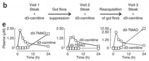
In the left panel of part (e), subjects have been fed a steak (eaten in 10 minutes) plus a 250 mg deuterated carnitine supplement – in total, the carnitine equivalent of 1.5 pounds of meat. Deuterated TMAO levels in blood rise to about 1.8 parts per million after 24 hours.

Then subjects are given antibiotics for a week to suppress their gut flora, and fed steak and deuterated carnitine again. On antibiotics, their blood has no deuterated TMAO at all 24 hours after the meal.

In the right panel, 3 weeks after coming off antibiotics to allow gut flora to regrow, subjects are challenged again with steak and deuterated carnitine. Their blood level of deuterated TMAO now exceeds 12 parts per million – 7 times higher than before the antibiotics.

They go on to test the mix of flora in subjects, and show that flora composition is closely correlated with blood levels of deuterated TMAO after consumption of deuterated carnitine. Some types of gut bacteria produce a lot of TMA from food carnitine, others produce little.

So the amount of TMAO entering the blood from bacterial metabolism of food carnitine is highly dependent on the nature of the gut flora. If you kill off normal flora with antibiotics, then eat meat and carnitine, you will get an overgrowth of bacteria specialized to feed on meat and carnitine. That might not be good for you.

The Vegan vs Omnivore Comparison

Food carnitine is far from the only source of blood TMAO. In fact, TMA is a natural breakdown product of choline, one of the most abundant molecules in the body, and the body has evolved an enzyme for converting TMA into TMAO — the gene is FMO3. So we should ask, how much does metabolism of carnitine by gut bacteria affect blood TMAO levels? For that we need measurements of normal TMAO, not just deuterated TMAO.

We can see what that data looks like in a plot comparing blood TMAO levels between vegans and omnivores (panel c of their Figure 2):
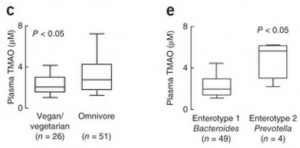
These are the TMAO levels normally circulating in the fasting blood of SAD omnivores and vegetarians. As you can see, there’s considerable overlap between the two distributions. 75% of the omnivores had TMAO levels within the same range as 90% of the vegetarians.

So about 75% of omnivores and 90% of vegetarians have normal TMAO levels. What about the 25% of omnivores and 10% of vegetarians whose TMAO levels are elevated?

In panel e, you can see that the enterotype of the gut flora is a much better predictor of blood TMAO levels than whether someone eats meat. Those with high Prevotella, low Bacteroides averaged about triple the TMAO levels of those with low Prevotella, high Bacteroides flora.

So it really is the gut flora that determine blood TMAO levels.

What Drives the Gut Flora?

What determines whether you have the bad gut flora?

The general picture is this. The immune system regulates the number of microbes living in the gut. When levels become high, antimicrobial peptides are released into the gut to kill some off. When levels are low, antimicrobial peptide production is reduced to let microbes multiply.

This means that if the proportion of bacteria who feed on protein, carnitine, and choline is too high, it’s probably because there is insufficient food for the competing bacteria who feed on carbohydrate forms of fiber. If you have a lot of gut bacteria feeding on fiber, there’s no room in the gut for large amounts of bacteria who feed on meat.

So the 25% of omnivores and 10% of vegan/vegetarians with high TMAO levels are probably the people on low-fiber diets – the ones who get their carbs from flour and sugar. On such a diet, the good bacteria are starved and the bad bacteria that produce TMA multiply.

How Does TMAO Produce Atherosclerosis?

The explanation offered by the Hazen group is that TMAO suppresses “reverse cholesterol transport” conceived broadly as the process of migrating excess cholesterol out of macrophages for transport to the liver and excretion in feces via the bile.

Basically, the idea here is:

  1. Atherosclerosis begins with metabolic syndrome, a state characterized by high LDL levels and caused by endotoxemia (high levels of endotoxins entering the body from the gut).
  2. As we’ve discussed (“Blood Lipids and Infectious Disease, Part II,” July 12, 2011), LDL particles have an immune function. They are oxidized by microbial cell wall components. The resulting oxLDL particles are taken up by macrophages, which then present the microbial cell wall components to other immune cells for antibody formation.
  3. Endotoxemia initiates the process of atherosclerosis by (a) poisoning the liver to cause metabolic syndrome which raises LDL levels, and (b) oxidizing LDL – since endotoxins are bacterial cell wall components that can oxidize LDL – and driving the oxLDL into macrophages.
  4. After macrophages have separated the microbial cell wall components from their accompanying LDL particle, the cholesterol and fat have to be exported to keep them from building up in the cell.
  5. If cholesterol and fat cannot be exported quickly enough, the macrophage is injured and becomes a “foam cell.” Disabled foam cells accumulate in specific locations and form atherosclerotic plaques.
  6. TMAO suppresses bile acid creation, reducing the excretion of cholesterol from the body and leading to higher LDL levels and a greater likelihood that macrophages will become foam cells.

If this is true, then TMAO is not intrinsically atherosclerotic. TMAO in blood only becomes atherosclerotic in the context of metabolic syndrome brought on by endotoxemia.

What causes endotoxemia? A dysbiotic flora generated by a diet high in sugar, flour, and omega-6 fats (see our book, pp 220-222).

Conclusion: Lessons Learned

The lessons of this study are:

  1. Don’t eat a high-sugar, high-flour, low-fiber diet.
  2. Do eat natural whole foods that have the kind of fiber we and our probiotic gut flora co-evolved eating; mainly, resistant starch from in-ground starches like potatoes and soluble fiber from fruits and vegetables.
  3. Don’t eat excessive amounts of meat. As we noted in the book, excess protein is available to gut bacteria for fermentation and that produces a number of toxic byproducts.
  4. Do eat PHD levels of meat – one-half to one pound per day. This level of meat consumption will provide healthful and nourishing amounts of protein, choline, and carnitine, and will not cause any harm if accompanied by PHD levels of healthy plant foods.

None of these lessons is new. This study doesn’t overturn any established dietary wisdom. It is just one more piece of data reminding us to eat a balanced diet consisting of the foods we evolved eating – plant as well as animal.

References

[1] Koeth RA et al. Intestinal microbiota metabolism of l-carnitine, a nutrient in red meat, promotes atherosclerosis. Nat Med. 2013 Apr 7. http://pmid.us/23563705.

[2] Wang Z et al. Gut flora metabolism of phosphatidylcholine promotes cardiovascular disease. Nature. 2011 Apr 7;472(7341):57-63. http://pmid.us/21475195.

[3] Bennett BJ et al. Trimethylamine-N-oxide, a metabolite associated with atherosclerosis, exhibits complex genetic and dietary regulation. Cell Metab. 2013 Jan 8;17(1):49-60. http://pmid.us/23312283.


More Evidence for Low-Carb Diets

In our book we point out a number of dietary tactics that appear to substantially decrease risk of cardiovascular disease. They include:

  • Optimizing tissue omega-6 to omega-3 balance by minimizing intake of omega-6 fats and eating an oily marine fish like salmon or sardines once a week.
  • Optimizing various micronutrients including vitamins D and K2, choline, magnesium, iodine, and selenium.
  • Reducing carbohydrate intake to the body’s natural level of glucose utilization, about 30% of total calories.

We cited two main sources for the claim that reducing carbohydrate intake reduces risk of cardiovascular disease:

–          The Nurses Health Study found that risk of coronary heart disease went down steadily as dietary carbohydrates were reduced and replaced by fat. Those eating a 59% carb diet were 42% more likely to have heart attacks than those eating a 37% carb diet. [1]

–          Replacing dietary carbohydrate with saturated or monounsaturated fat raises HDL and lowers triglycerides, changes that are associated with low rates of cardiovascular disease. Blood lipids are optimized when carb intake drops to 30% of energy or less. [2]

I think this is pretty strong evidence. It is not completely bulletproof, because associations don’t prove causation and improving risk factors doesn’t necessarily improve disease risk; but, combined with supportive evidence from cellular biology and clear evidence that evolutionary selection favors a carbohydrate intake around 30%, I consider it convincing.

However, it’s always good to have more evidence; and two new studies provide some. One directly relates utilization of carbohydrates for energy to atherosclerosis, and the other conducted a 12-month clinical trial of a carbohydrate restricted diet.

Carbohydrate Utilization is Associated With Atherosclerosis

Via Stephan Guyenet comes a study that directly links carbohydrate metabolism to atherosclerosis: “Metabolic fuel utilization and subclinical atherosclerosis in overweight/obese subjects.” [3]

The study used intima-media thickness in the carotid artery, which serves the head and neck, as a measure of atherosclerosis. As Wikipedia notes,

Since the 1990s, both small clinical and several larger scale pharmaceutical trials have used carotid artery IMT as a surrogate endpoint for evaluating the regression and/or progression of atherosclerotic cardiovascular disease. Many studies have documented the relation between the carotid IMT and the presence and severity of atherosclerosis.

To assess metabolism it measured the “respiratory quotient” or RQ. RQ is the ratio of carbon dioxide (CO2) generated in the body to oxygen (O2) consumed in the body.

RQ indicates which fuels are being burned for energy in the body. When carbohydrates are burned, the reaction involves carbon exclusively, so for every O2 molecule consumed there is a CO2 molecule created. This makes the RQ 1.0 when carbohydrates are burned.

Fats, however, donate both carbon and hydrogen, and the hydrogens react with oxygen to make water (H2O). So some of the oxygen consumed when fats are burned goes into water, not carbon dioxide, and the RQ when fats are burned is about 0.7. Ketones also have an RQ around 0.7.

Amino acids from protein have variable amounts of hydrogen and carbon, some amino acids are ketogenic and some are glucogenic, and so the RQ of protein depends on its amino acid mix. Typically RQ from different types of food protein is between 0.8 and 0.9.

However, most people eat a fairly consistent amount of protein, around 15% of energy, so the variable that generally determines RQ in practice is the ratio of carbs to fat in the diet. Higher RQ indicates a higher-carb diet.

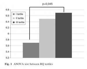
Another study had previously shown that calorie restriction, which also reduces RQ by replacing dietary carbohydrate with fat released from adipose tissue, reduces the thickness of the carotid intima-media. [4] This study was the first testing whether the RQ-CIMT relationship holds also in subjects not known to be restricting calories.

The study found that indeed it does: the lower RQ, the less atherosclerosis the subjects had. Unfortunately they don’t present data in a visually useful way (a scatter plot of RQ vs CIMT would have been helpful); here is what they do show:

RQ was better than waist circumference or BMI at predicting degree of atherosclerosis. Only age was a stronger predictor of atherosclerosis than RQ.

RQ predicted atherosclerosis equally well in subjects with and without obesity. This tells us two things:

  1. It supports the idea that it was habitual diet rather than recent calorie restriction (which decreases RQ by replacing food-sourced calories with fat from adipose tissue) that generated low RQ and low CIMT.
  2. As the authors say, it indicates “the main role of metabolic factors rather than BMI” in generating atherosclerosis – metabolic factors meaning burning glucose for energy rather than fat.

It is also supporting evidence for one of the more controversial lines of our book, that “mitochondria prefer fat.”

One caution: Most of the subjects in this study were eating diets that were around 50% to 55% carbohydrate, so the study was testing whether it’s better to eat a little above or below this carb intake. It tells us, I think, that a 45% carb diet is healthier than a diet with more than 50% carbs. It doesn’t tell us what carb intake is optimal.

The Clinical Trial

In a trial lasting 12 months, restricting carbohydrates to 600 to 850 calories per day – that is, about the 30% of energy that we recommend – in the context of a slightly hypocaloric diet improved cardiovascular risk factors. [5]

Overweight and obese subjects in the trial lost 2.8 kg (6 pounds) over the year-long trial, so it couldn’t have been severely calorie restricted. Changes in other risk factors:

–          Blood pressure dropped from 121/79 to 112/72;

–          Fasting blood glucose dropped from prediabetic 106 mg/dl to normal 96 mg/dl;

–          Lipids improved, with triglycerides decreasing from 217 to 155 mg/dl and HDL increasing from 39 to 45 mg/dl.

They conclude:

The results of this study indicate that a moderately restricted calorie and carbohydrate diet has a positive effect on body weight loss and improves the elements of metabolic syndrome in patients with overweight or obesity and prediabetes. These results underscore the need to provide dietary recommendations focusing on calorie and carbohydrate restrictions … Our results are in agreement with reports produced by other authors who also assessed a carbohydrate-reduced diet …

Conclusion

A number of simple dietary and nutritional changes appear to reduce the risk of atherosclerosis and cardiovascular disease generally. One of them is reducing carbohydrate intake.

I believe the optimum carbohydrate intake is around 30% of energy. Many studies generate clear evidence of benefits as carbs are brought down into the range of 20% to 30% of energy, especially in metabolic disorders like metabolic syndrome, diabetes, and obesity. It’s good to see that evidence from other diseases, such as CVD, also supports the same carb intake.

Because most people’s diets are flawed in so many different ways, and fixing an individual factor is often associated with a reduction in CVD risk of 40% to 70%, it’s possible that we could reduce CVD risk by 90% or more by implementing all of the dietary optimizations described in our book.

It’s well worth pursuing all these little optimizations!

References

[1] Halton TL et al. Low-carbohydrate-diet score and the risk of coronary heart disease in women.  N Engl J Med. 2006 Nov 9;355(19):1991-2002. http://pmid.us/17093250.

[2] Krauss RM. Atherogenic lipoprotein phenotype and diet-gene interactions. J Nutr. 2001 Feb;131(2):340S-3S. http://pmid.us/11160558.

[3] Montalcini T et al. Metabolic fuel utilization and subclinical atherosclerosis in overweight/obese subjects. Endocrine. 2012 Nov 28. [Epub ahead of print] http://pmid.us/23188694.

[4] Iannuzzi A et al. Comparison of two diets of varying glycemic index on carotid subclinical atherosclerosis in obese children. Heart Vessels. 2009 Nov;24(6):419-24. http://pmid.us/20108073.

[5] Velázquez-López L et al. Low calorie and carbohydrate diet: to improve the cardiovascular risk indicators in overweight or obese adults with prediabetes. Endocrine. 2012 Sep 1. [Epub ahead of print] http://pmid.us/22941424.

HDL and Immunity

HDL – high-density lipoprotein – particles are good for you: High HDL levels are associated with lower mortality overall and lower mortality from many diseases – not only cardiovascular disease but also cancer and infection.

People with high HDL are only one-sixth as likely to develop pneumonia [1], and in the Leiden 85-Plus study, those with high HDL experienced 35% lower mortality from infection [2].

Each rise of 16.6 mg/dl in HDL reduced the risk of bowel cancer by 22% in the EPIC study. [3]

In terms of overall mortality, in the VA Normative Aging Study, “Each 10-mg/dl increment in HDL cholesterol was associated with a 14% [decrease] in risk of mortality before 85 years of age.” [4]

This must be surprising to those who think HDL is only a carrier of cholesterol. The lipid hypothesis presumed that the function of HDL is to clear toxic cholesterol from arteries, cholesterol having evolved for the purpose of giving us heart attacks. HDL then brings cholesterol to the liver which disposes of it returns it to the blood via LDL (which evolved for the purpose of poisoning arteries with cholesterol, and giving HDL something to do). (Hat tip to Peter for this formulation of the lipid hypothesis.)

But there is an alternative hypothesis: that infections cause disease, and that HDL has an immune function. This hypothesis would explain why HDL protects against infections and against all diseases of aging.

Immune Functions of HDL

I got interested in immune functions of HDL upon reading an article in ScienceDaily last year (“How Disease-Causing Parasite Gets Around Human Innate Immunity,” Sept 13, 2010). The article states:

Several species of African trypanosomes infect non-primate mammals and cause important veterinary disease yet are unable to infect humans. The trypanosomes that cause human disease, Trypanosoma brucei gambiense and T. b. rhodensiense, have evolved mechanisms to avoid the native human defense molecules in the circulatory system that kill the parasites that cause animal disease….

Human innate immunity against most African trypanosomes is mediated by a subclass of HDL (high density lipoprotein, which people know from blood tests as “good cholesterol”) called trypanosome lytic factor-1, or TLF-1….

The parasite that causes fast-onset, acute sleeping sickness in humans, T. b. rhodensiense, is able to cause disease because it has evolved an inhibitor of TLF-1 called Serum Resistance Associated (SRA) protein…. T. b. gambiense resistance to TLF-1 is caused by a marked reduction of TLF-1 uptake by the parasite….

To survive in the bloodstream of humans, these parasites have apparently evolved mutations in the gene encoding a surface protein receptor. These mutations result in a receptor with decreased TLF-1 binding, leading to reduced uptake and thus allow the parasites to avoid the toxicity of TLF-1.

“Humans have evolved TLF-1 as a highly specific toxin against African trypanosomes by tricking the parasite into taking up this HDL because it resembles a nutrient the parasite needs for survival,” said Hajduk, “but T. b. gambiense has evolved a counter measure to these human ‘Trojan horses’ simply by barring the door and not allowing TLF-1 to enter the cell, effectively blocking human innate immunity and leading to infection and ultimately disease.”

So HDL is actually an immune particle carrying proteins that poison pathogens. The TLF-1 HDL subclass consists of those HDL particles carrying two anti-trypanosome proteins, apolipoprotein L-1 and haptoglobin-related protein. [5]

Any HDL particle can become an anti-trypanosome defender simply by acquiring and carrying these proteins.

It turns out that HDL can carry a great assortment of immune proteins. The orchestrator of HDL’s immune functions seems to be a circulating plasma protein called phospholipid transfer protein (PLTP), which forms complexes with immune molecules and then associates with apolipoprotein A-I (the primary HDL protein). PLTP brings 24 different immune molecules into HDL particles, including apolipoproteins such as clusterin (apoJ), coagulation factors, and complement factors. [6] These immune protein complexes add protein but not fat to HDL particles:

Unexpectedly, lipids accounted for only 3% of the mass of the PLTP complexes. Collectively, our observations indicate that PLTP in human plasma resides on lipid-poor complexes dominated by clusterin and proteins implicated in host defense and inflammation. [6]

It looks like HDL may not be primarily a carrier of cholesterol, but rather a carrier of antimicrobial proteins. Its cholesterol and lipids may serve, as the ScienceDaily article suggests, to make the HDL particle attractive to pathogens so that it may enter as a “Trojan Horse.”

HDL-associated immune proteins under strong selection

As pathogens evolve, immune proteins have to evolve. It turns out that apolipoprotein L-1, the immune protein that protects against trypanosomes, is under strong selection in both Africa and Europe.

The version selected in Europe does not protect against Trypanosoma brucei rhodesiense, cause of one of the African sleeping sickness diseases, but the version selected in Africa does. Unfortunately, the African version also increases risk of kidney disease – which may explain why African-Americans have higher rates of kidney disease than white Americans. [7]

So Africans have sacrificed kidney health for greater immunity against sleeping sickness. This suggests that African sleeping sickness may be a relatively recently evolved human disease.

HDL neutralizes toxins

HDL binds bacterial endotoxins, especially lipopolysaccharide (LPS), and neutralizes their toxicity. As a result, people with high HDL have substantially less release of tumor necrosis factor-alpha (TNF-α) during infection. [8]

TNF-α is an inflammatory molecule that stimulates the acute phase response to infections. Levels of C-reactive protein are a good index of TNF-α levels, so generally speaking high HDL will lead to low TNF-α and low CRP.

What’s the best HDL profile?

It should be desirable to have more HDL particles. Since each HDL particle is capable of poisoning a pathogen, the more you have, the stronger your immune defenses.

However, the weight of each HDL particle is likely to be an indicator of infection severity. An infection-free person will have few immune proteins to pick up; the HDL particles will be fat-rich and buoyant. But a person with extensive infections will have heavier HDL particles freighted with immune proteins.

Conventional tests in the doctor’s office measure the weight of HDL in mg per deciliter of blood. Since having more HDL particles (which raises the weight) is good, but having heavy HDL particles indicates infection which is bad, mass is not the best measure of HDL status. We would expect the number or concentration of HDL particles to provide a better indicator of health.

Indeed, this appears to be what is observed. The most important determinant of HDL status is the number of HDL particles:

The association between HDL size and CAD risk was abolished on adjustment for apolipoprotein B and triglyceride levels (adjusted odds ratio, 1.00 [95% CI, 0.71 to 1.39] for top vs. bottom quartile), whereas HDL particle concentration remained independently associated with CAD risk (adjusted odds ratio, 0.50 [CI, 0.37 to 0.66]). [9]

Conclusion

HDL particles are “Trojan Horses” that attack pathogens and neutralize their toxins.

If you want to remain free from infectious diseases – which is to say, all diseases – to a ripe old age, it’s important to make your HDL particles numerous.

On Thursday, I’ll discuss ways to do that.

References

[1] Gruber M et al. Prognostic impact of plasma lipids in patients with lower respiratory tract infections – an observational study. Swiss Med Wkly. 2009 Mar 21;139(11-12):166-72. http://pmid.us/19330560.

[2] Berbée JF et al. Plasma apolipoprotein CI protects against mortality from infection in old age. J Gerontol A Biol Sci Med Sci. 2008 Feb;63(2):122-6. http://pmid.us/18314445

[3] van Duijnhoven FJ et al. Blood lipid and lipoprotein concentrations and colorectal cancer risk in the European Prospective Investigation into Cancer and Nutrition. Gut. 2011 Mar 7. [Epub ahead of print] http://pmid.us/21383385.

[4] Rahilly-Tierney CR et al. Relation Between High-Density Lipoprotein Cholesterol and Survival to Age 85 Years in Men (from the VA Normative Aging Study). Am J Cardiol. 2011 Apr 15;107(8):1173-7. http://pmid.us/21296318.

[5] Kieft R et al. Mechanism of Trypanosoma brucei gambiense (group 1) resistance to human trypanosome lytic factor. Proc Natl Acad Sci U S A. 2010 Sep 14;107(37):16137-16141. http://pmid.us/20805508.

[6] Cheung MC et al. Phospholipid transfer protein in human plasma associates with proteins linked to immunity and inflammation. Biochemistry. 2010 Aug 31;49(34):7314-22. http://pmid.us/20666409.

[7] Genovese G et al. Association of trypanolytic ApoL1 variants with kidney disease in African Americans. Science. 2010 Aug 13;329(5993):841-5. http://pmid.us/20647424.

[8] Henning MF et al. Contribution of the C-terminal end of apolipoprotein AI to neutralization of lipopolysaccharide endotoxic effect. Innate Immun. 2010 May 25. [Epub ahead of print] http://pmid.us/20501516.

[9] El Harchaoui K et al. High-density lipoprotein particle size and concentration and coronary risk. Ann Intern Med. 2009 Jan 20;150(2):84-93. http://pmid.us/19153411.

Answer Day: What Causes High LDL on Low-Carb Paleo?

First, thank you to everyone who commented on the quiz. I enjoyed reading your thoughts.

Is High LDL Something to Worry About?

Perhaps this ought to be the first question. Jack Kronk says “I don’t believe that high LDL is necessarily a problem” and Poisonguy writes “Treat the symptoms, Larry, not the numbers.” Poisonguy’s comment assumes that the LDL number is not a symptom of trouble. Is it?

I think so. It helps to know a little about the biology of cholesterol and of blood vessels.

When cells in culture plates are separated from their neighbors and need to move, they make a lot of cholesterol and transport it to their membranes. When cells find good neighbors and settle down, they stop producing cholesterol.

The same thing happens in the body. Any time there is a wound or injury that needs to be healed, cholesterol production gets jacked up.

When people have widespread vascular injuries, cholesterol is produced in large quantities by cells lining blood vessels. Now, to repair injuries cells have to coordinate their functions. Endothelial cells are the coordinators of vascular repair: they direct other cell types, like smooth muscle cells and fibroblasts, in the healing of vascular injuries.

To heal vascular injuries, these cells not only need more cholesterol for movement; they also need to multiply. It turns out that LDL, which carries cholesterol, also causes vascular cells to reproduce (“mitogenesis”):

The best-characterized function of LDLs is to deliver cholesterol to cells. They may, however, have functions in addition to transporting cholesterol. For example, they seem to produce a mitogenic effect on endothelial cells, smooth muscle cells, and fibroblasts, and induce growth-factor production, chemotaxis, cell proliferation, and cytotoxicity (3). Moreover, an increase of LDL plasma concentration, which is observed during the development of atherosclerosis, can activate various mitogen-activated protein kinase (MAPK) pathways …

We also show … LDL-induced fibroblast spreading … [1]

If endothelial cells are the coordinators of vascular repair, and LDL particles their messengers to fibroblasts and smooth muscle cells, then ECs should be able to generate LDL particles locally. Guess what:  ECs make a lipase whose main effect is to decrease HDL levels but can also convert VLDL and IDL particles into LDL particles and remove fat from LDL particles to make them into small, dense LDL:

Endothelial lipase (EL) has recently been identified as a new member of the triglyceride lipase gene family. EL shares a relatively high degree of homology with lipoprotein lipase and hepatic lipase …

In vitro, EL has hydrolyzed phospholipids in chylomicrons, very low density lipoprotein (VLDL), intermediate density lipoprotein and LDL. [2]

Immune cells, of course, are essential for wound healing and they should be attracted to any site of vascular injury. It turns out that immune cells have LDL receptors and these receptors may help them congregate at sites of vascular injury. [3]

I don’t want to exaggerate the state of the literature here:  this is a surprisingly poorly investigated area. But I believe these things:

1.      Cholesterol and LDL particles are part of the vascular wound repair process.

2.      Very high LDL levels are a marker of widespread vascular injury.

Now this is not the “lipid hypothesis.” Compare the two views:

  • The lipid hypothesis:  LDL cholesterol causes vascular injury.
  • My view:  LDL cholesterol is the ambulance crew that arrives at the scene of the crime to help the victims. The lipid hypothesis is the view that ambulance drivers should be arrested for homicide because they are commonly found at murder scenes.

So, to Poisonguy, on my view high LDL numbers are a symptom of vascular injury and are a cause for concern.

Big-Picture View of the Cause of High LDL

So, on a micro-level, I think vascular damage causes high LDL. But what causes vascular damage?

Here I notice a striking difference in commenters’ perspectives and mine. I tend to take a big-picture, top-down view of biology. There are three basic causes of nearly all pathologies:

1.      Toxins, usually food toxins.

2.      Malnutrition.

3.      Pathogens.

The whole organization of our book is dictated by this view. It is organized in four Steps. Step One is about re-orienting people’s views of macronutrients away from high-grain, fat-phobic, vegetable-oil-rich diets toward diets rich in animal fats. The other steps are about removing the causes of disease:

1.      Step Two is “Eat Paleo, Not Toxic” – remove food toxins.

2.      Step Three is “Be Well Nourished” – eliminate malnutrition.

3.      Step Four is “Heal and Prevent Disease” – address pathogens by enhancing immunity and, where appropriate, taking advantage of antibiotic therapies.

So when someone offers a pathology, any pathology, my first question is: Which cause is behind this, and which step do they need to focus on?

In Larry’s case, he had been eating low-carb Paleo for years. So toxins were not a problem.

Pathogens might be a problem – after all, he’s 64, and everybody collects chronic infections which tend to grow increasingly severe with age – but Larry hadn’t reported any other symptoms. More to the point, low-carb Paleo diets typically enhance immunity, yet Larry had fine LDL numbers before adopting low-carb Paleo and then his LDL got worse. So it wouldn’t be infectious in origin unless his diet had suppressed immunity through malnutrition – in which case the first step would be to address the malnutrition.

Step Three, malnutrition, was the only logical answer. The conversion to Paleo removes a lot of foods from the diet and could easily have removed the primary sources of some micronutrients.

So I was immediately convinced, just from the time-course of the pathology, that the cause was malnutrition.

Micronutrient Deficiencies are Very Common

In the book (Step Three) we explain why nearly everyone is deficient in micronutrients. The problems are most severe for minerals:  water treatment removes minerals from water, and mineral depletion of soil by industrial agriculture leads to mineral deficiencies in farmed plants and grain-fed animals.

This is why our “essential supplements” include a multimineral supplement plus additional quantities of five minerals – magnesium, copper, chromium, iodine, and selenium. Vitamins get a lot of attention, but minerals are where the big health gains are.

Copper Deficiency and LDL

Some micronutrient deficiencies are known to cause elevated LDL.

Readers of our book know that copper causes vascular disease; blog readers may be more familiar with an excellent post by Stephan, “Copper and Cardiovascular Disease”, discussing evidence that copper deficiency causes cardiovascular disease. As I’ve just argued that cardiovascular disease causes high LDL, it shouldn’t be a surprise that copper deficiency also causes hypercholesterolemia:

Copper and iron are essential nutrients in human physiology as their importance is linked to their role as cofactors of many redox enzymes involved in a wide range of biological processes, as well as in oxygen and electron transport. Mild dietary deficiencies of both metals … may cause long-term deleterious effects in cardiovascular system and alterations in lipid metabolism (3)….

Several studies showed a clear correlation among copper deficiency and dyslipidemia. The main alterations concern higher plasma CL and triglyceride (TG) concentrations, increased VLDL-LDL to HDL lipoproteins ratio, and the shape alteration of HDL lipoproteins.  [4]

The essentiality of copper (Cu) in humans is demonstrated by various clinical features associated with deficiency, such as anaemia, hypercholesterolaemia and bone malformations. [5]

Over the last couple of decades, dietary copper deficiency has been shown to cause a variety of metabolic changes, including hypercholesterolemia, hypertriglyceridemia, hypertension, and glucose intolerance. [6]

Copper deficiency is, I believe, the single most likely cause of elevated LDL on low-carb Paleo diets. The solution is to eat beef liver or supplement.

So, was my advice to Larry to supplement copper?  Yes, but that was not my only advice.

Other Micronutrient Deficiencies and Elevated LDL

Another common micronutrient deficiency that causes elevated LDL cholesterol is choline deficiency that is NOT accompanied by methionine deficiency. That is discussed in my post “Choline Deficiency and Plant Oil Induced Diabetes”:

Choline deficiency (CD) by itself induces metabolic syndrome (indicated by insulin resistance and elevated serum triglycerides and cholesterol) and obesity.

A combined methionine and choline deficiency (MCD) actually causes weight loss and reduces serum triglycerides and cholesterol …

I quote both these effects because it illustrates the complexity of nutrition. A deficiency of a micronutrient can present with totally different symptoms depending on the status of other micronutrients.

Julianne had a really nice comment, unfortunately caught in the spam filter for a while, with a number of links. She mentions vitamin C deficiency and, with other commenters, noted the link between hypothyroidism and elevated LDL. As one cause of hypothyroidism is iodine or selenium deficiency, this is another pathway by which mineral deficiencies can elevate LDL.

UPDATE: Mike Gruber reduced his LDL by 200 mg/dl by supplementing iodine. Clearly iodine can have big effects!

Other commenters brought up fish oil. They may be interested to know that fish oil not only balances omega-6 to modulate inflammatory pathways, it also suppresses endothelial lipase and thus moderates the LDL-raising and HDL-lowering effect of vascular damage:

On the other hand, physical exercise and fish oil (a rich source of eicosapentaenoic acid and docosahexaenoic acid) suppress the activity of EL and this, in turn, enhances the plasma concentrations of HDL cholesterol. [7]

Whether this effect is always desirable is a topic for another day.

My December Advice to Larry

So what was my December advice to Larry?

It was simple. In adopting a low-carb Paleo diet, he had implemented Steps One and Two of our book. My advice was to implement Step Three (“Be well nourished”) by taking our recommended supplements. Eating egg yolks and beef liver for copper and choline is a good idea too.

Just to cover all bases, I advised to include most of our “therapeutic supplements” as well as all the “essential supplements.”

Since December, Larry has been taking all the recommended supplements and eating 5 ounces per week of beef liver. As I noted yesterday, Larry’s LDL decreased from 295 mg/dl to 213 mg/dl, HDL rose from 74 mg/dl to 92 mg/dl, and triglycerides fell from 102 to 76 mg/dl since he started Step Three. This is all consistent with a healthier vasculature and reduced production of endothelial lipase.

Conclusion

Some people think there is something wrong with a diet if supplements are recommended. They believe that a well-designed diet should provide sufficient nutrition from food alone, and that if supplements are advised then the diet must be flawed.

I think this is quite mistaken. The reality is that Paleolithic man was often mildly malnourished, and modern man – due to the absence of minerals from treated water and agriculturally produced food, and the reduced diversity and higher caloric density of our foods – is severely malnourished compared to Paleolithic man.

We recommend eating a micronutrient-rich diet, including nourishing foods like egg yolks, liver, bone broth soups, seaweed, fermented vegetables, and so forth. But I think it’s only prudent to acknowledge and compensate for the widespread nutrient depletion that is so prevalent today. Even when nutrient-rich food is regularly eaten, micronutrient deficiencies are still possible.

Eating Paleo-style is not enough to guarantee perfect health. Luckily, supplementation of the key nutrients that we need for health and that are often missing from foods will often get us the rest of the way.

References

[1] Dobreva I et al. LDLs induce fibroblast spreading independently of the LDL receptor via activation of the p38 MAPK pathway. J Lipid Res. 2003 Dec;44(12):2382-90. http://pmid.us/12951358.

[2] Paradis ME, Lamarche B. Endothelial lipase: its role in cardiovascular disease. Can J Cardiol. 2006 Feb;22 Suppl B:31B-34B. http://pmid.us/16498510.

[3] Giulian D et al. The role of mononuclear phagocytes in wound healing after traumatic injury to adult mammalian brain. J Neurosci. 1989 Dec;9(12):4416-29. http://pmid.us/2480402.

[4] Tosco A et al. Molecular bases of copper and iron deficiency-associated dyslipidemia: a microarray analysis of the rat intestinal transcriptome. Genes Nutr. 2010 Mar;5(1):1-8. http://pmid.us/19821111.

[5] Harvey LJ, McArdle HJ. Biomarkers of copper status: a brief update. Br J Nutr. 2008 Jun;99 Suppl 3:S10-3. http://pmid.us/18598583.

[6] Aliabadi H. A deleterious interaction between copper deficiency and sugar ingestion may be the missing link in heart disease. Med Hypotheses. 2008;70(6):1163-6. http://pmid.us/18178013.

[7] Das UN. Long-chain polyunsaturated fatty acids, endothelial lipase and atherosclerosis. Prostaglandins Leukot Essent Fatty Acids. 2005 Mar;72(3):173-9. http://pmid.us/15664301.