Category Archives: Nutrients

Around the Web; and Why Is Aspirin Toxic to Cats?

[1] Interesting posts this week: Melissa McEwen assures us: Robb Wolf is not Satan. Kurt Harris’s reader Tara makes the most persuasive case I’ve seen for grass-fed meat through pictures. Emily Deans compares eating disorders to addictions.

[2] Kurt Harris re-re-brands: Paleonu became PaNu became Paleo 2.0 becomes Archevore.com, archevore being a neologism for “one who eats of the essentials.”

Well, it’s more euphonious than “EM2vore,” for “one who eats of the evolutionary metabolic milieu.” A more descriptive name might have been “nontoxivore,” since Kurt’s primary theme is avoidance of “neolithic agents of disease – wheat, excess fructose and excess linoleic acid.”

It will be interesting to see where he’s taking this. Are Archevorean essentials the same as PaNu?

[3] Posts of the week: Chris Masterjohn posts always deserve special notice. On Tuesday he continued his important series investigating whether wheat causes leaky gut, which will trigger a few edits in the next edition of our book. I was asked about this last Saturday and said:

There’s no question that gluten causes problems in non-celiacs – that’s the main result of the Fasano paper Chris cites, and also of papers cited by Andrew Badenoch in a post I linked today. It’s just that leaky gut does not appear to be one of those problems.

It certainly doesn’t mean that wheat is safe to eat.

I may add that pathogens and other food toxins – even perhaps other wheat toxins besides gluten – can cause a leaky gut, providing a way for wheat toxins to enter the body. Moreover, some wheat toxins don’t even need a leaky gut to enter the body. As we discuss in the book (p 134), wheat germ agglutinin can cross barriers via transcytosis, enabling them to enter the body even if the intestinal barrier is intact. Finally, wheat toxins can damage the gut without entering the body at all. So there are many pathways through which wheat toxicity can matter.

Chris had another outstanding post on Friday, about fatty liver disease.

[4] Rosacea is an infection of the skin and vessels: That’s why it can be transmitted through facial skin grafts.

Source: Kanitakis J. Transmission of Rosacea from the Graft in Facial Allotransplantation. Am J Transplant. 2011 Mar 28. [Epub ahead of print] http://pmid.us/21443678.

[5] Special offer: The folks at Emerald Forest Xylitol noticed that we recommend their product and would like to give a special offer to PerfectHealthDiet.com readers. Use the coupon code FIRST to get 10% off all products at www.emeraldforestxylitol.com.

Also, Matt Willer of Emerald Forest Xylitol is looking for recipes that include Xylitol for use in his newsletter. If you have a recipe, send it to matt@xylitolusa.com.

[6] Animal photos: If you saw a grizzly charging straight toward you, would you stop to take this photo?

Photographer Alex Wypyszinski did in Yellowstone. The grizzly was chasing an injured bison, and the pair went right past him:

For the full story, see Grizzly versus Bison: the rest of the story (Drew Trafton, 10/29/10, KRTV, Great Falls, Montana). Hat tip Orrin Judd.

[7] Don’t hate the sun: From Britain comes the sad story of a 21-year-old who “hated the sun” and died of skin cancer at 21.

Dr. John Briffa has a summary of the relevant science.

[8] I couldn’t disagree more: Mike the Mad Biologist and Newt Gingrich are dead wrong in their prescription for research funding. We don’t need more concentrated funding, we need more distributed, decentralized funding that is patient-driven, not top-scientist driven.

Discovering cures can be cheap – if you’re looking in the right place. If you’re looking in the wrong direction, the cost of a cure may be infinite.

[9] I hate when that happens:

(Via Stephen Wangen)

[10] Are choline supplements toxic?: At the very beginning of the book (p 3) we state that “the perfect diet should … deliver … no excess nutrients for pathogens.”

Later in the book we give examples of nutrients that, in excess, primarily benefit pathogens: niacin (the primary vitamin for bacteria), iron (critical for metabolism of most pathogens, and a component of bacterial biofilms), and calcium (a component of bacterial biofilms). These are on our list of micronutrients we recommend not supplementing (beyond a multivitamin).

Two readers, Leonardo and Patricia (thank you both!), emailed us about a ScienceDaily article suggesting that choline, one of the micronutrients we most frequently recommend, should be added to this list:

When fed to mice, lecithin and choline were converted to a heart disease-forming product by the intestinal microbes, which promoted fatty plaque deposits to form within arteries (atherosclerosis); in humans, higher blood levels of choline and the heart disease forming microorganism products are strongly associated with increased cardiovascular disease risk.

The story didn’t have enough information, so I downloaded the paper. The paper notes that choline is metabolized by gut bacteria to a gas with a fishy odor called TMA, which is then oxidized in the liver to a compound called TMAO:

Briefly, initial catabolism of choline and other trimethylamine-containing species (for example, betaine) by intestinal microbes forms the gas trimethylamine (TMA), which is efficiently absorbed and rapidly metabolized by at least one member of the hepatic flavin monooxygenase (FMO) family of enzymes, FMO3, to form trimethylamine N-oxide (TMAO).

They showed that (a) feeding phosphatidylcholine from egg yolk to mice led to increased blood levels of TMAO and that (b) in a separate study, people with atherosclerosis have elevated blood levels of TMAO, choline, and trimethylglycine.

Supplementing choline at 10 times normal levels to Apoe-knockout mice led to increased TMAO but not choline in blood:

Atherosclerosis-prone mice (C57BL/6J Apoe-/-) at time of weaning were placed on either normal chow diet (contains 0.08–0.09% total choline, wt/wt) or normal chow diet supplemented with intermediate (0.5%) or high amounts of additional choline (1.0%) or TMAO (0.12%)….

Analysis of plasma levels of choline and TMAO in each of the dietary arms showed nominal changes in plasma levels of choline, but significant increases of TMAO in mice receiving either choline or TMAO supplementation (Supplementary Fig. 10).

Serum TMAO levels were correlated with atherosclerotic plaque size and with macrophages turning into foam cells:

[A]ll dietary groups of mice revealed a significant positive correlation between plasma levels of TMAO and atherosclerotic plaque size (Fig. 3e and Supplementary Fig. 9b).

TMA (a gas with a fish odor) has to be converted in the liver to the toxic TMAO in order to produce these bigger atherosclerotic lesions. This conversion happened mainly in mice with low HDL:

Interestingly, a highly significant negative correlation with plasma high-density lipoprotein (HDL) cholesterol levels was noted in both male and female mice (Fig. 4b and Supplementary Fig. 12, middle row).

So if you’re an Apoe(-/-) mouse and eat ten times normal choline, if you have high HDL your arteries are safe but you smell fishy; if you have low HDL you smell fine but your arteries get injured.

What does this tell us about choline supplementation?

For humans with working ApoE alleles, I doubt we can infer anything yet.

For Apoe(-/-) mice fed ten times normal choline, I would suggest shooting for low HDL while dating, then high HDL after marriage.

Reference: Wang Z et al. Gut flora metabolism of phosphatidylcholine promotes cardiovascular disease. Nature, 2011; 472 (7341): 57 DOI: 10.1038/nature09922

[11] One-upping the standing desk: Jamie Scott has a walking desk:

[12] Why is aspirin toxic to cats?: In the book we mention that plant foods always contain toxins, but animal foods don’t – because in poisoning us, animals would poison themselves. As we point out in the book, Bruce Ames and Lois Gold estimate that over 99% of the toxins humans ingest come from plant foods – not industrial or environmental toxins.

One of the main functions of the liver is detoxification. A healthy liver enables us to consume plant foods.

But what happens to the livers of animals that never eat plant foods? If they and their descendants avoid plant foods for millions of years, how would their livers evolve?

The answer is in a fascinating piece by Ed Yong at Discover blogs: “Why Is Aspirin Toxic to Cats?”. The puzzle:

[C]ats are extremely sensitive to aspirin, and even a single extra-strength pill can trigger a fatal overdose.

Some scientists have been investigating this puzzle since the early 1990s. It turns out that all 18 of 18 species of cat studied, including housecats, cheetahs, servals, and tigers, have crippling mutations in a gene involved in liver detoxification. The same gene is also lost in other hypercarnivores, including the brown hyena and the northern elephant seal.

Mr. Yong explains:

Like many other “detoxifying” proteins, UGT1A6 evolved to help animals cope with the thousands of dangerous chemicals in the plants they eat….

But if an animal’s menu consists largely of meat, it has little use for these anti-plant defences. The genes are dispensable…. [T]he ancestral cats gradually built up mutations that disabled their UGT1A6 gene. Evolution is merciless that way – it works on a “use it or lose it” basis.

So – millions of years of hypercarnivory will disable the liver’s ability to metabolize toxins.

Pet owners, be kind to your cats: Don’t feed them plants!

And a new zero-carb danger: After ten thousand generations, your descendants may be unable to take aspirin.

Reference: Shrestha B et al. Evolution of a Major Drug Metabolizing Enzyme Defect in the Domestic Cat and Other Felidae: Phylogenetic Timing and the Role of Hypercarnivory. PLoS One. 2011 Mar 28;6(3):e18046. http://pmid.us/21464924.

[13] Not the weekly video: Best mobile phone commercial I’ve seen:

[14] Weekly video: I grew up near the University of Connecticut campus and have been a fan of their men’s basketball team since the late 1970’s. What Jim Calhoun has done there, building a minor program to national prominence and three championships, is one of the great accomplishment in coaching history. And this year’s team was a minor miracle: with unheralded and under-recruited freshmen playing half the minutes, they won a national championship.

Every year CBS makes a video montage of the tournament. Here it is, One Shining Moment:

Why Did We Evolve a Taste for Sweetness?

After I did my post on Seth Roberts’s new therapies for circadian rhythm disorders, Seth learned of my experience with scurvy and blogged about a similar experience of his own.

Seth made the important point that food cravings are driven by nutritional deficiencies – a point I heartily agree with, which is why it’s so important for those seeking to lose weight to be well nourished – and asked, “Why do we like sweet foods?” His suggested answer was that the taste for sweetness encouraged Paleo man “to eat more fruit so that we will get enough Vitamin C.”

This led to a fascinating contribution from Tomas in the comment thread:

I have read several books on the Traditional Chinese Medicine and they attributed that increased craving for sweets is in fact signaling some serious nutritious deficiencies. They said that it’s in fact meat or starches or other nutritionally dense foods that will soothe the craving, but sweets are more readily available. The taste of meat is in fact sweet as well.

In my experience this seems (the TCM view) to be true. I always have been very skinny, but eating enormous amounts of sweets. After I switched to a proper, paleo-like diet, the situation changed in many aspects and I no longer have such strong cravings and slowly I am gaining some weight.

Shou-Ching and I have great respect for the empirical claims of Traditional Chinese Medicine, and so I found this a fascinating idea. Is our modern taste for sweets actually derived from a taste that evolved to encourage meat eating?

Human tastes

It is generally agreed that animals evolved the sense of taste to detect nutrients and toxins:

Taste helps animals to decide whether a food is beneficial for them and should be consumed or whether it is dangerous for them and should be rejected. Probably, taste evolved to insure animals choose food appropriate for body needs. [1]

The five basic human tastes are sweet, salty, sour, bitter, and umami. Each taste detects either a nutrient class we need or toxins we should avoid:

  • Sweet – carbohydrate.
  • Salty – electrolytes.
  • Sour – acids.
  • Bitter – toxins.
  • Umami – glutamate and nucleotides.

Electrolytes are essential to life, and toxins best avoided, so the evolution of salty and bitter tastes is easy to understand. The umami taste is mainly a sensor for natural (healthy) protein. The sour taste is interesting, in that it is attractive in small doses but aversive in large. Seth argues that low-dose sourness is desirable because it leads us to seek out fermented foods, which supply probiotic bacteria and their fermentation products such as vitamin K2. If so, it is natural that strong sourness, indicating high bacterial populations, would be aversive.

But what of the sweet taste? Is it really a sensor for carbohydrates? If so it does a rather poor job. The healthiest carbohydrate source – starch, which is fructose-free – hardly activates this taste, while fructose, a toxin, activates it in spades. If this taste evolved to be a carbohydrate sensor, it should have made us aversive to the carbohydrates it detects, as the bitter taste makes us avoid toxins. But sweet tastes are attractive!

Sweetness activators

It turns out that the sweetness receptors are complex; many things activate them, and they appear to serve multiple functions.

Wikipedia (“Sweetness”) notes:

A great diversity of chemical compounds, such as aldehydes and ketones, are sweet.

Some of the amino acids are mildly sweet: alanine, glycine, and serine are the sweetest. Some other amino acids are perceived as both sweet and bitter.

The sweetness of some amino acids would seem to support Tomas’s assertions that sweetness detect meat: perhaps it is detecting amino acids. But this seems a bit odd: there is another taste, umami, that detects protein. Would we really need two taste receptors for protein? And lean meats don’t taste sweet.

A possible clue is that the sweet tasting amino acids are hydrophobic, while hydrophilic (or polar) amino acids are not sweet.

Proteins that are hydrophobic end up lodging in cell membranes alongside lipids; proteins that are hydrophilic dissolve in water and reside apart from the fat. Glutamate and nucleotides, which are detected by the umami taste, are hydrophilic and water-soluble.

So maybe the umami taste detects proteins that aren’t associated with fat, while the sweet taste detects proteins that are associated with fat.

Indeed, a leading theories of sweetness holds that compounds must be hydrophobic, or fat-associated, in order to invoke the sweetness taste:

B-X theory proposed by Lemont Kier in 1972. While previous researchers had noted that among some groups of compounds, there seemed to be a correlation between hydrophobicity and sweetness, this theory formalized these observations by proposing that to be sweet, a compound must have a third binding site (labeled X) that could interact with a hydrophobic site on the sweetness receptor via London dispersion forces. Wikipedia (“Sweetness”)

The sweet taste seems to work in collaboration with the bitter taste to regulate toxin avoidance. Wikipedia (“Sweetness”) again:

Sweetness appears to have the highest taste recognition threshold, being detectable at around 1 part in 200 of sucrose in solution. By comparison, bitterness appears to have the lowest detection threshold, at about 1 part in 2 million for quinine in solution.[4] In the natural settings that human primate ancestors evolved in, sweetness intensity should indicate energy density, while bitterness tends to indicate toxicity[5][6][7] The high sweetness detection threshold and low bitterness detection threshold would have predisposed our primate ancestors to seek out sweet-tasting (and energy-dense) foods and avoid bitter-tasting foods. Even amongst leaf-eating primates, there is a tendency to prefer immature leaves, which tend to be higher in protein and lower in fibre and poisons than mature leaves.[8]

This makes some sense: we need a certain number of calories per day, and since “the dose makes the poison,” what determines the toxicity of the diet as a whole is not the amount of toxins in a food, but the ratio of toxins to calories. In an evolutionary setting, our ancestors needed to eat foods with a low toxin-to-calorie ratio in order to minimize daily toxin intake.

So if sweetness is an “energy density” detector, it should be especially strongly activated by fatty foods. If it detects fat-associated compounds, then it would do so.

Why not detect fats directly? In natural foods, fats are bound in triglycerides or phospholipids which are chemically inert. So they won’t bond to taste receptors. Free fatty acids will, but these are not present in fresh foods and would probably indicate some kind of degradation of the food. In fact there seems to be a taste receptor for free fatty acids, CD36 [2], but this may be an aversive sensor for decayed food.

Interestingly, color also affects sweetness:

The color of food can affect sweetness perception. Adding more red color to a drink increases its sweetness with darker colored solutions being rated 2–10% higher than lighter ones even though it had 1% less sucrose concentration.[26] Wikipedia (“Sweetness”)

So red meats are sweetest. Richard Nikoley would approve.

Summary and A Puzzle

A plausible inference would be:

1.      The sweet taste evolved primarily to encourage the eating of fatty, energy-dense meats; and of essential fat-associated micronutrients such as choline and inositol.

2.      The sweetness of fruit may result from plants having evolved a way to hijack the sweetness receptors, and animal food preferences, for their own purposes.

This still leaves a few puzzles. Why, Seth asks, do we tend to neglect sweet tastes when we are hungry, but after dinner is done crave sweet desserts?

Here’s something to consider. Fats are a special macronutrient. We have unlimited storage space for fats, in our adipose tissue, but very limited storage space for other calories. Once we’re full, of course we should lose our appetite for calories we cannot store. But for fats, why not get a little extra in case food is scarce in days to come? There’s always room for a little more fat.

Implications for Binge Eaters

Correct me if I’m wrong, but when people go on an eating binge, they go for sweets.

Presumably, they have a craving for the sweet taste – which, evolutionarily, may be a craving for fatty meats and fat-associated micronutrients.

But if they’ve imbibed the anti-fat propaganda of recent decades and are afraid to eat fat, binge eaters must follow their taste buds to sugars – which unfortunately fail to satisfy any of the micronutrient deficiencies the sweet craving is designed to redress.

Perhaps, then, a good fatty steak, preferably accompanied by some liver and cream sauce, would be the best cure for binge eating. It would satisfy the craving, but also satisfy the underlying nutritional need that generated the craving.

Implications for Weight Loss

If, as I believe, the key to weight loss and curing obesity is eliminating appetite, then it’s important to eliminate any deficiencies of fat-associated micronutrients. Micronutrient deficiencies trigger food cravings, and deficiencies of fat-associated micronutrients will trigger a craving for sweets.

In the modern world, we know how a craving for sweets is likely to be satisfied – by eating sugary, nutrient-poor foods. Unfortunately these foods do not contain the fat-associated nutrients (such as choline) whose deficiency is probably driving the craving. So the craving persists unabated no matter how many sugars are eaten.

Persistent food cravings despite an excess of caloric intake is probably a necessary (though not sufficient) condition for obesity to develop. Unsatisfied cravings probably make weight loss extremely difficult.

What of Vitamin C?

Vitamin C – ascorbic acid – is an acid so it directly activates the sour taste.

So perhaps the sour taste evolved to help us get vitamin C. This would actually complement Seth’s idea that the sour taste encourages us to eat fermented foods. Fermented foods are high in vitamin C.

I had a fairly severe case of scurvy and don’t recall being attracted to sweet flavors. Instead, I was ravenously hungry. My appetite generally, not craving for any particular taste, was promoted. If anything, I was less attracted to sweet tastes. So I think it’s plausible that vitamin C deficiencies may lead to a general appetite upregulation, or to cravings for sour foods, rather than a craving for sweets.

Conclusion

Our evolved taste receptors can tell us a lot about what our bodies need. Food cravings are a pretty good sign of an unsatisfied nutrient deficiency.

But sometimes, it’s less than obvious what a craving signifies. Our modern food environment is so different from the Paleolithic: We have many industrially produced foods designed to fool our Paleolithic taste buds into eating nutritionally unsatisfying calories.

Humans evolved, not in the forests where fruit was available, but in open woodlands where tubers and other tasteless starch sources were abundant but fruit rare. In this context, our cravings for sweet foods may have been directing us to eat animal fats.

It may be that the cravings for sweets often experienced by binge eaters and the obese are really a craving for animal fats. If you feel drawn to sugar, perhaps you should ask yourself: Steak or salmon?

References

[1] Bachmanov AA, Beauchamp GK. Taste receptor genes. Annu Rev Nutr. 2007;27:389-414. http://pmid.us/17444812.

[2] Laugerette F et al. CD36 involvement in orosensory detection of dietary lipids, spontaneous fat preference, and digestive secretions. J Clin Invest. 2005 Nov;115(11):3177-84. http://pmid.us/16276419.

Answer Day: What Causes High LDL on Low-Carb Paleo?

First, thank you to everyone who commented on the quiz. I enjoyed reading your thoughts.

Is High LDL Something to Worry About?

Perhaps this ought to be the first question. Jack Kronk says “I don’t believe that high LDL is necessarily a problem” and Poisonguy writes “Treat the symptoms, Larry, not the numbers.” Poisonguy’s comment assumes that the LDL number is not a symptom of trouble. Is it?

I think so. It helps to know a little about the biology of cholesterol and of blood vessels.

When cells in culture plates are separated from their neighbors and need to move, they make a lot of cholesterol and transport it to their membranes. When cells find good neighbors and settle down, they stop producing cholesterol.

The same thing happens in the body. Any time there is a wound or injury that needs to be healed, cholesterol production gets jacked up.

When people have widespread vascular injuries, cholesterol is produced in large quantities by cells lining blood vessels. Now, to repair injuries cells have to coordinate their functions. Endothelial cells are the coordinators of vascular repair: they direct other cell types, like smooth muscle cells and fibroblasts, in the healing of vascular injuries.

To heal vascular injuries, these cells not only need more cholesterol for movement; they also need to multiply. It turns out that LDL, which carries cholesterol, also causes vascular cells to reproduce (“mitogenesis”):

The best-characterized function of LDLs is to deliver cholesterol to cells. They may, however, have functions in addition to transporting cholesterol. For example, they seem to produce a mitogenic effect on endothelial cells, smooth muscle cells, and fibroblasts, and induce growth-factor production, chemotaxis, cell proliferation, and cytotoxicity (3). Moreover, an increase of LDL plasma concentration, which is observed during the development of atherosclerosis, can activate various mitogen-activated protein kinase (MAPK) pathways …

We also show … LDL-induced fibroblast spreading … [1]

If endothelial cells are the coordinators of vascular repair, and LDL particles their messengers to fibroblasts and smooth muscle cells, then ECs should be able to generate LDL particles locally. Guess what:  ECs make a lipase whose main effect is to decrease HDL levels but can also convert VLDL and IDL particles into LDL particles and remove fat from LDL particles to make them into small, dense LDL:

Endothelial lipase (EL) has recently been identified as a new member of the triglyceride lipase gene family. EL shares a relatively high degree of homology with lipoprotein lipase and hepatic lipase …

In vitro, EL has hydrolyzed phospholipids in chylomicrons, very low density lipoprotein (VLDL), intermediate density lipoprotein and LDL. [2]

Immune cells, of course, are essential for wound healing and they should be attracted to any site of vascular injury. It turns out that immune cells have LDL receptors and these receptors may help them congregate at sites of vascular injury. [3]

I don’t want to exaggerate the state of the literature here:  this is a surprisingly poorly investigated area. But I believe these things:

1.      Cholesterol and LDL particles are part of the vascular wound repair process.

2.      Very high LDL levels are a marker of widespread vascular injury.

Now this is not the “lipid hypothesis.” Compare the two views:

  • The lipid hypothesis:  LDL cholesterol causes vascular injury.
  • My view:  LDL cholesterol is the ambulance crew that arrives at the scene of the crime to help the victims. The lipid hypothesis is the view that ambulance drivers should be arrested for homicide because they are commonly found at murder scenes.

So, to Poisonguy, on my view high LDL numbers are a symptom of vascular injury and are a cause for concern.

Big-Picture View of the Cause of High LDL

So, on a micro-level, I think vascular damage causes high LDL. But what causes vascular damage?

Here I notice a striking difference in commenters’ perspectives and mine. I tend to take a big-picture, top-down view of biology. There are three basic causes of nearly all pathologies:

1.      Toxins, usually food toxins.

2.      Malnutrition.

3.      Pathogens.

The whole organization of our book is dictated by this view. It is organized in four Steps. Step One is about re-orienting people’s views of macronutrients away from high-grain, fat-phobic, vegetable-oil-rich diets toward diets rich in animal fats. The other steps are about removing the causes of disease:

1.      Step Two is “Eat Paleo, Not Toxic” – remove food toxins.

2.      Step Three is “Be Well Nourished” – eliminate malnutrition.

3.      Step Four is “Heal and Prevent Disease” – address pathogens by enhancing immunity and, where appropriate, taking advantage of antibiotic therapies.

So when someone offers a pathology, any pathology, my first question is: Which cause is behind this, and which step do they need to focus on?

In Larry’s case, he had been eating low-carb Paleo for years. So toxins were not a problem.

Pathogens might be a problem – after all, he’s 64, and everybody collects chronic infections which tend to grow increasingly severe with age – but Larry hadn’t reported any other symptoms. More to the point, low-carb Paleo diets typically enhance immunity, yet Larry had fine LDL numbers before adopting low-carb Paleo and then his LDL got worse. So it wouldn’t be infectious in origin unless his diet had suppressed immunity through malnutrition – in which case the first step would be to address the malnutrition.

Step Three, malnutrition, was the only logical answer. The conversion to Paleo removes a lot of foods from the diet and could easily have removed the primary sources of some micronutrients.

So I was immediately convinced, just from the time-course of the pathology, that the cause was malnutrition.

Micronutrient Deficiencies are Very Common

In the book (Step Three) we explain why nearly everyone is deficient in micronutrients. The problems are most severe for minerals:  water treatment removes minerals from water, and mineral depletion of soil by industrial agriculture leads to mineral deficiencies in farmed plants and grain-fed animals.

This is why our “essential supplements” include a multimineral supplement plus additional quantities of five minerals – magnesium, copper, chromium, iodine, and selenium. Vitamins get a lot of attention, but minerals are where the big health gains are.

Copper Deficiency and LDL

Some micronutrient deficiencies are known to cause elevated LDL.

Readers of our book know that copper causes vascular disease; blog readers may be more familiar with an excellent post by Stephan, “Copper and Cardiovascular Disease”, discussing evidence that copper deficiency causes cardiovascular disease. As I’ve just argued that cardiovascular disease causes high LDL, it shouldn’t be a surprise that copper deficiency also causes hypercholesterolemia:

Copper and iron are essential nutrients in human physiology as their importance is linked to their role as cofactors of many redox enzymes involved in a wide range of biological processes, as well as in oxygen and electron transport. Mild dietary deficiencies of both metals … may cause long-term deleterious effects in cardiovascular system and alterations in lipid metabolism (3)….

Several studies showed a clear correlation among copper deficiency and dyslipidemia. The main alterations concern higher plasma CL and triglyceride (TG) concentrations, increased VLDL-LDL to HDL lipoproteins ratio, and the shape alteration of HDL lipoproteins.  [4]

The essentiality of copper (Cu) in humans is demonstrated by various clinical features associated with deficiency, such as anaemia, hypercholesterolaemia and bone malformations. [5]

Over the last couple of decades, dietary copper deficiency has been shown to cause a variety of metabolic changes, including hypercholesterolemia, hypertriglyceridemia, hypertension, and glucose intolerance. [6]

Copper deficiency is, I believe, the single most likely cause of elevated LDL on low-carb Paleo diets. The solution is to eat beef liver or supplement.

So, was my advice to Larry to supplement copper?  Yes, but that was not my only advice.

Other Micronutrient Deficiencies and Elevated LDL

Another common micronutrient deficiency that causes elevated LDL cholesterol is choline deficiency that is NOT accompanied by methionine deficiency. That is discussed in my post “Choline Deficiency and Plant Oil Induced Diabetes”:

Choline deficiency (CD) by itself induces metabolic syndrome (indicated by insulin resistance and elevated serum triglycerides and cholesterol) and obesity.

A combined methionine and choline deficiency (MCD) actually causes weight loss and reduces serum triglycerides and cholesterol …

I quote both these effects because it illustrates the complexity of nutrition. A deficiency of a micronutrient can present with totally different symptoms depending on the status of other micronutrients.

Julianne had a really nice comment, unfortunately caught in the spam filter for a while, with a number of links. She mentions vitamin C deficiency and, with other commenters, noted the link between hypothyroidism and elevated LDL. As one cause of hypothyroidism is iodine or selenium deficiency, this is another pathway by which mineral deficiencies can elevate LDL.

UPDATE: Mike Gruber reduced his LDL by 200 mg/dl by supplementing iodine. Clearly iodine can have big effects!

Other commenters brought up fish oil. They may be interested to know that fish oil not only balances omega-6 to modulate inflammatory pathways, it also suppresses endothelial lipase and thus moderates the LDL-raising and HDL-lowering effect of vascular damage:

On the other hand, physical exercise and fish oil (a rich source of eicosapentaenoic acid and docosahexaenoic acid) suppress the activity of EL and this, in turn, enhances the plasma concentrations of HDL cholesterol. [7]

Whether this effect is always desirable is a topic for another day.

My December Advice to Larry

So what was my December advice to Larry?

It was simple. In adopting a low-carb Paleo diet, he had implemented Steps One and Two of our book. My advice was to implement Step Three (“Be well nourished”) by taking our recommended supplements. Eating egg yolks and beef liver for copper and choline is a good idea too.

Just to cover all bases, I advised to include most of our “therapeutic supplements” as well as all the “essential supplements.”

Since December, Larry has been taking all the recommended supplements and eating 5 ounces per week of beef liver. As I noted yesterday, Larry’s LDL decreased from 295 mg/dl to 213 mg/dl, HDL rose from 74 mg/dl to 92 mg/dl, and triglycerides fell from 102 to 76 mg/dl since he started Step Three. This is all consistent with a healthier vasculature and reduced production of endothelial lipase.

Conclusion

Some people think there is something wrong with a diet if supplements are recommended. They believe that a well-designed diet should provide sufficient nutrition from food alone, and that if supplements are advised then the diet must be flawed.

I think this is quite mistaken. The reality is that Paleolithic man was often mildly malnourished, and modern man – due to the absence of minerals from treated water and agriculturally produced food, and the reduced diversity and higher caloric density of our foods – is severely malnourished compared to Paleolithic man.

We recommend eating a micronutrient-rich diet, including nourishing foods like egg yolks, liver, bone broth soups, seaweed, fermented vegetables, and so forth. But I think it’s only prudent to acknowledge and compensate for the widespread nutrient depletion that is so prevalent today. Even when nutrient-rich food is regularly eaten, micronutrient deficiencies are still possible.

Eating Paleo-style is not enough to guarantee perfect health. Luckily, supplementation of the key nutrients that we need for health and that are often missing from foods will often get us the rest of the way.

References

[1] Dobreva I et al. LDLs induce fibroblast spreading independently of the LDL receptor via activation of the p38 MAPK pathway. J Lipid Res. 2003 Dec;44(12):2382-90. http://pmid.us/12951358.

[2] Paradis ME, Lamarche B. Endothelial lipase: its role in cardiovascular disease. Can J Cardiol. 2006 Feb;22 Suppl B:31B-34B. http://pmid.us/16498510.

[3] Giulian D et al. The role of mononuclear phagocytes in wound healing after traumatic injury to adult mammalian brain. J Neurosci. 1989 Dec;9(12):4416-29. http://pmid.us/2480402.

[4] Tosco A et al. Molecular bases of copper and iron deficiency-associated dyslipidemia: a microarray analysis of the rat intestinal transcriptome. Genes Nutr. 2010 Mar;5(1):1-8. http://pmid.us/19821111.

[5] Harvey LJ, McArdle HJ. Biomarkers of copper status: a brief update. Br J Nutr. 2008 Jun;99 Suppl 3:S10-3. http://pmid.us/18598583.

[6] Aliabadi H. A deleterious interaction between copper deficiency and sugar ingestion may be the missing link in heart disease. Med Hypotheses. 2008;70(6):1163-6. http://pmid.us/18178013.

[7] Das UN. Long-chain polyunsaturated fatty acids, endothelial lipase and atherosclerosis. Prostaglandins Leukot Essent Fatty Acids. 2005 Mar;72(3):173-9. http://pmid.us/15664301.

Choline Deficiency and Plant Oil Induced Diabetes

I’m going to deviate from my original plan for the “Dangers of a Zero-Carb Diet” series to discuss a topic that came up in the comments to the first post.

Leonie’s Diabetes and the Rose Corn Oil Trial

What prompted this diversion is Leonie’s interesting comment from Wednesday’s post:

I developed diabetes several years after being on a low carb diet. Continuing low carb to manage the diabetes did not halt its progress. It has taken about 18 months of adding more carbs (60 – 100 gr/day) to my diet to bring my fasting glucose down by a couple of mmol and eating more carbs has also lowered my Hba1c and post meal spikes significantly. I wonder if the liver is another organ that may be affected by carbohydrate deficiency.

I had not heard of such cases before, or so I thought, but Dr. Deans in the comments reminded us that Peter at Hyperlipid had noticed two similar cases in the Rose Corn Oil trial. [1] (The Rose Corn Oil trial, of course, figures prominently in our book’s discussion of PUFA toxicity.)

In the Rose Corn Oil trial, there were three arms – a normal diet arm, a high corn oil arm, and a high olive oil arm. The normal dieters were expected to eat “fried foods, fatty meat, sausages, … ice cream, cheese, … milk, eggs, and butter” while the oil arms were supposed to restrict these foods and replace them with corn or olive oil.

Here’s what happened:

Four patients were removed from the trial for other reasons. Two developed non-cardiac thromboembolism and were given anticoagulant therapy. The other two were removed because of diabetes mellitus. One of them already had mild diabetes, but glycosuria increased considerably soon after he started oil. Oil was stopped and glycosuria disappeared. Oil was restarted, but was stopped a month later because heavy glycosuria recurred. The other patient, not a previously recognized diabetic, developed glycosuria with a diabetic glucose-tolerance test a few weeks after starting oil. [1]

The patients who developed diabetes came one from the corn oil arm and one from the olive oil arm. Likewise, the patients who developed thromboembolisms came one from the corn oil arm and one from the olive oil arm. No such disasters occurred on the “fatty meat” arm.

Since all three diets were similarly fatty, it doesn’t appear to be the quantity of fat that was the issue. Rather it was the type of lipid, or some micronutrient that was present in the animal and dairy foods but lacking in the plant oils.

For insight into what the problem might be, let’s look at how scientists poison lab animals.

Insights from Diet Animal Poisoning Research

You have to pity diet researchers. It takes 60 years for bad diets to poison humans enough to significantly raise mortality rates. Yet a diet researcher is supposed to gain a Ph.D. in 4 years (or in 5 while simultaneously obtaining an MD!), do a postdoc in 2 years, win a grant in the first years of an entry-level position with PI status, and then demonstrate productive results within the term of a 2-to-5 year grant. Deadlines are pressing: A study needs to start rats or mice on two diets, and have one diet produce much better health than the other, in considerably less than a two-year time frame.

Just comparing McDonald’s fast food with a Mediterranean diet won’t do. Two years later both sets of mice will die happily of old age, with no significant differences between groups. Peer reviewers judge you to have discovered no new results. No new results means no paper, no grant, no job.

So “diet” researchers first have to become experts at quickly inducing disease in rats and mice. Find a diet that poisons animals in a few months, compare it to another diet that doesn’t, and you have a paper. Look for variations that slow or hasten the poisoning, and you have more papers. To be a highly productive scientist, one must be a skilled animal poisoner.

Various techniques have been developed for this purpose, including: knocking out some crucial gene; breeding a mutant strain that naturally develops disease; giving the animals poison with their food; or depriving them of crucial nutrients. Almost every study of diet in mice or rats uses one of these techniques.

If a missing nutrient can cause diabetes within a few years for Leonie and 12 to 18 months for the Rose Corn Oil trial volunteers, it’s likely to be pretty good at inducing disease in animals too. There’s a good chance diet animal poisoning researchers have already stumbled upon it in rats or mice.

Choline Deficiency Diseases

One of the most popular deficiency diets among researchers is the choline-deficient diet. A useful paper by Dutch scientists [2] gives a nice look at the impact of choline deficiency on rats.

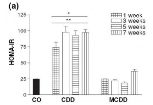
Choline deficiency (CD) by itself induces metabolic syndrome (indicated by insulin resistance and elevated serum triglycerides and cholesterol) and obesity.

A combined methionine and choline deficiency (MCD) actually causes weight loss and reduces serum triglycerides and cholesterol, but induces more severe liver damage. The MCD diet prevents the body from manufacturing choline from methionine, vitamin B12, and folate, so MCD diets severely reduce choline levels; and without choline VLDL particles are not produced. Without VLDL particles, fats and cholesterol are trapped in the liver and never reach the blood and adipose cells.

Here is a measure of insulin resistance on the two diets:

The induction of insulin resistance by the CD diet is very rapid, requiring less than a week.

Induction of insulin resistance is thought to be mediated by elevated TNF-alpha production by adipose cells and by hypertriglyceridemia. Since the MCD diet neither raised serum triglycerides nor caused obesity which induces TNF-alpha production in adipose cells, it did not cause insulin resistance.

What Does This Have to Do With Diabetes?

Insulin resistance is a key step in the development of diabetes:

  • Insulin resistance in the liver causes the liver to release more glucose into the blood (since insulin inhibits glucose release by the liver). This is discussed in a nice paper [3] found by LynMarie Daye and cited in the comments by CarbSane.
  • Peripheral insulin resistance means that the rest of the body is less sensitive to insulin. The pancreas has to produce more insulin to dispose of the excess glucose that the liver is releasing.

This elevation of insulin and glucose levels is a crucial step toward diabetes; it is “pre-diabetes.”

Persistently elevated glucose levels can then poison the beta cells of the pancreas, diminishing insulin secretion capability and causing diabetes. [4]

The Rose Corn Oil trial was not a low-carb diet, so postprandial glucose levels could easily have risen to toxic levels.

If a CD diet can cause insulin resistance in a week, it’s plausible that it might cause diabetes in 12 to 18 months, which is when the Rose Corn Oil trial patients developed it.

What About the Thromboembolism Cases?

MCD diets induce fibrinogenesis. In the blood, excess fibrin formation leads to clotting, and clots can block vessels to cause thromboembolisms. It may be that the thromboembolism cases in the Rose Corn Oil trial had methionine, folate, or B12 deficiencies to go with their choline deficiency.

Why Do Plant Oils Induce Diabetes But Not Animal Fats?

So why did diabetes develop in the corn and olive oil arms of the Rose Corn Oil trial but not the “fatty meat and dairy” arm?

Well, look at the choline content of these foods:

Choline content of one cup (~200 g) oil or fat or 227 g (1/2 lb) meat

Beef liver 968.0 mg
Cube steak (beef) 290.0 mg
Beef tallow 164.0 mg
Butter 42.7 mg
Olive oil 0.6 mg
Corn oil 0.4 mg

Source: http://nutritiondata.com.

Take away meat and dairy and replace them with plant oils, and it’s very easy to have a choline deficiency.

What Does This Have to Do With Zero-Carb Diets?

Maybe nothing … without carb consumption, postprandial glucose levels are not as high, and beta cell poisoning is less likely … but it may be that a zero-carb diet aggravates a choline deficiency in some fashion. I will leave this as a topic for further research.

UPDATE: Leonie in a new comment gives us more information: she has PCOS, goiter with nodules, and auto-antibodies. This suggests autoimmunity as a more likely explanation for her zero-carb diabetes.

Conclusion

In the book, we recommend the use of animal fats such as beef tallow for cooking, and recommend that pregnant women and vegetarians supplement with choline. We thought seriously about recommending that everyone supplement choline, but were reluctant to recommend too many supplements.

In retrospect, we should have recommended choline supplements for everyone who is overweight, has elevated blood glucose or lipids, or has elevated liver enzymes.

We have been using beef tallow as our cooking oil for several months now. It might be good practice for everyone to favor animal fats like beef tallow over plant oils for cooking.

References

[1] Rose GA et al. Corn oil in the treatment of ischaemic heart disease.  Br Med J. 1965 Jun 12;1(5449):1531-3. http://pmid.us/14288105.

[2] Veteläinen R et al. Essential pathogenic and metabolic differences in steatosis induced by choline or methione-choline deficient diets in a rat model. J Gastroenterol Hepatol. 2007 Sep;22(9):1526-33. http://pmid.us/17716355.

[3] Sonksen P, Sonksen J. Insulin: understanding its action in health and disease. Br J Anaesth. 2000 Jul;85(1):69-79. http://pmid.us/10927996.

[4] Leibowitz G et al. Glucose regulation of ?-cell stress in type 2 diabetes. Diabetes Obes Metab. 2010 Oct;12 Suppl 2:66-75. http://pmid.us/21029302.