Category Archives: Nutrients - Page 8

Mice Who Tear Their Fur Out and The Psychiatrists Who Treat Them

Chris Highcock of Conditioning Research mentioned a fascinating paper yesterday, and then Dr. Emily Deans blogged about it. The paper tells about mice who tore their fur out – akin to the condition of “trichotillomania” in which humans tear their hair out – after being put on a high-tryptophan diet. [1]

Dr.Deans points to the paper’s importance:

As far as I know, it may be the only paper showing a definitive development of psychopathology with an adjustment of diet.  So that’s a big deal!

Since I suspect that most psychopathologies are induced by diet in the context of infection, I think this shows that psychiatric researchers have barely begun to understand their diseases.

As soon as I saw Chris’s post I knew I had to blog about it, because I had similar symptoms to these mice.

My Experience

Briefly, I had a chronic bacterial infection of the brain and nerves, probably from Chlamydophila pneumoniae, plus a few other problems which masked the bacterial infection until I fixed my diet.

C. pneumoniae is a parasitic intracellular bacteria whose main activities are reproduction and diversion of the immune system. Its main effects are:

  • Neuronal hypoglycemia. C. pneumoniae steals glucose products like pyruvate for energy. This can create hypoglycemia in neurons even if blood glucose levels are normal.
  • Serotonin deficiency. C. pneumoniae steals key amino acids like tryptophan, tyrosine, and phenylalanine for protein and niacin synthesis. Of these tryptophan is most important. To block C. pneumoniae activity, the innate immune response triggered by interferon gamma sequesters tryptophan. This denudes neurons of the neurotransmitter serotonin, which is made from tryptophan.
  • Inflammation. C. pneumoniae is able to trigger inflammation which re-directs the immune response away from itself toward extracellular pathogens.

Thus common symptoms of a bacterial infection of the brain are those of cognitive hypoglycemia and serotonin deficiency. Symptoms include:

  • Hypoglycemia : Feeling nervous or jittery; mood changes such as irritability, anxiety, restlessness; confusion, difficulty in thinking, and inability to concentrate; poor coordination.
  • Serotonin deficiency: Anxiety, depression, impaired memory or cognition, low self-esteem, loss of pleasure, poor impulse control, insomnia.

These lists don’t fully capture the experience however.

I started having these symptoms in 1992 during a year-long course of antibiotics, and they would get worse for about the next 15 years. I experienced a dramatic loss of happiness and positive emotions. I had always been happy; now suddenly I wasn’t. Along with this came a weird mental state which is hard to describe, because it has no normal analog. Irritability or anger come closest, so I’ll use those words. But understand that it was a generalized state, not irritation or anger directed at anyone in particular; being naturally phlegmatic, I doubt in 20 years I was uncivil to anyone on more than a few occasions. It was just a persistent irritated/angry emotional state that I was well aware was unnatural and could consciously control.

It seemed like this negative emotional state would build up, and could be discharged a bit by a few expressive habits. I would wring my hands; I still have some slightly twisted finger bones and calluses from over a decade of hand-wringing. And, when alone, I would sometimes scratch my head. This sometimes led to hair loss and bare patches.

Trichotillomania

This kind of behavior turns out to be not that rare. About 4% of the population is said to have “trichotillomania,” compulsive pulling or twisting of the hair causing hair loss. Trichotillomania strikes women more frequently than men. [Wikipedia, “Trichotillomania” ]

Serotonin depletion is a common feature of mood disorders. I wouldn’t be surprised if most of these disorders are due to brain infections, and the serotonin deficiency is due either to theft of tryptophan by bacteria or to the immune response to intracellular infections, which increases interferon gamma and decreases serotonin.

Evidence, such as it is, is consistent with that idea. People with mood disorders or depression are far more likely than normal people to test positive for antibodies to chronic intracellular pathogens like coronaviruses. [2]

Drugs Help At First, But Often Do Long-Term Harm

The first impulse of modern medicine is to fight the body’s response to disease. If the body has downregulated serotonin, doctors look for drugs that upregulate it.

That is why people with depression and mood disorders are commonly given SSRI’s, drugs that raise serotonin levels.

If these diseases are due to infections, then we would expect the SSRI’s to improve mood immediately, but also to defeat the body’s immune response, supply the pathogens with tryptophan, and promote their replication. As a result, the disease should progress faster. In time, the patient will become worse than would have been the case without the drugs.

And, more often than not, this is what actually happens. Drugs are often “unsafe at any dose”. Antidepressant treatment increases mortality in men by 30%.

The Mice Who Tear Their Hair Out

One of the common breeds of mice used in laboratory research is the C57BL/6 breed. This breed has “an easily irritable temperament … [and] a tendency to bite … [and] display barbering behavior.” [Wikipedia, “C57BL/6”] In barbering, “individuals pluck whiskers and/or fur from their cage-mates and/or themselves.” [1]

C57BL/6 mice also have a modified immune response:

The immune response of mice from the C57BL/6 strain distinguish it from other inbred strains like BALB/c. For example the immunological response to the same pathogen in C57BL/6 mice is often of an opposite spectrum compared to BALBb/c mice, namely C57BL/6 shows Th1 and BALB/c shows Th2 response in response to intracellular pathogen Leishmania major, where a Th1 response results in a resistant ie healer phenotype (since the pathogen is intracellular), whereas a Th2 response results in a susceptible (nonhealer) phenotype. [Wikipedia, “C57BL/6”]

This Th1 response increases interferon gamma levels:

The Th1 response is characterized by the production of Interferon-gamma … [Wikipedia, “Adaptive Immune System”]

Interferon gamma, of course, sequesters tryptophan and diminishes neuronal serotonin levels.

All this sounds familiar: C57BL/6 have lower serotonin; they become irritable and will bite and tear fur out.

Like trichotillomania in humans, tearing of fur is more common in female mice than males:  “Barbering is more frequently seen in female mice; male mice are more likely to display dominance through fighting.” [Wikipedia, “C57BL/6”]

Research Idea: Treat the Mice As We Do Humans

If these mice went to a human psychiatrist (and had health insurance), they’d be prescribed SSRIs to raise their serotonin levels.

A group at Purdue led by professor of animal sciences Joseph Garner decided to see if they could cure barbering through an alternative dietary therapy that would raise serotonin levels just like SSRIs.

[W]e wished to test the hypothesis that a diet which increases serotonin metabolism would decrease the hair-plucking behavior of barbering mice. [1]

The treatment diet was essentially identical to the control diet, except for these differences: Tryptophan levels were four times higher, methionine levels were the same, and other amino acids were halved. Overall protein levels were cut from 24% to 13.3% of calories. Since tryptophan competes with other amino acids for entry to the brain, this shift in amino acid composition led to much larger tryptophan entry to the brain. [1, Table 2] The lost protein calories were made up by increasing carb intake from 57.3% to 68.0%, which I consider a relatively marginal change. In both diets fructose was minimal, 2.5% of calories, and glucose, mostly from starch or dextrose, provided the bulk of the carbs.

The tryptophan was converted to serotonin in the brain, but not for long. Serotonin levels were 55.5 ng/ml in brains of mice on the control diet, 57.6 ng/ml in brains of mice on the high-tryptophan diet [1, Table 3]. However, levels of serotonin metabolites – the leftovers after serotonin destruction – were much higher in the treatment mice.

The results weren’t good:

[E]levating brain serotonin metabolism by tryptophan and carbohydrate supplementation increased the severity of barbering, and induced ulcerative dermatitis. In humans, the induction of compulsive skin-picking by serotonergic agents (SSRIs) has been reported. (24,43) Thus, the current data suggest a homologous outcome in mice, achieved nutritionally instead of pharmacologically. [1]

If you don’t like scientific-ese, here’s Professor Garner in the press release:

[The] diet … was expected to reduce abnormal hair-pulling. Instead, mice that were already ill worsened their hair-pulling behaviors or started a new self-injurious scratching behavior, and the seemingly healthy mice developed the same abnormal behaviors….

“We put them on this diet, and it made them much, much worse,” Garner said.

This does indeed sound like a “homologous outcome” to the experience of human patients treated with SSRIs!

Very likely if the mice had been interviewed at Day 1 after initiation of the high-tryptophan, they would have reported mood improvements, just as human patients do on SSRIs. As with humans on SSRIs, the negative effects took some time to appear. The increase in scratching behavior was not apparent at 6 weeks after initiation of the high-tryptophan diet, but was apparent at 12 weeks (3 months) [1, Figure 4]. Ulcerative dermatitis tended to appear after about 10 weeks on the high-tryptophan diet [1, Figure 3]. 

Conclusion

I’ll follow up in my next post, on Monday, with speculation about what is happening in these mice.

In the meantime, I think it is worth remarking how an intervention thought to be beneficial – restoring serotonin levels to “normal” – has health-impairing consequences over time in both mice and people.

In many ways, contemporary medical practitioners resemble the Sorcerer’s Apprentice. They have at their disposal powerful magic drugs, whose long-term consequences they do not fully understand. The drugs come into wide use, and only years later do we learn that they do more harm than good. And the data showing they don’t work is always “surprising” and “paradoxical.”

In my view, the philosophy behind drug-based medicine is misplaced. Too often drugs are designed to fight or defeat the body’s natural mechanisms. As my parable argued, I believe it is much more effective to cooperate with the body through diet and nutrition.

References

[1] Dufour BD et al. Nutritional up-regulation of serotonin paradoxically induces compulsive behavior. Nutr Neurosci. 2010 Dec;13(6):256-64. http://pmid.us/21040623.

[2] Okusaga O et al. Association of seropositivity for influenza and coronaviruses with history of mood disorders and suicide attempts. J Affect Disord. 2010 Oct 26. [Epub ahead of print]. http://pmid.us/21030090. Hat tip Dr. Deans, http://evolutionarypsychiatry.blogspot.com/2010/11/depression-flu-and-to-do.html.

Micronutrient Deficiencies: An Underappreciated Cause of Hypothyroidism

A significant number of our readers have hypothyroidism with normal T4 but low T3. For instance, Kratos:

I followed a strict low carb diet with around 50g of carb per day for over 1 year and I think I have developed hypothyroidism …

TSH 3.4 (0.3-4.0)

FT3 2.2 (2.1-4.9)

FT4 11.4 (6.8-18.0)

This situation can have many causes. Our last post discussed how shift work and disrupted circadian rhythms can cause hypothyroidism. Another often-overlooked cause of hypothyroidism is nutrient deficiencies.

As noted in the book, selenium and iodine deficiencies are classic causes of hypothyroidism. Here I want to look at a few other possiblities.

Copper and Iron Deficiency

Copper deficiency, iron deficiency, and iodine deficiency during pregnancy or infancy generate similar neurological defects, and during adulthood generate similar hypothyroid symptoms:

Cu, Fe, and iodine/TH deficiencies result in similar defects in rodent brain development, including hypomyelination of axons, aberrant hippocampal structure and function, altered brain energy metabolism, and altered neuronal signaling (8–13). In addition, the behavioral and neurochemical abnormalities associated with perinatal Cu, Fe, and iodine/TH deficiencies are irreversible and persist into adulthood (14–16). These similarities suggest that there may be a common underlying mechanism associated with all three deficiencies contributing to the observed neurodevelopmental defects.

Several studies in postweanling rodents show that Cuand Fe deficiencies impair thyroid metabolism. Fe deficiency reduces circulating thyroxine (T4) and triiodothyronine (T3) concentrations (17–20), peripheral conversion of T4 to T3 (18, 19), TSH response to TRH (19), and thyroid peroxidase (TPO) activity (20). Cu deficiency also reduces circulating T4 andT3 concentrations and peripheral conversion of T4 to T3  (21, 22). In addition, Cu deficiency reduces serum and brain Fe levels, which may contribute to the Cu-dependent effect on thyroidal status (23). [1]

In infant rats, deficiencies of either copper or iron cause hypothyroidism:

Cu deficiency reduced serum total T(3) by 48%, serum total T(4) by 21%, and whole-brain T(3) by 10% at P12. Fe deficiency reduced serum total T(3) by 43%, serum total T(4) by 67%, and whole-brain T(3) by 25% at P12. [1]

Note that copper deficiency hypothyroidism reduces serum T3 levels more strongly than T4 levels, the same pattern that Kratos displays.

While We’re On the Topic of Micronutrients and Hypothyroidism …

Hypothyroidism induces the symptoms of riboflavin deficiency. This is because thyroid hormone is needed for production of the enzyme flavin kinase, which is in turn needed to generate flavin adenine dinucleotide (FAD). Riboflavin deficiency and thyroid hormone deficiency lead to the same low FAD levels in both rats and humans. [2]

This suggests that hypothyroid persons may wish to supplement with riboflavin, so that extra riboflavin may help make up for deficient flavin kinase.

Conclusion

I believe that those with health problems should strive to “overnourish” themselves. Micronutrient deficiencies can have insidious disabling effects, yet be impossible to diagnose. In disease conditions, needs for many micronutrients are increased. Many micronutrients are non-toxic up to fairly large doses and can be safely supplemented.

An effort to eat micronutritious foods and supplement micronutrients into their “plateau ranges” to eliminate deficiencies might generate startling health improvements.

Minerals like copper, selenium, and iodine are among the most important nutrients – they are among our eight essential supplements – yet also among the most widely deficient. Most supplementers neglect key minerals; but optimizing their intake can pay large health dividends.

References

[1] Bastian TW et al. Perinatal iron and copper deficiencies alter neonatal rat circulating and brain thyroid hormone concentrations. Endocrinology. 2010 Aug;151(8):4055-65. http://pmid.us/20573724.

[2] Cimino JA et al. Riboflavin metabolism in the hypothyroid newborn. Am J Clin Nutr. 1988 Mar;47(3):481-3. http://pmid.us/3348160.

Danger of Zero-Carb Diets III: Scurvy

I started low-carb Paleo dieting in late 2005. I ate a lot of vegetables but no starches and hardly any fruit. In retrospect, I would call it a near zero-carb diet. At that time I was 12 years into a chronic illness that got a little worse each year and was quite mysterious to me. Adopting a low-carb diet brought immediate changes: it made what I would much later recognize as a chronic bacterial infection better (in parts of the body, not the brain) and made a chronic fungal infection worse.

Within about a year I had developed scurvy. It took me an embarrassingly long time to figure out what it was. By the time knew what it was, I had 3 cavities; had lost 25 pounds; had developed diverticulitis and an abdominal aorta that visibly swelled with every heartbeat; and had minor skin wounds – scrapes and scratches – that hadn’t healed in 6 months.

Developing scurvy was a surprise, because I was eating many vegetables plus taking a multivitamin containing 90 mg of vitamin C. I had never had any signs of vitamin C deficiency before adopting a low-carb diet.

Four grams a day of vitamin C for two months cured all the scurvy symptoms. It would be several more years before I figured out the infections, but this experience taught me the importance of micronutrition. The experience persuaded me that I needed to research diets and nutrition closely, and started us down the path of writing Perfect Health Diet.

Scurvy on a Ketogenic Diet

My experience is not unique. Here’s one case we mention in the book: the story of a young girl with epilepsy.

KM was a 9-year old girl … diagnosed with epilepsy at six months old. She started a ketogenic diet in October 2003, as her multiple antiepileptic drugs were proving to be less than effective; indeed she was having as many as 12 tonic seizures per day with prolonged periods of non-convulsive status epilepticus. After the diet was prescribed the seizure frequency reduced markedly and there were a number of long periods of time in which she had no seizures.

KM’s mother gave a history of her daughter having had bleeding gums since the beginning of September 2006; she described them as being very dark red, swollen and bleeding. In addition, she explained that her daughter had dry, crusted blood peri-orally. The family’s general dental practitioner had explained that this was probably caused by erupting teeth and instructed her to use 0.2% chlorhexidine gluconate gel and to continue her regular oral hygiene regimen; however this had no effect. About a month later the patient’s right arm became swollen. It was thought that she had sustained a fracture or a dislocation; however she was discharged from the local hospital’s fracture clinic because there was clinical improvement and radiographs showed no callus formation.

In early November KM inhaled a primary molar tooth while she was having her teeth cleaned (Fig. 1). This required an emergency bronchoscopy to retrieve it; at the same time the surgeons extracted her remaining primary teeth in order to avoid a recurrence of the problem….

At that time an appointment was made to attend a paediatric dentistry consultant clinic at the Leeds Dental Institute; however this was never kept as about three weeks after the extractions the patient was admitted to hospital with low grade fever, persistently bleeding gums, oedema of her hands and feet and a petechial rash on her legs. [1]

This girl was eating a typical amount of vitamin C: her dietary intake was calculated at 73 mg/day, well above the US RDA for 9-13 year olds of 45 mg/day. Yet her blood level was only 0.7 µmol/l. Scurvy is diagnosed at levels below 11 µmol/l.

The symptoms of scurvy are sufficiently insidious that it is easy to miss the diagnosis. In KM’s case, it happened that a “senior house officer” – a junior doctor in training – from India recognized the symptoms of scurvy. Otherwise, it might have never have occurred to the doctors to test her vitamin C level. [2]

What Is the Cause of Low-Carb Scurvy?

So what causes scurvy to develop on low-carb diets even with vitamin C intake well above the US RDA?

It seems to be a confluence of two factors:

  • An infection or some other stress (e.g. injury, cancer) leads to the oxidation of extracellular vitamin C; and
  • On a low-insulin or glutathione-deficiency-inducing diet, oxidized vitamin C is not recycled.

Infection and Vitamin C

The immune response to infections generates reactive oxygen species, which oxidize vitamin C. Oxidation removes a hydrogen atom from vitamin C, turning it into “dehydroascorbic acid,” or DHAA. If DHAA remains in the blood, it degrades with a half-life of 6 minutes. [3]

Infections can cause vitamin C deficiency on any diet. During the “acute phase response” to infection or injury, vitamin C often becomes deficient. Here is a nice paper in which French doctors surveyed their hospital patients for scurvy:

We determined serum ascorbic acid level (SAAL) and searched for clinical and biological signs of scurvy in 184 patients hospitalized during a 2-month period.

RESULTS: The prevalence of hypovitaminosis C (depletion: SAAL<5 mg/l or deficiency: SAAL<2 mg/l) was 47.3%. Some 16.9% of the patients had vitamin C deficiency. There was a strong association between hypovitaminosis C and the presence of an acute phase response (p=0.002). [4]

So half were at least depleted in vitamin C and 17% had outright deficiency, which if it persisted would produce scurvy.

We’ve previously written of how important it is to supplement with vitamin C during infections:

I might add here that in sepsis, an extremely dangerous inflammatory state brought on by bacterial infections, intravenous vitamin C reverses some of the worst symptoms. [5] If you have a loved one suffering from a dangerous infection, it might not be a bad idea to get them some vitamin C.

Insulin Dependence of Vitamin C Recycling

DHAA can be recycled back into vitamin C, but only inside cells.

In order to enter cells, DHAA needs to be transported by glucose transporters. GLUT1, GLUT3, and GLUT4 are the three human DHAA transporters; GLUT1 does most of the work. [6]

DHAA transport is crucial for brain vitamin C status. There is no direct transport of vitamin C into the brain, yet the brain is one of the most vitamin C-dependent tissues in the body. The brain relies entirely on GLUT1-mediated transport of DHAA from the blood for its vitamin C supply. Within the brain, DHAA is restored to vitamin C by glutathione.

Supplying DHAA to stroke victims (of the mouse persuasion) as late as 3 hours after the stroke can reduce the stroke-damaged volume by up to 95%:

DHA (250 mg/kg or 500 mg/kg) administered at 3 h postischemia reduced infarct volume by 6- to 9-fold, to only 5% with the highest DHA dose (P < 0.05). [7]

This is a fascinating reminder of the importance of vitamin C for wound repair and protection from injury.

Glucose transporters are activated by insulin. Thus, DHAA import into cells is increased by insulin, leading to more effective recycling of vitamin C [8]:

Insulin and IGF-1 promote recycling of DHAA into ascorbate. Source.

Confirming the role of insulin in promoting vitamin C recycling, Type I diabetics (who lack insulin) have lower blood levels of vitamin C, higher blood levels of DHAA, increased urinary loss of vitamin C metabolites, and greater need for dietary vitamin C. [9, 10]

Now we have a mechanism by which zero-carb diets reduce vitamin C recycling: by lowering insulin levels they inhibit the transport of DHAA into cells, preventing its recycling into vitamin C. Instead, DHAA is degraded and excreted. As a result, vitamin C is lost from the body.

Glutathione and Vitamin C Recycling

Once inside the cell, DHAA is recycled back to ascorbate, mainly by glutathione inside mitochondria:

Dehydroascorbate, the fully oxidized form of vitamin C, is reduced spontaneously by glutathione, as well as enzymatically in reactions using glutathione or NADPH. [11]

A GLUT1 transporter on the mitochondrial membrane is needed to bring DHAA into mitochondria, possibly squaring the effect of insulin on vitamin C recycling.

Since glutathione recycles vitamin C, glutathione deficiency is another possible cause of vitamin C deficiency.

Glutathione is recycled by the enzyme glutathione peroxidase, a selenium-containing enzyme whose abundance is sensitive to selenium status. One difficulty with zero-carb diets is that they seem to deplete selenium levels.

Selenium deficiency is a common side effect of ketogenic diets. Some epileptic children on ketogenic diets have died from selenium deficiency! [12]

So here we have a second mechanism contributing to the development of scurvy on a zero-carb diet. The diet produces a selenium deficiency, which produces a glutathione deficiency, which prevents DHAA from being recycled into vitamin C, which leads to DHAA degradation and permanent loss of vitamin C.

Conclusion

Zero-carb dieters are at high risk for vitamin C deficiency, glutathione deficiency, and selenium deficiency. Anyone on a zero-carb diet should remedy these by supplementation.

These deficiencies are exacerbated by chronically low insulin levels. Insulin helps recycle vitamin C, which supports glutathione status. Lack of insulin increases vitamin C degradation and loss.

The failure of the body to efficiently recycle vitamin C and maintain antioxidant stores on a zero-carb diet is evidence of an evolutionary maladaption to the zero-carb diet.

There was no reason why our ancestors should have become adapted to a zero-carb diet; after, all they’ve been eating starches for at least 2 million years. It seems a risky step to try to live this way.

Related Posts

Other posts in this series:

  1. Dangers of Zero-Carb Diets, I: Can There Be a Carbohydrate Deficiency? Nov 10, 2010.
  2. Dangers of Zero-Carb Diets, II: Mucus Deficiency and Gastrointestinal Cancers A Nov 15, 2010.
  3. Dangers of Zero-Carb Diets, IV: Kidney Stones Nov 23, 2010.

References

[1] Willmott NS, Bryan RA. Case report: Scurvy in an epileptic child on a ketogenic diet with oral complications.  Eur Arch Paediatr Dent. 2008 Sep;9(3):148-52. http://pmid.us/18793598.

[2] Willmott NS, personal communication.

[3] “Dehydroascorbate,” Wikipedia, http://en.wikipedia.org/wiki/Dehydroascorbate.

[4] Fain O et al. Hypovitaminosis C in hospitalized patients. Eur J Intern Med. 2003 Nov;14(7):419-425. http://pmid.us/14614974.

[5] Tyml K et al. Delayed ascorbate bolus protects against maldistribution of microvascular blood flow in septic rat skeletal muscle. Crit Care Med. 2005 Aug;33(8):1823-8. http://pmid.us/16096461.

[6] Rivas CI et al. Vitamin C transporters. J Physiol Biochem. 2008 Dec;64(4):357-75. http://pmid.us/19391462.

[7] Huang J et al. Dehydroascorbic acid, a blood-brain barrier transportable form of vitamin C, mediates potent cerebroprotection in experimental stroke. Proc Natl Acad Sci U S A. 2001 Sep 25;98(20):11720-4. http://pmid.us/11573006.

[8] Qutob S et al. Insulin stimulates vitamin C recycling and ascorbate accumulation in osteoblastic cells. Endocrinology. 1998 Jan;139(1):51-6. http://pmid.us/9421397.

[9] Will JC, Byers T. Does diabetes mellitus increase the requirement for vitamin C? Nutr Rev. 1996 Jul;54(7):193-202. http://pmid.us/8918139.

[10] Seghieri G et al. Renal excretion of ascorbic acid in insulin dependent diabetes mellitus. Int J Vitam Nutr Res. 1994;64(2):119-24. http://pmid.us/7960490.

[11] Linster CL, Van Schaftingen E. Vitamin C. Biosynthesis, recycling and degradation in mammals. FEBS J. 2007 Jan;274(1):1-22. http://pmid.us/17222174.

[12] Bank IM et al. Sudden cardiac death in association with the ketogenic diet. Pediatr Neurol. 2008 Dec;39(6):429-31. http://pmid.us/19027591. (Hat tip Dr. Deans.)

Choline Deficiency and Plant Oil Induced Diabetes

I’m going to deviate from my original plan for the “Dangers of a Zero-Carb Diet” series to discuss a topic that came up in the comments to the first post.

Leonie’s Diabetes and the Rose Corn Oil Trial

What prompted this diversion is Leonie’s interesting comment from Wednesday’s post:

I developed diabetes several years after being on a low carb diet. Continuing low carb to manage the diabetes did not halt its progress. It has taken about 18 months of adding more carbs (60 – 100 gr/day) to my diet to bring my fasting glucose down by a couple of mmol and eating more carbs has also lowered my Hba1c and post meal spikes significantly. I wonder if the liver is another organ that may be affected by carbohydrate deficiency.

I had not heard of such cases before, or so I thought, but Dr. Deans in the comments reminded us that Peter at Hyperlipid had noticed two similar cases in the Rose Corn Oil trial. [1] (The Rose Corn Oil trial, of course, figures prominently in our book’s discussion of PUFA toxicity.)

In the Rose Corn Oil trial, there were three arms – a normal diet arm, a high corn oil arm, and a high olive oil arm. The normal dieters were expected to eat “fried foods, fatty meat, sausages, … ice cream, cheese, … milk, eggs, and butter” while the oil arms were supposed to restrict these foods and replace them with corn or olive oil.

Here’s what happened:

Four patients were removed from the trial for other reasons. Two developed non-cardiac thromboembolism and were given anticoagulant therapy. The other two were removed because of diabetes mellitus. One of them already had mild diabetes, but glycosuria increased considerably soon after he started oil. Oil was stopped and glycosuria disappeared. Oil was restarted, but was stopped a month later because heavy glycosuria recurred. The other patient, not a previously recognized diabetic, developed glycosuria with a diabetic glucose-tolerance test a few weeks after starting oil. [1]

The patients who developed diabetes came one from the corn oil arm and one from the olive oil arm. Likewise, the patients who developed thromboembolisms came one from the corn oil arm and one from the olive oil arm. No such disasters occurred on the “fatty meat” arm.

Since all three diets were similarly fatty, it doesn’t appear to be the quantity of fat that was the issue. Rather it was the type of lipid, or some micronutrient that was present in the animal and dairy foods but lacking in the plant oils.

For insight into what the problem might be, let’s look at how scientists poison lab animals.

Insights from Diet Animal Poisoning Research

You have to pity diet researchers. It takes 60 years for bad diets to poison humans enough to significantly raise mortality rates. Yet a diet researcher is supposed to gain a Ph.D. in 4 years (or in 5 while simultaneously obtaining an MD!), do a postdoc in 2 years, win a grant in the first years of an entry-level position with PI status, and then demonstrate productive results within the term of a 2-to-5 year grant. Deadlines are pressing: A study needs to start rats or mice on two diets, and have one diet produce much better health than the other, in considerably less than a two-year time frame.

Just comparing McDonald’s fast food with a Mediterranean diet won’t do. Two years later both sets of mice will die happily of old age, with no significant differences between groups. Peer reviewers judge you to have discovered no new results. No new results means no paper, no grant, no job.

So “diet” researchers first have to become experts at quickly inducing disease in rats and mice. Find a diet that poisons animals in a few months, compare it to another diet that doesn’t, and you have a paper. Look for variations that slow or hasten the poisoning, and you have more papers. To be a highly productive scientist, one must be a skilled animal poisoner.

Various techniques have been developed for this purpose, including: knocking out some crucial gene; breeding a mutant strain that naturally develops disease; giving the animals poison with their food; or depriving them of crucial nutrients. Almost every study of diet in mice or rats uses one of these techniques.

If a missing nutrient can cause diabetes within a few years for Leonie and 12 to 18 months for the Rose Corn Oil trial volunteers, it’s likely to be pretty good at inducing disease in animals too. There’s a good chance diet animal poisoning researchers have already stumbled upon it in rats or mice.

Choline Deficiency Diseases

One of the most popular deficiency diets among researchers is the choline-deficient diet. A useful paper by Dutch scientists [2] gives a nice look at the impact of choline deficiency on rats.

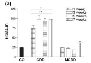
Choline deficiency (CD) by itself induces metabolic syndrome (indicated by insulin resistance and elevated serum triglycerides and cholesterol) and obesity.

A combined methionine and choline deficiency (MCD) actually causes weight loss and reduces serum triglycerides and cholesterol, but induces more severe liver damage. The MCD diet prevents the body from manufacturing choline from methionine, vitamin B12, and folate, so MCD diets severely reduce choline levels; and without choline VLDL particles are not produced. Without VLDL particles, fats and cholesterol are trapped in the liver and never reach the blood and adipose cells.

Here is a measure of insulin resistance on the two diets:

The induction of insulin resistance by the CD diet is very rapid, requiring less than a week.

Induction of insulin resistance is thought to be mediated by elevated TNF-alpha production by adipose cells and by hypertriglyceridemia. Since the MCD diet neither raised serum triglycerides nor caused obesity which induces TNF-alpha production in adipose cells, it did not cause insulin resistance.

What Does This Have to Do With Diabetes?

Insulin resistance is a key step in the development of diabetes:

  • Insulin resistance in the liver causes the liver to release more glucose into the blood (since insulin inhibits glucose release by the liver). This is discussed in a nice paper [3] found by LynMarie Daye and cited in the comments by CarbSane.
  • Peripheral insulin resistance means that the rest of the body is less sensitive to insulin. The pancreas has to produce more insulin to dispose of the excess glucose that the liver is releasing.

This elevation of insulin and glucose levels is a crucial step toward diabetes; it is “pre-diabetes.”

Persistently elevated glucose levels can then poison the beta cells of the pancreas, diminishing insulin secretion capability and causing diabetes. [4]

The Rose Corn Oil trial was not a low-carb diet, so postprandial glucose levels could easily have risen to toxic levels.

If a CD diet can cause insulin resistance in a week, it’s plausible that it might cause diabetes in 12 to 18 months, which is when the Rose Corn Oil trial patients developed it.

What About the Thromboembolism Cases?

MCD diets induce fibrinogenesis. In the blood, excess fibrin formation leads to clotting, and clots can block vessels to cause thromboembolisms. It may be that the thromboembolism cases in the Rose Corn Oil trial had methionine, folate, or B12 deficiencies to go with their choline deficiency.

Why Do Plant Oils Induce Diabetes But Not Animal Fats?

So why did diabetes develop in the corn and olive oil arms of the Rose Corn Oil trial but not the “fatty meat and dairy” arm?

Well, look at the choline content of these foods:

Choline content of one cup (~200 g) oil or fat or 227 g (1/2 lb) meat

Beef liver 968.0 mg
Cube steak (beef) 290.0 mg
Beef tallow 164.0 mg
Butter 42.7 mg
Olive oil 0.6 mg
Corn oil 0.4 mg

Source: http://nutritiondata.com.

Take away meat and dairy and replace them with plant oils, and it’s very easy to have a choline deficiency.

What Does This Have to Do With Zero-Carb Diets?

Maybe nothing … without carb consumption, postprandial glucose levels are not as high, and beta cell poisoning is less likely … but it may be that a zero-carb diet aggravates a choline deficiency in some fashion. I will leave this as a topic for further research.

UPDATE: Leonie in a new comment gives us more information: she has PCOS, goiter with nodules, and auto-antibodies. This suggests autoimmunity as a more likely explanation for her zero-carb diabetes.

Conclusion

In the book, we recommend the use of animal fats such as beef tallow for cooking, and recommend that pregnant women and vegetarians supplement with choline. We thought seriously about recommending that everyone supplement choline, but were reluctant to recommend too many supplements.

In retrospect, we should have recommended choline supplements for everyone who is overweight, has elevated blood glucose or lipids, or has elevated liver enzymes.

We have been using beef tallow as our cooking oil for several months now. It might be good practice for everyone to favor animal fats like beef tallow over plant oils for cooking.

References

[1] Rose GA et al. Corn oil in the treatment of ischaemic heart disease.  Br Med J. 1965 Jun 12;1(5449):1531-3. http://pmid.us/14288105.

[2] Veteläinen R et al. Essential pathogenic and metabolic differences in steatosis induced by choline or methione-choline deficient diets in a rat model. J Gastroenterol Hepatol. 2007 Sep;22(9):1526-33. http://pmid.us/17716355.

[3] Sonksen P, Sonksen J. Insulin: understanding its action in health and disease. Br J Anaesth. 2000 Jul;85(1):69-79. http://pmid.us/10927996.

[4] Leibowitz G et al. Glucose regulation of ?-cell stress in type 2 diabetes. Diabetes Obes Metab. 2010 Oct;12 Suppl 2:66-75. http://pmid.us/21029302.