HDL and Immunity

HDL – high-density lipoprotein – particles are good for you: High HDL levels are associated with lower mortality overall and lower mortality from many diseases – not only cardiovascular disease but also cancer and infection.

People with high HDL are only one-sixth as likely to develop pneumonia [1], and in the Leiden 85-Plus study, those with high HDL experienced 35% lower mortality from infection [2].

Each rise of 16.6 mg/dl in HDL reduced the risk of bowel cancer by 22% in the EPIC study. [3]

In terms of overall mortality, in the VA Normative Aging Study, “Each 10-mg/dl increment in HDL cholesterol was associated with a 14% [decrease] in risk of mortality before 85 years of age.” [4]

This must be surprising to those who think HDL is only a carrier of cholesterol. The lipid hypothesis presumed that the function of HDL is to clear toxic cholesterol from arteries, cholesterol having evolved for the purpose of giving us heart attacks. HDL then brings cholesterol to the liver which disposes of it returns it to the blood via LDL (which evolved for the purpose of poisoning arteries with cholesterol, and giving HDL something to do). (Hat tip to Peter for this formulation of the lipid hypothesis.)

But there is an alternative hypothesis: that infections cause disease, and that HDL has an immune function. This hypothesis would explain why HDL protects against infections and against all diseases of aging.

Immune Functions of HDL

I got interested in immune functions of HDL upon reading an article in ScienceDaily last year (“How Disease-Causing Parasite Gets Around Human Innate Immunity,” Sept 13, 2010). The article states:

Several species of African trypanosomes infect non-primate mammals and cause important veterinary disease yet are unable to infect humans. The trypanosomes that cause human disease, Trypanosoma brucei gambiense and T. b. rhodensiense, have evolved mechanisms to avoid the native human defense molecules in the circulatory system that kill the parasites that cause animal disease….

Human innate immunity against most African trypanosomes is mediated by a subclass of HDL (high density lipoprotein, which people know from blood tests as “good cholesterol”) called trypanosome lytic factor-1, or TLF-1….

The parasite that causes fast-onset, acute sleeping sickness in humans, T. b. rhodensiense, is able to cause disease because it has evolved an inhibitor of TLF-1 called Serum Resistance Associated (SRA) protein…. T. b. gambiense resistance to TLF-1 is caused by a marked reduction of TLF-1 uptake by the parasite….

To survive in the bloodstream of humans, these parasites have apparently evolved mutations in the gene encoding a surface protein receptor. These mutations result in a receptor with decreased TLF-1 binding, leading to reduced uptake and thus allow the parasites to avoid the toxicity of TLF-1.

“Humans have evolved TLF-1 as a highly specific toxin against African trypanosomes by tricking the parasite into taking up this HDL because it resembles a nutrient the parasite needs for survival,” said Hajduk, “but T. b. gambiense has evolved a counter measure to these human ‘Trojan horses’ simply by barring the door and not allowing TLF-1 to enter the cell, effectively blocking human innate immunity and leading to infection and ultimately disease.”

So HDL is actually an immune particle carrying proteins that poison pathogens. The TLF-1 HDL subclass consists of those HDL particles carrying two anti-trypanosome proteins, apolipoprotein L-1 and haptoglobin-related protein. [5]

Any HDL particle can become an anti-trypanosome defender simply by acquiring and carrying these proteins.

It turns out that HDL can carry a great assortment of immune proteins. The orchestrator of HDL’s immune functions seems to be a circulating plasma protein called phospholipid transfer protein (PLTP), which forms complexes with immune molecules and then associates with apolipoprotein A-I (the primary HDL protein). PLTP brings 24 different immune molecules into HDL particles, including apolipoproteins such as clusterin (apoJ), coagulation factors, and complement factors. [6] These immune protein complexes add protein but not fat to HDL particles:

Unexpectedly, lipids accounted for only 3% of the mass of the PLTP complexes. Collectively, our observations indicate that PLTP in human plasma resides on lipid-poor complexes dominated by clusterin and proteins implicated in host defense and inflammation. [6]

It looks like HDL may not be primarily a carrier of cholesterol, but rather a carrier of antimicrobial proteins. Its cholesterol and lipids may serve, as the ScienceDaily article suggests, to make the HDL particle attractive to pathogens so that it may enter as a “Trojan Horse.”

HDL-associated immune proteins under strong selection

As pathogens evolve, immune proteins have to evolve. It turns out that apolipoprotein L-1, the immune protein that protects against trypanosomes, is under strong selection in both Africa and Europe.

The version selected in Europe does not protect against Trypanosoma brucei rhodesiense, cause of one of the African sleeping sickness diseases, but the version selected in Africa does. Unfortunately, the African version also increases risk of kidney disease – which may explain why African-Americans have higher rates of kidney disease than white Americans. [7]

So Africans have sacrificed kidney health for greater immunity against sleeping sickness. This suggests that African sleeping sickness may be a relatively recently evolved human disease.

HDL neutralizes toxins

HDL binds bacterial endotoxins, especially lipopolysaccharide (LPS), and neutralizes their toxicity. As a result, people with high HDL have substantially less release of tumor necrosis factor-alpha (TNF-α) during infection. [8]

TNF-α is an inflammatory molecule that stimulates the acute phase response to infections. Levels of C-reactive protein are a good index of TNF-α levels, so generally speaking high HDL will lead to low TNF-α and low CRP.

What’s the best HDL profile?

It should be desirable to have more HDL particles. Since each HDL particle is capable of poisoning a pathogen, the more you have, the stronger your immune defenses.

However, the weight of each HDL particle is likely to be an indicator of infection severity. An infection-free person will have few immune proteins to pick up; the HDL particles will be fat-rich and buoyant. But a person with extensive infections will have heavier HDL particles freighted with immune proteins.

Conventional tests in the doctor’s office measure the weight of HDL in mg per deciliter of blood. Since having more HDL particles (which raises the weight) is good, but having heavy HDL particles indicates infection which is bad, mass is not the best measure of HDL status. We would expect the number or concentration of HDL particles to provide a better indicator of health.

Indeed, this appears to be what is observed. The most important determinant of HDL status is the number of HDL particles:

The association between HDL size and CAD risk was abolished on adjustment for apolipoprotein B and triglyceride levels (adjusted odds ratio, 1.00 [95% CI, 0.71 to 1.39] for top vs. bottom quartile), whereas HDL particle concentration remained independently associated with CAD risk (adjusted odds ratio, 0.50 [CI, 0.37 to 0.66]). [9]

Conclusion

HDL particles are “Trojan Horses” that attack pathogens and neutralize their toxins.

If you want to remain free from infectious diseases – which is to say, all diseases – to a ripe old age, it’s important to make your HDL particles numerous.

On Thursday, I’ll discuss ways to do that.

References

[1] Gruber M et al. Prognostic impact of plasma lipids in patients with lower respiratory tract infections – an observational study. Swiss Med Wkly. 2009 Mar 21;139(11-12):166-72. http://pmid.us/19330560.

[2] Berbée JF et al. Plasma apolipoprotein CI protects against mortality from infection in old age. J Gerontol A Biol Sci Med Sci. 2008 Feb;63(2):122-6. http://pmid.us/18314445

[3] van Duijnhoven FJ et al. Blood lipid and lipoprotein concentrations and colorectal cancer risk in the European Prospective Investigation into Cancer and Nutrition. Gut. 2011 Mar 7. [Epub ahead of print] http://pmid.us/21383385.

[4] Rahilly-Tierney CR et al. Relation Between High-Density Lipoprotein Cholesterol and Survival to Age 85 Years in Men (from the VA Normative Aging Study). Am J Cardiol. 2011 Apr 15;107(8):1173-7. http://pmid.us/21296318.

[5] Kieft R et al. Mechanism of Trypanosoma brucei gambiense (group 1) resistance to human trypanosome lytic factor. Proc Natl Acad Sci U S A. 2010 Sep 14;107(37):16137-16141. http://pmid.us/20805508.

[6] Cheung MC et al. Phospholipid transfer protein in human plasma associates with proteins linked to immunity and inflammation. Biochemistry. 2010 Aug 31;49(34):7314-22. http://pmid.us/20666409.

[7] Genovese G et al. Association of trypanolytic ApoL1 variants with kidney disease in African Americans. Science. 2010 Aug 13;329(5993):841-5. http://pmid.us/20647424.

[8] Henning MF et al. Contribution of the C-terminal end of apolipoprotein AI to neutralization of lipopolysaccharide endotoxic effect. Innate Immun. 2010 May 25. [Epub ahead of print] http://pmid.us/20501516.

[9] El Harchaoui K et al. High-density lipoprotein particle size and concentration and coronary risk. Ann Intern Med. 2009 Jan 20;150(2):84-93. http://pmid.us/19153411.

Pacific Sweet and Sour Salmon

You’ve just come home from work, you’re tired and hungry, and would like to eat in 20 minutes. How do you make a meal that’s easy and quick, but new and different?

The solution is to fry a meat and boil a starch, but use a tasty sauce to give variety to this most easily-cooked meal. One of our favorite sauces is something we call Pacific Sweet&Sour Sauce.

The Sauce

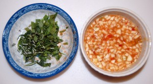
The essential ingredients are rice syrup for sweetness, lemon juice or rice vinegar for sourness, tarragon, rosemary, and garlic. An optional ingredient is any kind of chili powder for spiciness; we used the Vietnamese spicy sauce at top – this one happens to be Huy Fong Chili-Garlic Sauce and is made from chili, salt, garlic, and distilled vinegar.

Of course you can substitute: basil works well as a herb, and coconut milk is an excellent addition to the sauce. Fish sauce gives it a more Vietnamese flavor.

Dice the herbs and set them aside; mince the garlic and mix with the sweet, sour, and spicy ingredients:

The sauce is ready. We used about 1 tbsp rice syrup and juice of a full lemon.

Pacific Sweet&Sour Salmon

Removing the central bone from the salmon steaks speeds up cooking significantly, and finishes the meat when the skin is cooked to just the right crispiness. Here we’ve filleted, and put salt and pepper on, two salmon steaks:

We put a bit of coconut oil in the pan and cooked at medium-high heat.

When the color has changed about 2/3 of the way up the sides of the steaks, which may take 3-4 minutes, they’re ready to flip. After flipping cook another 2 minutes and add the sauce:

With the sauce in cook another 1 minute on one side, and flip again to finish for another minute. Add the herbs in this final minute:

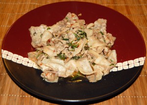
Be sure to keep all the sauce when you transfer to a serving plate:

Pacific Sweet&Sour Pork Bellies

We like pork belly a lot: it is a tasty, fatty cut of pork, and we like the natural flavor better than processed bacon.

If you’re nervous about timing, you don’t have to cook everything together: you can pre-cook the meat, then add it back to the sauce.

Here’s an example. After cooking pork belly slices, remove the meat and add the Pacific Sweet&Sour sauce to the pan oil:

After a minute of heating and stirring, add the herbs:

Return the meat to the pan, mix, and serve:

Pacific Sweet&Sour Beef Liver

We find this sauce works very well with beef liver too:

Shou-Ching, who doesn’t like the taste of beef liver, is happy to eat this.

If you really dislike the taste of liver, you can further remove the liver taste by boiling the beef liver slices briefly before putting them in the pan.

Conclusion

This sort of meal is easily cooked in 20 minutes or so. Alongside the meat, we prepare a starch – usually rice cooked in our rice cooker or warmed in the microwave (one batch lasts us 2-3 days), or potatoes or taro boiled or re-heated in the microwave (again, we prepare enough for 2-3 days) – and serve assorted vegetables – for us, usually various flavors of kimchi and seasoned seaweed. Kimchi and seaweed require no cooking.

We’re having Pacific Sweet&Sour Mussels tonight, with coconut milk in the sauce. Create your own variations! They’ll all taste great.

Around the Web; and Why Is Aspirin Toxic to Cats?

[1] Interesting posts this week: Melissa McEwen assures us: Robb Wolf is not Satan. Kurt Harris’s reader Tara makes the most persuasive case I’ve seen for grass-fed meat through pictures. Emily Deans compares eating disorders to addictions.

[2] Kurt Harris re-re-brands: Paleonu became PaNu became Paleo 2.0 becomes Archevore.com, archevore being a neologism for “one who eats of the essentials.”

Well, it’s more euphonious than “EM2vore,” for “one who eats of the evolutionary metabolic milieu.” A more descriptive name might have been “nontoxivore,” since Kurt’s primary theme is avoidance of “neolithic agents of disease – wheat, excess fructose and excess linoleic acid.”

It will be interesting to see where he’s taking this. Are Archevorean essentials the same as PaNu?

[3] Posts of the week: Chris Masterjohn posts always deserve special notice. On Tuesday he continued his important series investigating whether wheat causes leaky gut, which will trigger a few edits in the next edition of our book. I was asked about this last Saturday and said:

There’s no question that gluten causes problems in non-celiacs – that’s the main result of the Fasano paper Chris cites, and also of papers cited by Andrew Badenoch in a post I linked today. It’s just that leaky gut does not appear to be one of those problems.

It certainly doesn’t mean that wheat is safe to eat.

I may add that pathogens and other food toxins – even perhaps other wheat toxins besides gluten – can cause a leaky gut, providing a way for wheat toxins to enter the body. Moreover, some wheat toxins don’t even need a leaky gut to enter the body. As we discuss in the book (p 134), wheat germ agglutinin can cross barriers via transcytosis, enabling them to enter the body even if the intestinal barrier is intact. Finally, wheat toxins can damage the gut without entering the body at all. So there are many pathways through which wheat toxicity can matter.

Chris had another outstanding post on Friday, about fatty liver disease.

[4] Rosacea is an infection of the skin and vessels: That’s why it can be transmitted through facial skin grafts.

Source: Kanitakis J. Transmission of Rosacea from the Graft in Facial Allotransplantation. Am J Transplant. 2011 Mar 28. [Epub ahead of print] http://pmid.us/21443678.

[5] Special offer: The folks at Emerald Forest Xylitol noticed that we recommend their product and would like to give a special offer to PerfectHealthDiet.com readers. Use the coupon code FIRST to get 10% off all products at www.emeraldforestxylitol.com.

Also, Matt Willer of Emerald Forest Xylitol is looking for recipes that include Xylitol for use in his newsletter. If you have a recipe, send it to matt@xylitolusa.com.

[6] Animal photos: If you saw a grizzly charging straight toward you, would you stop to take this photo?

Photographer Alex Wypyszinski did in Yellowstone. The grizzly was chasing an injured bison, and the pair went right past him:

For the full story, see Grizzly versus Bison: the rest of the story (Drew Trafton, 10/29/10, KRTV, Great Falls, Montana). Hat tip Orrin Judd.

[7] Don’t hate the sun: From Britain comes the sad story of a 21-year-old who “hated the sun” and died of skin cancer at 21.

Dr. John Briffa has a summary of the relevant science.

[8] I couldn’t disagree more: Mike the Mad Biologist and Newt Gingrich are dead wrong in their prescription for research funding. We don’t need more concentrated funding, we need more distributed, decentralized funding that is patient-driven, not top-scientist driven.

Discovering cures can be cheap – if you’re looking in the right place. If you’re looking in the wrong direction, the cost of a cure may be infinite.

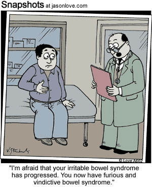
[9] I hate when that happens:

(Via Stephen Wangen)

[10] Are choline supplements toxic?: At the very beginning of the book (p 3) we state that “the perfect diet should … deliver … no excess nutrients for pathogens.”

Later in the book we give examples of nutrients that, in excess, primarily benefit pathogens: niacin (the primary vitamin for bacteria), iron (critical for metabolism of most pathogens, and a component of bacterial biofilms), and calcium (a component of bacterial biofilms). These are on our list of micronutrients we recommend not supplementing (beyond a multivitamin).

Two readers, Leonardo and Patricia (thank you both!), emailed us about a ScienceDaily article suggesting that choline, one of the micronutrients we most frequently recommend, should be added to this list:

When fed to mice, lecithin and choline were converted to a heart disease-forming product by the intestinal microbes, which promoted fatty plaque deposits to form within arteries (atherosclerosis); in humans, higher blood levels of choline and the heart disease forming microorganism products are strongly associated with increased cardiovascular disease risk.

The story didn’t have enough information, so I downloaded the paper. The paper notes that choline is metabolized by gut bacteria to a gas with a fishy odor called TMA, which is then oxidized in the liver to a compound called TMAO:

Briefly, initial catabolism of choline and other trimethylamine-containing species (for example, betaine) by intestinal microbes forms the gas trimethylamine (TMA), which is efficiently absorbed and rapidly metabolized by at least one member of the hepatic flavin monooxygenase (FMO) family of enzymes, FMO3, to form trimethylamine N-oxide (TMAO).

They showed that (a) feeding phosphatidylcholine from egg yolk to mice led to increased blood levels of TMAO and that (b) in a separate study, people with atherosclerosis have elevated blood levels of TMAO, choline, and trimethylglycine.

Supplementing choline at 10 times normal levels to Apoe-knockout mice led to increased TMAO but not choline in blood:

Atherosclerosis-prone mice (C57BL/6J Apoe-/-) at time of weaning were placed on either normal chow diet (contains 0.08–0.09% total choline, wt/wt) or normal chow diet supplemented with intermediate (0.5%) or high amounts of additional choline (1.0%) or TMAO (0.12%)….

Analysis of plasma levels of choline and TMAO in each of the dietary arms showed nominal changes in plasma levels of choline, but significant increases of TMAO in mice receiving either choline or TMAO supplementation (Supplementary Fig. 10).

Serum TMAO levels were correlated with atherosclerotic plaque size and with macrophages turning into foam cells:

[A]ll dietary groups of mice revealed a significant positive correlation between plasma levels of TMAO and atherosclerotic plaque size (Fig. 3e and Supplementary Fig. 9b).

TMA (a gas with a fish odor) has to be converted in the liver to the toxic TMAO in order to produce these bigger atherosclerotic lesions. This conversion happened mainly in mice with low HDL:

Interestingly, a highly significant negative correlation with plasma high-density lipoprotein (HDL) cholesterol levels was noted in both male and female mice (Fig. 4b and Supplementary Fig. 12, middle row).

So if you’re an Apoe(-/-) mouse and eat ten times normal choline, if you have high HDL your arteries are safe but you smell fishy; if you have low HDL you smell fine but your arteries get injured.

What does this tell us about choline supplementation?

For humans with working ApoE alleles, I doubt we can infer anything yet.

For Apoe(-/-) mice fed ten times normal choline, I would suggest shooting for low HDL while dating, then high HDL after marriage.

Reference: Wang Z et al. Gut flora metabolism of phosphatidylcholine promotes cardiovascular disease. Nature, 2011; 472 (7341): 57 DOI: 10.1038/nature09922

[11] One-upping the standing desk: Jamie Scott has a walking desk:

[12] Why is aspirin toxic to cats?: In the book we mention that plant foods always contain toxins, but animal foods don’t – because in poisoning us, animals would poison themselves. As we point out in the book, Bruce Ames and Lois Gold estimate that over 99% of the toxins humans ingest come from plant foods – not industrial or environmental toxins.

One of the main functions of the liver is detoxification. A healthy liver enables us to consume plant foods.

But what happens to the livers of animals that never eat plant foods? If they and their descendants avoid plant foods for millions of years, how would their livers evolve?

The answer is in a fascinating piece by Ed Yong at Discover blogs: “Why Is Aspirin Toxic to Cats?”. The puzzle:

[C]ats are extremely sensitive to aspirin, and even a single extra-strength pill can trigger a fatal overdose.

Some scientists have been investigating this puzzle since the early 1990s. It turns out that all 18 of 18 species of cat studied, including housecats, cheetahs, servals, and tigers, have crippling mutations in a gene involved in liver detoxification. The same gene is also lost in other hypercarnivores, including the brown hyena and the northern elephant seal.

Mr. Yong explains:

Like many other “detoxifying” proteins, UGT1A6 evolved to help animals cope with the thousands of dangerous chemicals in the plants they eat….

But if an animal’s menu consists largely of meat, it has little use for these anti-plant defences. The genes are dispensable…. [T]he ancestral cats gradually built up mutations that disabled their UGT1A6 gene. Evolution is merciless that way – it works on a “use it or lose it” basis.

So – millions of years of hypercarnivory will disable the liver’s ability to metabolize toxins.

Pet owners, be kind to your cats: Don’t feed them plants!

And a new zero-carb danger: After ten thousand generations, your descendants may be unable to take aspirin.

Reference: Shrestha B et al. Evolution of a Major Drug Metabolizing Enzyme Defect in the Domestic Cat and Other Felidae: Phylogenetic Timing and the Role of Hypercarnivory. PLoS One. 2011 Mar 28;6(3):e18046. http://pmid.us/21464924.

[13] Not the weekly video: Best mobile phone commercial I’ve seen:

[14] Weekly video: I grew up near the University of Connecticut campus and have been a fan of their men’s basketball team since the late 1970’s. What Jim Calhoun has done there, building a minor program to national prominence and three championships, is one of the great accomplishment in coaching history. And this year’s team was a minor miracle: with unheralded and under-recruited freshmen playing half the minutes, they won a national championship.

Every year CBS makes a video montage of the tournament. Here it is, One Shining Moment:

What Telomeres Tell Us About Human Disease

We believe that almost all diseases are caused by food toxins, malnutrition, and infections. Toxic and malnourishing diets depress immunity and make infections worse.

Once you have this point of view in mind, supporting evidence is everywhere.

Take, for example, a story today in ScienceDaily about depression. Depression is not just a mental illness, but a whole body illness:

Previously considered a mental illness affecting only the brain, major depressive disorder, or MDD, now is believed to be tied to significant physical damage outside the brain, explained Wolkowitz. For example, depressed individuals are more likely to develop the diseases of advanced age, including diabetes, heart disease, osteoporosis, stroke and dementia. [1]

The ScienceDaily article summarizes new research showing a link between depression and telomere length in white blood cells. Telomeres are the end-caps on chromosomes. If telomeres become too short, DNA becomes unstable, genetic integrity is lost during cell division, and cells become senescent (crippled beyond hope of recovery) or commit apoptosis (suicide).

An enzyme called telomerase lengthen telomeres. Normally, most cell types maintain a balance between telomerase levels and replication so that telomeres are maintained at healthy lengths throughout normal cell life.

It turns out that in depressed people, white blood cell telomeres are shorter than in normal people, even though telomerase is more active. [2] Moreover, for a given telomere length, the more telomerase activity, the more depressed the patient. [3] Finally, telomerase activity predicts which patients will recover: patients who recovered from depression had the highest telomerase activity along with their short telomeres. [3]

This suggests that some exogenous factor, not part of normal human biology, is shortening telomeres in the depressed; and that the body’s capacity to resist this factor determines its ability to recover from depression. If the body can overcome the exogenous factor, eliminating its ability to shorten telomeres, then the depression goes away.

What could this exogenous factor be?

Telomeres and Viral Infections

Well, it happens that a number of viruses shorten telomeres in white blood cells.

Cytomegalovirus reduces telomere length in T cells:

After primary CMV infection, we observed … a steep drop in telomere length. Moreover, we found in a cohort of 159 healthy individuals that telomere shortening was more rapid in CMV-seropositive individuals and correlated with the amount of differentiated T cells in both CD4(+) T cells and CD8(+) T cells. [4]

The Epstein-Barr virus (EBV) is carried by more than 90% of the adult world population and has been implicated in several human cancers. [5]  EBV disrupts the caps of telomeres, creating dysfunctional telomeres: “The telomere capping protein TRF2 was partially displaced from telomeres in EBV-infected cells, suggesting an EBV-mediated uncapping problem.” [5]

HIV also shortens telomeres: “Analysis of telomere length in HIV-1 exposed U373 showed a statistically significant telomere shortening” [6]. Interestingly, telomere shortening by HIV was reversed by providing N-acetylcysteine, suggesting that NAC should be beneficial for AIDS and possibly other chronic viral infections.

Connections between viruses and telomere loss run deep. In fact, it has been proposed that cellular senescence, the usual outcome of telomere loss, evolved as an anti-viral defense mechanism. [7]

If viruses cause major depression, then they probably also cause the diseases associated with depression. After all, they have to infect the rest of the body before they can infiltrate the brain. So we should look at viruses and other systemic diseases, and see if the connection with telomere shortening holds in those diseases.

Cancer and Blood Cell Telomeres

There is steadily increasing evidence implicating viruses as causes of cancers. Wikipedia (“Infectious causes of cancer”) has a summary:

Worldwide approximately 18% of cancers are related to infectious diseases…. Viruses are usual infectious agents that cause cancer but bacteria and parasites may also have an effect.

A virus that can cause cancer is called an oncovirus. These include human papillomavirus (cervical carcinoma), Epstein-Barr virus (B-cell lymphoproliferative disease and nasopharyngeal carcinoma), Kaposi’s sarcoma herpesvirus (Kaposi’s Sarcoma and primary effusion lymphomas), hepatitis B and hepatitis C viruses (hepatocellular carcinoma), and Human T-cell leukemia virus-1 (T-cell leukemias). Bacterial infection may also increase the risk of cancer, as seen in Helicobacter pylori-induced gastric carcinoma.[2] Parasitic infections strongly associated with cancer include Schistosoma haematobium (squamous cell carcinoma of the bladder) and the liver flukes, Opisthorchis viverrini and Clonorchis sinensis (cholangiocarcinoma).[3]

According to some authors, viruses are one of the most important risks factor for cancer development in humans, second only to tobacco use.[4]

This summary overlooks some known associations (such as that between XMRV and prostate cancer, see our post Retroviruses and Chronic Fatigue Syndrome, Aug 24, 2010) and evidence that tobacco use raises cancer risk primarily in people with a high viral infectious burden (see ref. [10] below). Although only 18% of cancers may yet have been confidently linked to infectious pathogens, it is not impossible that 100% of cancers are caused by as-yet-mostly-unidentified infectious pathogens, probably mainly viruses.

If viruses cause cancers, and if viruses shorten white blood cell telomeres, then we would expect cancer patients to have shortened telomeres.

Well, gastric cancer patients have shorter white blood cell telomeres, and being in the bottom half of telomere length doubles gastric cancer risk:

GC patients had significantly shorter average telomere length than matched controls (mean +/- SD 0.89 +/- 0.19 vs 1.06 +/- 0.25, P < 0.001)…. We found that short telomere length was associated with a significantly increased GC risk (adjusted odds ratio = 2.14, 95% confidence interval = 1.52-2.93)…. Collectively, our findings provide the first evidence linking the short telomere length in peripheral blood lymphocytes to elevated GC risk. [8]

Lung cancer patients have shorter white blood cell telomeres, and being in the bottom half of telomere length triples lung cancer risk:

Telomere length was significantly shorter in lung cancer patients than in controls (mean +/- standard deviation: 1.59 +/- 0.75 versus 2.16 +/- 1.10, P < 0.0001). When the subjects were categorized into quartiles based on telomere length, the risk of lung cancer was found to increase as telomere length shortened (P(trend) < 0.0001)…. [I]ndividuals with short telomeres were at a significantly higher risk of lung cancer than those with long telomeres (adjusted odds ratio = 3.15, 95% confidence interval = 2.12-4.67, P < 0.0001). [9]

Bladder cancer patients also had short white blood cell telomeres. Being in the bottom quarter of telomere length increases risk 4.5-fold, 6.3-fold for smokers:

Patients with bladder cancer displayed significantly shorter telomeres than control subjects (P = 0.001). Median telomere length ratio was 0.95 (range 0.53-3.2) for cases and 1.1 (0.51-2.4) for controls. Moreover, the adjusted odds ratio (OR) for bladder cancer was significantly increased in the quartile with the shortest telomere length OR = 4.5 [95% confidence interval (CI) 1.7-12]. [10]

Same story with head and neck cancer [11], renal cancer [12], breast cancer [13], and probably also thyroid cancer [14].

Cardiovascular Disease

A weakness of those cancer studies is that they only looked at blood cell telomeres and the presence of cancer; they didn’t also measure viral burden, for instance by looking for antibody seropositivity.

So I was pleased to find a study that did that in coronary heart disease. Again, white blood cell telomeres were shorter in heart disease patients:

Telomere length (TL) was approximately 0.5 kilobases (kb) shorter in leukocytes from patients with CHD than in their age-matched control subjects….

TL shortening was particularly pronounced in CD8+CD28(-) T cells obtained from cytomegalovirus-seropositive CHD patients. [15]

So cytomegalovirus may be involved in coronary heart disease.

The reason all these studies have looked at white blood cells is because it is easy to get blood samples. But sometimes it is possible to get samples from diseased and normal tissues and do a direct comparison.

That was done in this study of atherosclerotic plaques:

Arterial segments which did not develop atherosclerosis such as the saphenous vein and internal mammary artery, had longer telomere length than aortic segments. On the other hand, telomere length was shorter in aortic tissues which presented atherosclerotic lesions compared to corresponding tissues without atherosclerotic lesions. These results also suggest tissue regulation of telomere size by local factors likely related to oxidative stress responses.

So the normal vessels have long telomeres, indicating an absence of viral infections, but the atherosclerotic plaques have short telomeres, suggesting of high infectious burden.

Conclusion

Telomere shortening is probably a marker of infectious burden, especially of viral infections. Telomere shortening in blood cells is associated with major depression, cancer, heart disease, and probably nearly every other disease.

Diseases probably result from a combination of factors, but a heavy burden of chronic infectious pathogens is probably almost always one of them. These pathogens are usually little more than parasites, sapping nutrients from human cells and disabling their immune defenses. But combined with toxic and malnourishing diets, they cripple the body and shorten lifespan.

The association of shortened telomeres with shortened lifespan may be due to the life-shortening effects of infections.

This is why the immunity-enhancing dietary steps discussed in Step Four of our book are so central to a long and healthy life. We cannot avoid exposure to these pathogens. But we can keep their numbers down, so that they do minimal harm to us throughout life.

References

[1] University of California – San Francisco (2011, April 6). Link between chronic depression and accelerated immune cell aging. ScienceDaily. Retrieved April 7, 2011, from http://www.sciencedaily.com/releases/2011/04/110405151223.htm.

[2] Wolkowitz OM et al. Leukocyte telomere length in major depression: correlations with chronicity, inflammation and oxidative stress – preliminary findings. PLoS One. 2011 Mar 23;6(3):e17837. http://pmid.us/21448457.

[3] Wolkowitz OM et al. Resting leukocyte telomerase activity is elevated in major depression and predicts treatment response. Mol Psychiatry. 2011 Jan 18. [Epub ahead of print] http://pmid.us/21242992.

[4] van de Berg PJ et al. Cytomegalovirus infection reduces telomere length of the circulating T cell pool. J Immunol. 2010 Apr 1;184(7):3417-23. http://pmid.us/20176738.

[5] Lacoste S et al. Chromosomal rearrangements after ex vivo Epstein-Barr virus (EBV) infection of human B cells. Oncogene. 2010 Jan 28;29(4):503-15. http://pmid.us/19881539.

[6] Pollicita M et al. Apoptosis and telomeres shortening related to HIV-1 induced oxidative stress in an astrocytoma cell line. BMC Neurosci. 2009 May 22;10:51. http://pmid.us/19463156.

[7] Reddel RR. Senescence: an antiviral defense that is tumor suppressive? Carcinogenesis. 2010 Jan;31(1):19-26. http://pmid.us/19887513.

[8] Liu X et al. Constitutive telomere length and gastric cancer risk: case-control analysis in Chinese Han population. Cancer Sci. 2009 Jul;100(7):1300-5. http://pmid.us/19432888.

[9] Jang JS et al. Telomere length and the risk of lung cancer. Cancer Sci. 2008 Jul;99(7):1385-9. http://pmid.us/18452563.

[10] Broberg K et al. Constitutional short telomeres are strong genetic susceptibility markers for bladder cancer. Carcinogenesis. 2005 Jul;26(7):1263-71.  http://pmid.us/15746160.

[11] Wu X et al. Telomere dysfunction: a potential cancer predisposition factor. J Natl Cancer Inst. 2003 Aug 20;95(16):1211-8. http://pmid.us/12928346.

[12] Shao L et al. Telomere dysfunction in peripheral lymphocytes as a potential predisposition factor for renal cancer. J Urol. 2007 Oct;178(4 Pt 1):1492-6. http://pmid.us/17707063.

[13] Shen J et al. Short telomere length and breast cancer risk: a study in sister sets. Cancer Res. 2007 Jun 1;67(11):5538-44. http://pmid.us/17545637.

[14] Capezzone M et al. Telomeres and thyroid cancer. Curr Genomics. 2009 Dec;10(8):526-33. http://pmid.us/20514214.

[15] Spyridopoulos I et al. Accelerated telomere shortening in leukocyte subpopulations of patients with coronary heart disease: role of cytomegalovirus seropositivity. Circulation. 2009 Oct 6;120(14):1364-72. http://pmid.us/19770396.

[16] Nzietchueng R et al. Telomere length in vascular tissues from patients with atherosclerotic disease. J Nutr Health Aging. 2011;15(2):153-6. http://pmid.us/21365170.