Category Archives: Alzheimer’s

Chocolate: What is the Optimal Dose?

Bret asked us how much chocolate is needed for good health:

I have a question about having dark chocolate daily. Does it need to be every day or what is the mininum grams per day. I have been having around 35g a day of 70% but I wondered if less would be ok or not having it at all.

This is a great time for this question, since Halloween candy will be running out soon, and those on tight budgets may be tempted to skimp on their chocolate. Should they?

Chocolate Is Not Considered Essential … Yet

Chocolate has not yet been recognized by the Food and Nutrition Board of the National Academies as an essential nutrient. We haven’t either: Our food plate lists it among “pleasure foods,” which are healthful but optional.

However, we are becoming ever-more chocolate friendly. In the new edition of our book, we list chocolate among our “supplemental foods” which we recommend consuming regularly. But our suggested dose is “as desired.” Perhaps we should narrow that down a little.

Chocolate Against Cardiovascular Disease

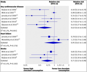
We’ve previously warned of the danger of chocolate deficiency, based on a systematic review that found: “The highest levels of chocolate consumption were associated with a 37% reduction in cardiovascular disease and a 29% reduction in stroke.” [1]

Here’s a visual summary of their findings:

The review authors report that every study accounted for chocolate intake in a different way, so they could only compare the groups with highest and lowest chocolate consumption in each study, not specific doses of chocolate.

Chocolate Against Diabetes

Bret was concerned about the sugar in chocolate, but if this is a problem, it’s outweighed by the benefits of chocolate. A Japanese study found that the rate of diabetes was reduced by 30% in those who consume the most chocolate. [2]

Chocolate Against Dementia and In Support of Cognitive Function

Several studies [3, 4] have found that chocolate consumption reduces risk of dementia and enhances performance on tests of cognitive function.

One of them found that cognitive function was optimized with a relatively low dose of chocolate – ten grams per day:

The associations between intake of these foodstuffs and cognition were dose dependent, with maximum effect at intakes of approximately 10 g/d for chocolate and approximately 75-100 mL/d for wine, but approximately linear for tea. [3]

The other found that cognition improved with intake of cocoa flavanols up to quite high doses – elderly given 1 g/day cocoa flavanols performed significantly better on cognitive tests than those given lower doses. [4]

Unfortunately I don’t know what fraction of chocolate is made of flavanols. I’m guessing it’s not more than a few percent, in which case this research suggests the optimal dose of chocolate may be 50 g/day or more.

Chocolate in Support of Circadian Rhythms

Most authors attribute the benefits of chocolate to their flavanols, which are thought to improve endothelial function and increase blood flow to the brain, among other effects.

However, there are other active compounds in chocolate, include peptides that interact with the opioid receptor. The opioid receptor has a role in circadian rhythms, which is one reason low-dose naltrexone (which blocks opioid function at night) works. It’s possible that eating chocolate during the day may support circadian rhythms via opioid receptor stimulation, especially if the peptides can reach the systemic circulation.

Indirect evidence that this may be beneficial comes from a Russian study in which exorphins (opioid receptor ligands) were injected into rats:

The chronic intraperitoneal administration of the peptide at the same dose of 5 mg/kg significantly increased exploratory activity, decreased anxiety, and improved learning. [5]

I don’t know how much chocolate would have to be eaten to achieve a similar exorphin dose in humans, but I imagine it’s large.

Chocolate in Support of Nobel Prizes

So how shall we resolve the issue of optimal chocolate dose? For me, the decisive evidence comes from a recent study by Franz Messerli published in the New England Journal of Medicine.

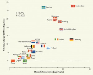
Based on chocolate’s support for cognitive function, he decided to see if chocolate consumption was related to another measure of cognition – Nobel Prize awards per capita. He counted Nobel Prizes and compared them to the recipient’s country’s chocolate consumption. These were his findings [6]:

There is clearly a strong correlation. The correlation coefficient is .79; p < 0.0001.

The correlation coefficient if Sweden is removed increases to .86 – which is suspicious:

Given its per capita chocolate consumption of 6.4 kg per year, we would predict that Sweden should have produced a total of about 14 Nobel laureates, yet we observe 32. Considering that in this instance the observed number exceeds the expected number by a factor of more than 2, one cannot quite escape the notion that either the Nobel Committee in Stockholm has some inherent patriotic bias when assessing the candidates for these awards or, perhaps, that the Swedes are particularly sensitive to chocolate, and even minuscule amounts greatly enhance their cognition. [6]

Those dastardly Swedes! Giving themselves more Nobel Prizes than their chocolate consumption warrants!

But I apologize, I’ve been diverted. The key point is, is there an optimum chocolate consumption?

the dose–response curve reveals no apparent ceiling on the number of Nobel laureates at the highest chocolate-dose level of 11 kg per year. [6]

11 kg/yr is an average of 30 g/day. So benefits are still increasing at that dose.

Of course, this was only a population level study. We still need to measure the doses in individual laureates to gain confidence. But anecdotally, there appears to be a correlation:

“I attribute essentially all my success to the very large amount of chocolate that I consume,” said Eric Cornell, an American physicist who received the Nobel Prize in 2001. “Personally I feel that milk chocolate makes you stupid. Now dark chocolate is the way to go. It’s one thing if you want like a medicine or chemistry Nobel Prize…but if you want a physics Nobel Prize it pretty much has got to be dark chocolate.”

Dark chocolate is, indeed, the PHD-approved form of this highly beneficial food.

Conclusion

This dose-response data might not be strong enough to define an RDA, but I’m going to take a stand: Bret’s intake of 35 g/day is healthy. Indeed, it’s right in line with the Nobel Prize-maximizing chocolate intake of the Swiss.

In regard to your last question, Bret – can you eat less chocolate, or none at all – the answer is clear. Yes, you can. But you must accept the consequences. You probably won’t be winning the next Nobel Prize for Physics.

References

[1] Buitrago-Lopez A et al. Chocolate consumption and cardiometabolic disorders: systematic review and meta-analysis. BMJ. 2011 Aug 26;343:d4488. doi: 10.1136/bmj.d4488. http://pmid.us/21875885.

[2] Oba S et al. Consumption of coffee, green tea, oolong tea, black tea, chocolate snacks and the caffeine content in relation to risk of diabetes in Japanese men and women. Br J Nutr. 2010 Feb;103(3):453-9. http://pmid.us/19818197.

[3] Nurk E et al. Intake of flavonoid-rich wine, tea, and chocolate by elderly men and women is associated with better cognitive test performance. J Nutr. 2009 Jan;139(1):120-7. http://pmid.us/19056649.

[4] Desideri G et al. Benefits in cognitive function, blood pressure, and insulin resistance through cocoa flavanol consumption in elderly subjects with mild cognitive impairment: the Cocoa, Cognition, and Aging (CoCoA) study. Hypertension. 2012 Sep;60(3):794-801. http://pmid.us/22892813.

[5] Belyaeva YA et al. Effects of acute and chronic administration of exorphin C on behavior and learning in white rat pups. Moscow University Biological Sciences Bulletin Volume 64, Number 2 (2009), 66-70, DOI: 10.3103/S0096392509020035. http://www.springerlink.com/content/qt537481061656gt/?MUD=MP.

[6] Messerli FH. Chocolate consumption, cognitive function, and Nobel laureates. N Engl J Med. 2012 Oct 18;367(16):1562-4. doi: 10.1056/NEJMon1211064. Epub 2012 Oct 10. http://pmid.us/23050509.

Omega-3s, Angiogenesis and Cancer: Part II

On Tuesday (Omega-3 Fats, Angiogenesis, and Cancer: Part I, April 26, 2011) I introduced the issue of possible relationships between omega-3 fatty acids, their lipid peroxidation products, and diseases of angiogenesis such as cancer, and promised to discuss a possible mechanism today.

It may be well, however, to start by saying a little bit more about the Brasky paper [1] linking prostate cancer to DHA.

Denise Minger’s Commentary on the Brasky Paper

Denise Minger wrote a commentary on this paper for Mark’s Daily Apple, which is excellent. Her conclusion – “given the oxidation-prone nature of all polyunsaturated fats, a massive intake of omega-3’s – despite their brilliance in moderation – could potentially do more harm than good” – is the proper one.

A few of Denise’s observations, however, could stand elaboration.

The study measured the fraction of serum phospholipid fatty acids in various polyunsaturated and trans-fat species, not dietary intake. This is the right parameter to measure, as fatty acid profiles can be measured precisely while dietary intakes assessed through questionnaires are notoriously unreliable. Also, phospholipids are the fats in cell membranes, and these are the ones involved in the inflammatory signaling pathways long thought to drive cancer risk. So cell membrane lipid measurements have the best chance to demonstrate a link to cancer risk.

Denise makes the important point, however, that the connection between dietary fish oil intake and serum fatty acid profile is not simple. Higher DHA intake raises phospholipid DHA levels, but lower intake of non-omega-3 fats also raises the DHA fraction. She points to a study [2] comparing a low-fat diet (20% fat, 6.7% PUFA, n-6:n-3 ratio 11.1) to a high-fat diet (45% fat, 15% PUFA, n-6:n-3 ratio 12.3).  The low-fat diet had more of its fat in the form of long-chain omega-3s, but the specific DHA intake on the diets was not reported. Membrane DHA ended up 28% higher on the low-fat diet.

So if DHA is dangerous, low-fat dieters will be in the most trouble. Another reason to eat a high-fat diet!

Does this affect our interpretation of the Brasky study? I don’t think it affects it much, because study participants were healthy at the start of the study with no history of cancer and macronutrient intakes don’t vary a lot among the general public. Americans vary surprisingly little from the median of about 50% carbs, 15% protein, and 35% fat – so it’s likely that the quartile with high tissue DHA levels were also high fish oil consumers.

However, study participants were followed for 7 years, at which point their prostate cancer status was assessed. Incidence of low-grade prostate cancers had no association with start-of-the-study DHA intake, but incidence of high-grade prostate cancers was strongly associated.

Here are a couple of possible explanations for this pattern:

  1. DHA is bad: DHA doesn’t drive early cancer development but does drive late-stage cancer growth – i.e. the transition from low-grade to high-grade cancer. So the DHA consumers got the high-grade cancers. Angiogenesis does, in fact, drive the shift from low-grade to high-grade cancer, so a DHA-angiogenesis association would be consistent with this explanation.
  2. Hospital diet advice is bad: DHA was a marker at the start of the study for conscientious, educated, disciplined persons who followed health advice and ate fish oil. When these people were diagnosed with low-grade cancer, they followed the dietary advice of their cancer dietitian. The dietitian’s advice?  Eat lots of wheat, whole grains, legumes, and vegetable oils. It could be the conscientious folks who followed bad diet advice from the hospital dietitian who got the high-grade cancers.

So there is a possible confounding effect.

Another of Denise’s assertions is that there is an “otherwise consistent train of research showing that DHA seems protective at best (and neutral at worst).” Now it is true that there are more studies showing DHA to have benefits against cancer than harm. But this trend is hardly consistent, and the vast majority of studies have failed to detect a relationship.

In the comments to Tuesday’s post, eric linked to a 2005 meta-review of studies on omega-3 fats and cancer. [3] The reviewers looked at 1,210 journal articles and found a mixed bag of mostly insignificant evidence:

Significant associations between omega-3 consumption and cancer risk were reported for lung cancer in two studies; for breast cancer in one; for prostate cancer in one; and for skin cancer in one. However, for lung cancer, one of the significant associations was for increased cancer risk and the other was for decreased risk (four other risk ratios were not significant for lung cancer). For breast cancer, five other estimates did not show a significant association. Only one study assessed skin cancer risk. No effects were reported for cancers of the aerodigestive tract, bladder cancer, colorectal cancer, lymphoma, ovarian cancer, pancreatic cancer, or stomach cancer. Thus, omega-3 fatty acids do not appear to decrease overall cancer risk.

Data were insufficient to permit assessment of a temporal or dose-response relationship. [3]

So the score was 4 studies finding that DHA is associated with less cancer, 1 that it is associated with more, and a boatload that it had no association.

Now there are two ways of interpreting this general insignificance of DHA against cancer. One is to note that there are slightly more studies showing DHA to have benefits than harm, and therefore to judge that DHA might be helpful against cancer.

But another, equally plausible, interpretation is this. Most Americans eat far too much omega-6, and their omega-6 to omega-3 tissue ratio is too high, which is pro-inflammatory via the COX-2 pathway. Eating omega-3s including DHA reduces inflammation by downregulating the COX-2 pathway. This accounts for the well-attested benefits of DHA against cardiovascular disease. Now, cancer is promoted by COX-2 pathway inflammation, which is why COX-2 inhibitors such as aspirin and ibuprofen are protective against cancer. [4] DHA’s action to downregulate this pathway must generate an anti-cancer effect. But, unlike aspirin and ibuprofen, DHA has no observable effect on overall cancer risk. This suggests that DHA has other effects, unrelated to its anti-inflammatory activity, that are cancer promoting. These counterbalance the benefits from its anti-inflammatory effect. If DHA has pro-angiogenic effects that are independent of COX-2 mediated inflammation, then this could account for the observations.

One reason an association of DHA with high-grade cancer may have been missed is that it would be detected only in large studies able to segregate cancers by grade. Brasky et al note:

In the European Prospective Investigation into Cancer and Nutrition (EPIC) (12), the highest quintile of percent DHA was associated with elevated risks of both low-grade (relative risk (RR) = 1.53, 95% CI: 0.96, 2.44) and high-grade (RR = 1.41, 95% CI: 0.76, 2.62) prostate cancer. They also reported significant positive associations of the percent EPA with high-grade prostate cancer (RR = 2.00, 95% CI: 1.07, 3.76). Given that the Prostate Cancer Prevention Trial and the European Prospective Investigation into Cancer and Nutrition, the 2 largest studies of blood levels of phospholipid fatty acids, reported increased risks of high-grade prostate cancer with high levels of ω-3 fatty acids, it remains a possibility that these fatty acids promote tumorigenesis. [1]

If there were no other evidence linking DHA to angiogenesis, the Brasky and EPIC study associations would be interesting, but unlikely to change anyone’s mind. Denise points out the need for other evidence – especially, mechanistic evidence – to make the connection more plausible:

We haven’t sleuthed out any mechanism that could explain why DHA (but not other polyunsaturated fats) promotes rapid tumor growth.

And this is where today’s post comes in. In fact, there is a known mechanism by which DHA but not other polyunsaturated fats can promote rapid tumor growth. Shou-Ching told me about it a few months ago.

DHA and Angiogenesis in Macular Degeneration

Let’s start by going back to 2003 and a paper on the role of a compound called carboxyethylpyrrole (CEP) in age-related macular degeneration (AMD). [5] AMD is an eye disease caused by improper angiogenesis. Basically, malformed blood vessels overgrow the eye, causing retinal detachment and blindness. It afflicts 35% of those over age 75, and is the leading cause of blindness in developed countries. CEP? Well, the paper explains:

Free radical-induced oxidation of docosahexaenoate (DHA)-containing lipids generates ω-(2-carboxyethyl)pyrrole (CEP) protein adducts that are more abundant in ocular tissues from AMD than normal human donors…. The CEP adduct uniquely indicates oxidative modification from DHA derivatives because CEP protein modifications cannot arise from any other common polyunsaturated fatty acid. [5]

CEP is uniquely produced by oxidation of DHA, not other PUFAs. Its abundance depends on DHA abundance, availability of retinyl proteins, and the level of oxidative stress.

CEP is elevated in AMD. The correlation is strong: a person in whom the immune system is trying but failing to clear elevated CEP levels almost invariably has macular degeneration (AMD):

Of individuals (n = 13) exhibiting both antigen and autoantibody levels above the mean for non-AMD controls, 92% had AMD. [5]

So CEP is a great marker for AMD. Is it causal?

Well, first it’s worth noting that the retina is uniquely vulnerable to DHA oxidation:

Although rare in most human tissues, DHA is present in ~80 mol % of the polyunsaturated lipids in photoreceptor outer segments (13). The abundance of DHA in photoreceptors, the high photooxidative stress in retina, and the fact that DHA is the most oxidizable fatty acid in humans (13) suggests that DHA oxidation products may have possible utility as biomarkers for AMD susceptibility. [5]

Oxidation is linked to AMD, and antioxidants slow AMD progression:

Oxidative damage appears to contribute to the pathogenesis of AMD (4) based on epidemiological studies showing that smoking significantly increases the risk of AMD (1, 24). The molecular mechanism for how smoking enhances the risk for AMD is not known. We speculate that reactive oxygen and nitrogen species derived from tobacco smoke in the lungs leads to oxidative protein modifications in the blood that contribute to drusen formation and choroidal neovascularization. Results from a recent clinical trial (5) also demonstrate that the progression of AMD can be slowed in some individuals by high daily doses of antioxidant vitamins and zinc. Direct evidence of oxidative damage in AMD donor eye tissues include elevated levels of CEP adducts uniquely derived from the oxidative fragmentation of DHA (6). [5]

This is where things stood in 2003. By 2010 this group, led by Case Western Reserve University chemist Robert G. Salomon, had established that administering CEP to mice can cause AMD:

To test the hypothesis that this hapten is causally involved in initiating an inflammatory response in AMD, we immunized C57BL/6J mice with mouse serum albumin (MSA) adducted with CEP. Immunized mice develop antibodies to CEP, fix complement component-3 in Bruch’s membrane, accumulate drusen below the retinal pigment epithelium during aging, show decreased a- and b-wave amplitudes in response to light, and develop lesions in the retinal pigment epithelium mimicking geographic atrophy, the blinding end-stage condition characteristic of the dry form of AMD. Inflammatory cells are present in the region of lesions and may be actively involved in the pathology observed. [6]

This constitutes the first really good animal model for AMD. [6]

How does this relate to cancer? That leads us to a Nature paper from October 2010 [7], from the group of Tatiana Byzova at the Cleveland Clinic.

DHA, Immunity, and Angiogenesis

This is a rich paper. Briefly, CEP has a physiological function: it is transiently elevated in wounds and recruits immune cells from bone marrow to the site of the wound. These immune cells further increase oxidative stress and promote angiogenesis; CEP levels are highest at the time of peak angiogenesis. CEP is highly elevated in cancers. Unlike in wounds, where CEP is elevated for a few days, in cancers CEP elevation is chronic.

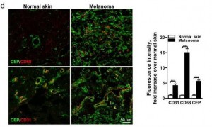
Here’s a staining comparing CEP in normal skin and in melanoma:

The CEP is co-localized with CD68, a glycoprotein which binds to LDL and is found on macrophages, and with CD31, a membrane marker of neutrophils, macrophages, and endothelial cells. CEP is marking endothelial cells and white blood cells in angiogenic vessels, and possibly LDL.

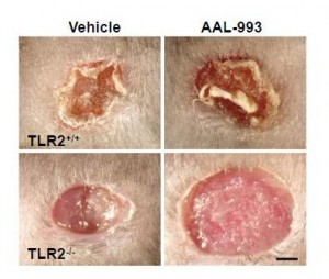
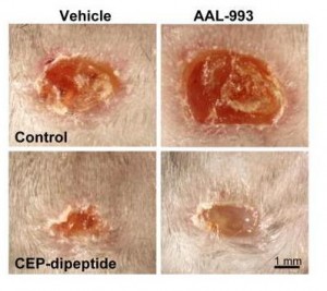
It turns out that CEP drives angiogenesis by attaching to an immune receptor, Toll-like receptor 2 (TLR2). There are two major pathways for angiogenesis: one driven by vascular endothelial growth factor (VEGF), which is dominant in conditions of hypoxia (oxygen starvation), and one by TLR2. Of these, the TLR2 pathway may in some contexts be more important. Here are pictures of wound healing in mice:

On the upper left is a normal mouse. On the upper right is a similar wound treated with the VEGF inhibitor AAL-993. This wound is rather like a cancer treated with the VEGF inhibitor Avastin. Wound healing is slightly impaired, but still works.

On the lower left is a similar wound with no VEGF inhibition, but the TLR2 pathway blocked by TLR2 knockout. The wound can’t scab and doesn’t heal successfully. If TLR2 is knocked out and VEGF inhibited, there is no wound healing at all (lower right).

You can accelerate angiogenesis and wound healing by adding CEP to the wound.

In the bottom row, CEP has been added. Left is without VEGF inhibition, right with.

If you administer CEP-neutralizing antibodies to a normal wound, wound healing takes more than twice as long. This confirms that angiogenesis driven specifically by CEP (and therefore by DHA oxidation) is part of healthy wound healing.

Tumors use these same pathways to generate vessels and feed their growth. As the paper notes:

[T]umors implanted in TLR2-/- mice exhibited dramatically decreased vascularization and increased areas of necrosis. [7]

Here’s the paper’s conclusion:

Altogether our results establish a novel mechanism of angiogenesis that is independent of hypoxia-triggered VEGF expression. The products of lipid oxidation are generated as a consequence of oxidative stress and are recognized by TLR2, possibly in a complex with TLR1 on ECs, and promote angiogenesis in vivo, thereby contributing to accelerated wound healing and tissue recovery. If high levels of CEP and its analogs accumulate in tissues, it may lead to excessive vascularization, e.g. in tumors. Contribution of the CEP/TLR2 axis to angiogenesis varies in different physiological settings possibly depending on the extent of oxidative stress. CEP-driven angiogenesis may be an attractive therapeutic target, especially in cancers resistant to anti-VEGF therapy. Inflammation and oxidation-driven angiogenesis may occur in other pathologies, for example atherosclerosis, where arterial thickening can depend on its microvasculature. In these settings, there is an extensive generation of oxidative products which might promote atherogenesis via TLR2. Indeed, it was shown that TLR2?/? mice are protected from atherosclerosis, and this effect could be mediated by cells other than bone marrow-derived29. Thus, along with pathogen- and danger-associated molecular patterns, TLR2 recognizes an oxidation-associated molecular pattern. This new function of TLR2 as a sensor of oxidative stress reveals the shortcut link between innate immunity, oxidation and angiogenesis. [7]

Connection to Vitamin A

DHA is oxidized to a compound called HOHA which then combines with a protein, generally a retinyl (vitamin A-derived) protein to form CEP.

Cancers generate lots of CEP from DHA, and perhaps one way they do that is by generating lots of retinyl proteins. Cancers are known to have disturbed vitamin A biology with lots of retinyl:

Disturbance in vitamin A metabolism seems to be an important attribute of cancer cells. Retinoids, particularly retinoic acid, have critical regulatory functions and appear to modulate tumor development and progression. The key step of vitamin A metabolism is the esterification of all-trans retinol, catalyzed by lecithin/retinol acyltransferase. In this work we show that malignant melanoma cells are able to esterify all-trans retinol and subsequently isomerise all-trans retinyl esters into 11-cis retinol, whereas their benign counterparts – melanocytes are not able to catalyze these reactions. Besides, melanoma cell lines express lecithin/retinol acyltranseferase both at the mRNA and protein levels. In contrast, melanocytes do not express this enzyme … [8]

I haven’t looked much into this literature but it may speak to higher cancer risk with excessive vitamin A intake. Thus high-vitamin A cod liver oil may be a double risk for cancer patients.

Conclusion

It looks like we have a recipe for angiogenesis:

DHA + retinyl + oxidative stress = angiogenesis

This recipe is invoked normally and properly during wound healing. But it is also invoked excessively in pathological contexts – notably in cancers and age-related macular degeneration, probably also in other angiogenesis-associated diseases such as arthritis, rosacea, obesity, psoriasis, endometriosis, dementia, and multiple sclerosis.

In the case of cancer, DHA oxidation to CEP might transform miniscule, harmless cancers to high-grade, life-threatening cancers.

Should this possibility affect our dietary omega-3 recommendations? Well, we need to know the relative importance of the three ingredients on the left side of the above equation in producing angiogenesis. Chris Kresser wondered in the comments Tuesday whether oxidation may be the key factor:

I question whether DHA supplementation would truly play a causative role in the absence of a *pro-oxidative environment*.

In other words, perhaps in someone eating a SAD, not exercising, under a lot of stress, etc. DHA is more easily oxidized and thus potentially carcinogenic.

But in someone that is keeping all other oxidative risk factors low (i.e. they’re avoiding n-6, exercising, managing stress, reducing exposure to chemical toxins, etc.) I tend to doubt that supplementing with DHA could cause significant harm.

That’s the last piece of the puzzle: how do we minimize the level of oxidized DHA?

As I replied to Chris in the comments, low-carb Paleo dieters are not out of the woods in regard to oxidative stress. Oxidative stress is generated normally during metabolism, immune function – and by cancers. If anti-oxidant minerals like zinc, copper, and selenium and vitamins like vitamin C are deficient, then oxidative stress can be very high on a low-carb Paleo diet.

At the moment, I think it’s prudent to eat no more than 1 pound of salmon or similar cold-water fish per week, to avoid further EPA/DHA supplements, and to avoid low-fat diets which tend to elevate membrane DHA levels. Moderate omega-3 consumption is especially important for those suffering from diseases of pathological angiogenesis – especially cancer. DHA is essential for good health – but in excess, it is probably dangerous.

References

[1] Brasky TM et al. Serum Phospholipid Fatty Acids and Prostate Cancer Risk: Results From the Prostate Cancer Prevention Trial. Am. J. Epidemiol. April 24, 2011 DOI: 10.1093/aje/kwr027 (Will be at http://pmid.us/21518693.)

[2] Raatz SK et al. Total fat intake modifies plasma fatty acid composition in humans. J Nutr. 2001 Feb;131(2):231-4. http://pmid.us/11160538.

[3] MacLean CH, Newberry SJ, Mojica WA, et al. Effects of Omega-3 Fatty Acids on Cancer. Summary, Evidence Report/Technology Assessment: Number 113. AHRQ Publication Number 05-E010-1, February 2005. Agency for Healthcare Research and Quality, Rockville, MD. http://www.ahrq.gov/clinic/epcsums/o3cansum.htm.

[4] Harris RE. Cyclooxygenase-2 (cox-2) and the inflammogenesis of cancer. Subcell Biochem. 2007;42:93-126. http://pmid.us/17612047.

[5] Gu X et al. Carboxyethylpyrrole protein adducts and autoantibodies, biomarkers for age-related macular degeneration. J Biol Chem. 2003 Oct 24;278(43):42027-35. http://pmid.us/12923198.

[6] Hollyfield JG et al. A hapten generated from an oxidation fragment of docosahexaenoic acid is sufficient to initiate age-related macular degeneration. Mol Neurobiol. 2010 Jun;41(2-3):290-8. http://pmid.us/20221855.

[7] West XZ et al. Oxidative stress induces angiogenesis by activating TLR2 with novel endogenous ligands. Nature. 2010 Oct 21;467(7318):972-6. http://pmid.us/20927103.

[8] Amann PM et al. Vitamin A metabolism in benign and malignant melanocytic skin cells: Importance of lecithin/retinol acyltransferase and RPE65. J Cell Physiol. 2011 Apr 4. doi: 10.1002/jcp.22779. [Epub ahead of print] http://pmid.us/21465477.

Jaminet’s Corollary to the Ewald Hypothesis

In Tuesday’s comments, Kriss brought up Paul Ewald, father of the “Ewald hypothesis.” (Also brought up by Dennis Mangan here.) Ewald did some of his work in collaboration with Gregory Cochran, who may be familiar to many for his appearances on blogs (notably at Gene Expression) and for his recent book The 10,000-Year Explosion.

In a 1999 Atlantic article, “A New Germ Theory,” Judith Hooper summarizes Ewald’s hypothesis:

Darwinian laws have led Ewald to a new theory: that diseases we have long ascribed to genetic or environmental factors — including some forms of heart disease, cancer, and mental illness — are in many cases actually caused by infections.

Regular readers won’t be surprised to hear that we wholeheartedly endorse the Ewald hypothesis. We believe that nearly all diseases are caused by infections and bad diet. Malnourishing, toxin-rich diets impair immune function and create vulnerability to infectious disease.

The Ewald Hypothesis

Ewald’s reasoning goes as follows. Quotations are from the Atlantic essay.

First, genetic causes of disease are unlikely. Any gene that led to impaired functioning of the human body would be selected against and removed from the genome. Therefore, genetic diseases should have the abundance of random mutations – about 1 in 100,000 people:

As noted, the background mutation rate — the ratate which a gene spontaneously mutates — is typically about one in 50,000 to one in 100,000. Not surprisingly, genetic diseases that are severely fitness-impairing (for example, achondroplastic dwarfism) tend to have roughly the same odds, depending on the gene.

Diseases that are fitness-impairing and reach higher prevalence – and this includes nearly all major diseases – must have a cause other than genetic mutations.

Germs, on the other hand, are plausible candidates as causes for disease. Germs can benefit from doing us harm. At a minimum, they would like to modify human functioning in order to make us better hosts for themselves — by suppressing immune function, for instance. Also, they wish to induce behaviors that help them spread to new hosts – like sneezing, coughing, diarrhea, or sexual promiscuity.

Germs evolve quickly. Gene exchange, and lack of error checking during gene replication, modifies genomes quickly. Short reproductive time scales – on the order of 20 minutes – mean that helpful mutations proliferate rapidly. Big evolutionary changes can occur in a few weeks:

“The time scale is so much shorter and the selective pressures so much more intense [in microbes]. You can get evolutionary change in disease organisms in months or weeks.”

This means that germs quickly optimize their disease characteristics through natural selection. For example, virulence, or the severity of the disease that a pathogen causes, is rapidly optimized.

One factor determining virulence is how easily the organism can spread to a new host. If the organism can spread easily, there’s little cost to harming the current host, and microbes produce severe disease. If it’s hard to spread, on the other hand, organisms will be mild and peaceable toward their hosts. It pays to keep their hosts alive and healthy.

Ewald and his students collected empirical data supporting their explanation for virulence:

The dots on Saunders’s graphs made it plain that cholera strains are virulent in Guatemala, where the water is bad, and mild in Chile, where water quality is good. “The Chilean data show how quickly it can become mild in response to different selective pressures,” Ewald explained…. Strains of the cholera agent isolated from Texas and Louisiana produce such small amounts of toxin that almost no one who is infected with them will come down with cholera.

In the last few decades, evidence has only grown for the infectious origins of most diseases. In 1999, over 80% of serious diseases were known to be caused by pathogens:

Of the top forty fitness-antagonistic diseases on the list, thirty-three are known to be directly infectious and three are indirectly caused by infection; Cochran believes that the others will turn out to be infectious too. The most fitness-antagonistic diseases must be infectious, not genetic, Ewald and Cochran reason, because otherwise their frequency would have sunk to the level of random mutations.

If this analysis were repeated today, the percentage would be still closer to 100%. More cancers are now known to be caused by viruses, and the links between microbes and cardiovascular disease, dementia, and multiple sclerosis are stronger than ever.

I think Ewald and Cochran are correct in asserting that mental and neurological illnesses are especially likely to be infectious in origin. These illnesses tend to have a big impact on number of descendants, supporting the evolutionary argument for an infectious origin. And, due to their dependence on glucose, neurons are unusually susceptible to infections.

Schizophrenia is a good example of a disease that must be infectious in origin:

From the fitness perspective, schizophrenia is a catastrophe. It is estimated that male schizophrenics have roughly half as many offspring as the general population has. Female schizophrenics have roughly 75 percent as many. Schizophrenia should therefore approach the level of a random mutation after many generations.

Ewald and Cochran suggest we need a “Human Germ Project”:

In Ewald and Cochran’s view, evolutionary laws dictate that infection must be a factor in schizophrenia. “They announced they had the gene for schizophrenia, and then it turned out not to be true,” Cochran said one day when I mentioned genetic markers. “I think they found and unfound the gene for depression about six times. Nobody’s found a gene yet for any common mental illness. Maybe instead of the Human Genome Project we should have the Human Germ Project.”

I concur. Medical research should make much bigger investments in detecting, understanding the effects of, and developing treatments for human infections. Many existing lines of research, including many of the “autoimmune” and genetic hypotheses for disease origins, are not panning out, but continue to monopolize funding.

Jaminet’s Corollary

In the last century, sewage and water treatment has cleaned up our water supply and removed sewage and water as a vector for disease transmission. Hygienic methods, such as daily bathing and the use of soap, also tend to inhibit disease transmission.

Just as cholera is an extremely mild constituent of gut flora in hygienic Texas, but creates acute disease in unclean Guatemala, so we can expect that germs that created acute disease in (unclean) 1900 will have evolved to create mild chronic infections in (hygienic) 2011.

This is Jaminet’s corollary to the Ewald hypothesis:  Microbes are evolving away from severe acute disease toward milder chronic disease.

The focus of modern medicine on acute conditions, and its neglect of chronic conditions, adds to the selective pressures on microbes. Any pathogen that creates acute disease is subject to the full arsenal of modern antimicrobial drugs. But pathogens that create mild chronic disease are generally left untreated.

Modern medicine has created a powerful selective pressure on pathogens to generate chronic illnesses that are just mild enough, and that resemble aging closely enough, to elude the attention and antimicrobial arsenal of medical doctors.

Why No Dementia in Kitava?

Staffan Lindeberg in the Kitava Study found no evidence of stroke, diabetes, dementia, heart disease, obesity, hypertension, or acne on Kitava.

Why were these diseases absent? Partly due to the Kitavans’ excellent toxin-free diet, no doubt, but partly also due to an absence of the pathogens that cause these diseases.

Why was there no multiple sclerosis in the Faeroe Islands until British troops were stationed there in World War II? Because the pathogen that causes MS was absent from the islands, until the Brits introduced it.

Why has the incidence of chronic diseases increased tremendously in the last century? Partly due to longer-lived populations, but also, I believe, due to evolution of pathogens toward these diseases.

I predict the incidence of chronic disease will increase further in decades to come; and we will gradually come to appreciate that nearly all forty year olds today are not fully healthy, but are mildly impaired by a collection of chronic infections.

Conclusion

Fifty thousand years ago there were a few hundred thousand humans in the world. Today there are over 6 billion.

If a pathogen today wants to adapt to a specific host, its best bet is to adapt to humans. And within humans, its best way to flourish is to develop a chronic infection that persists for many decades.

The evolutionary arms race is not over. It has simply moved to a new field of battle. And medicine will have to evolve as the microbes do. The microbes are developing a new style of fighting. Medicine needs to shift its focus toward this rising threat of mild chronic diseases.

The Vanderbilt Protocol for Multiple Sclerosis

The antibiotic approach to MS therapy was developed at Vanderbilt by Drs. Stratton and Mitchell. They have patented multiple versions of their protocol, most recently in 2005. (Since patents are publicly available and the “Description of the Invention” is often an excellent overview of the science, we’ve put links to patents at the end of this post.)

Since we have some readers with MS, including Alexander, I thought I would post a summary of the Vanderbilt protocol. This was written by Dr. Stratton in 2009:

Treatment Protocol for Chronic Infections Caused by C. pneumoniae

As far as the Cpn Antimicrobial Regimen is concerned, my thoughts (as of 2009) are as follows:

First, as a general rule, the sicker a patient is, the slower they should go. This is why our early protocol started out with only one antibiotic and one dose, and then gradually adding the next dose/antibiotic as the reactions to each dose/antibiotic became apparent. These reactions appear to be caused by destruction of Chlamydia organisms as well as by the death of some of the infected host cells. Even destruction of elementary bodies by reducing agents such as N-acetyl-cysteine (NAC) can cause these reactions as chlamydial major outer membrane protein (MOMP) is released. MOMP is known to interact with Toll-like receptors (TLRs) and can thus induce the production of cytokines. Moreover, chlamydial cell wall contains LPS, which also interacts with TLRs and induces the production of cytokines. The reaction to anti-chlamydial therapy is sometimes referred to as “die-off” as presumable both chlamydial organisms and host cells are dying. These reactions can be delayed by days to weeks and may include “flu-like symptoms”, arthralgias and myalgias, “hangover-like symptoms (“brain-fog”, nausea, malaise), gastroenteritis (including diarrhea), and (rarely) fever. These reactions are akin to the “leprosy reaction” well described with the therapy of leprosy. The use of prednisone (10-20 mg per day) and/or pentoxifylline (Trental, 400 mg bid or tid), as done for the leprosy reaction, may be beneficial.

I think that all patients should start with supplements/vitamins before they start any antibiotics. Baseline lab studies, including CBC and liver function studies, should be done and these parameters followed every 3-4 months, more frequently (i.e., monthly) for sicker patients. As C. pneumoniae can infect white blood cells and liver cells, potential death of these cells should be monitored in sicker patients. Our initial protocol recommended this. I would add NAC to the supplements. We used amoxicillin, which is degraded in the body to penicillamine (a reducing agent) in our regimen as an anti-elementary body agent, but NAC seems to work equally well and may offer additional benefits in boosting the immune system as well as protecting the liver. As far as supplements/vitamins are concerned, I think Professor David Wheldon’s supplement/vitamin suggestions are very complete and should be the benchmark. Once antibiotics are ready to be started, I would start with a macrolide. We have used Azithromycin because it is easy to give and has become somewhat cheaper since it went off patent. Clarithromycin (500 mg twice a day) or Roxithromycin (300 mg once a day) can be used instead of Azithromycin. I would still give just one 250 mg azithromycin tablet and then wait two weeks to see if there is any reaction to it. Then I would give two tablets, one on Monday and one on Wednesday. Once again I would wait two weeks. I’d continue in this way, adding each dose until the patient was taking 250 mg of azithromycin MWF. If the patient has severe reactions (meaning they can’t work – most people are trying to work and take care of a family while they are on this therapy), I’d slow down the process. After the azithromycin, I’d add doxycycline – again doing this very slowly. Once the patient was taking both azithromycin (250 mg MWF) and doxycycline (100 mg twice a day), I’d start the metronidazole pulses – again, doing these slowly and working up to a once a month pulse of 7 days of metronidazole (500 mg twice a day). Once the patient could do the monthly pulse of metronidazole, I’d add rifampin (300 mg twice a day). Once this was tolerated, I would increase the metronidazole pulse to 14 days, doing so slowly. Eventually, the patient should be able to tolerate metronidazole (500 mg twice a day) on a daily basis. Once a patient could do this regimen without any reactions, I would continue it for at least a year and probably three years for MS patients. It might take a year or two (or longer) to get to the point where there is no reaction to the daily metronidazole, depending on the chlamydial load, followed by 1-3 years of therapy. This might be a 5-year program, but should allow the patient to continue to work with minimal disruption. Patients should also be gradually improving during this time. The sicker the patient is, the longer the therapy is going to be. There is no shortcut. Younger patients who have not been sick as long tend to respond more quickly. ?

With MS patients, due to the possible CNS damage that might occur by going slowly, I would move more quickly unless there were major reactions. This means compressing what might have taken a year into several months. 

The reactions patients have are varied – some are severe enough that they stop the antibiotics. That, of course, defeats the purpose of the therapy. It is very tricky and each patient has to learn his own limitations. When we started our protocol, we were thinking of a hotline to answer questions that are now easily and better answered via the internet (http://Cpnhelp.org). Finally, I don’t think this is the only regimen that will work nor do I think it will work better or faster. It is just what I do in 2009 when treating a patient.

I’ve been informed that the late stages of the protocol have been further refined in 2010. Now, once die-off reactions to the metronidazole subside, Dr. Stratton tries rifabutin or rifampin.

I would agree with Dr. Stratton that nutritional supplements should begin 3-4 months before antibiotics, but would add that our diet should be adopted 3-4 months before antibiotics begin. This is necessary to improve immune response and healing capacity; and to avoid unnecessary cell death and die-off toxicity when antibiotics begin. The eleven ways to enhance immunity, discussed in Step Four of the book, should be routinely practiced.

Simply introducing diet and nutrition alone can lead to significant die-off effects. This shows that the adoption of diet and nutritional supplements is therapeutic in its own right.

On a bad diet, antibiotics are dangerous, as they risk gut dysbiosis and introduction of new co-infections.

Like Dr. Stratton, I think being active at http://cpnhelp.org is essential for anyone on this protocol. Many people at cpnhelp.org have used this protocol, often for years, and their experience can be very helpful.

As Dr. Stratton notes, it takes years to cure MS. It is necessary to be patient and to balance speed of killing pathogens against allowing the body time to recover from die-off effects – toxic bacterial proteins and human cell death. Remember, “the dose makes the poison” – doubling the rate of pathogen killing may quadruple the toxicity effects, so the optimum course is not the one with highest rate of pathogen killing. Every patient has to progress at his own pace. Go as fast as you can but no faster.

There are steps that can be taken to reduce die-off toxicity, such as drinking lots of water and eating salt to help urine excretion, and taking “moppers” such as charcoal, bentonite clay, cholestyramine, or chlorella to help assure that toxins released from the liver through the bile are excreted in feces, not re-absorbed.

Conclusion

MS recovery is not impossible; indeed, many have recovered on this protocol. However, it is long and arduous. Optimizing diet will shorten time to recovery, but it will still take years. 

MS is not the only disease that may be caused by C. pneumoniae. Alzheimer’s dementia, atherosclerosis, stroke, rheumatoid arthritis, and rosacea are all associated with C. pneumoniae infections and may be treatable by Dr. Stratton’s protocol.

Links

“Multiple Sclerosis:  A Curable Infectious Disease?”, July 7, 2010, https://perfecthealthdiet.com/?p=157.

“Is Multiple Sclerosis an Autoimmune Disease?”, July 5, 2010, https://perfecthealthdiet.com/?p=151.

“Eleven Steps for Overcoming Alzheimer’s and Other Chronic Infectious Diseases,” July 1, 2010, https://perfecthealthdiet.com/?p=134.

A list of the Vanderbilt patents.

The 2005 patent, ID 7,094,397.