Category Archives: Diabetes - Page 2

How to Minimize Hyperglycemic Toxicity

In my reply to Jimmy Moore’s safe starches symposium (see Jimmy Moore’s seminar on “safe starches”: My reply, Oct 12), I didn’t quite have time to fully address the issue of hyperglycemic toxicity.

As J Stanton commented, it would have been good to note that we recommend consuming “safe starches” as parts of meals, not as isolated snacks, and to discuss how meal design mitigates risk of hyperglycemic toxicity:

I’ve written entire articles on the fact that fat content is the primary driver of glycemic index. It’s silly to demonize white potatoes due to high GI when a couple pats of butter – or simply consuming it as part of a PHD-compliant high-fat meal – will drop it far more than substituting a sweet potato.

I thought I’d delve into the factors affecting blood glucose response to meals, and how to minimize the rise in blood sugar. It’s a topic of general interest, since hyperglycemia might have a mild detrimental health effect in nearly everyone; but of special importance to diabetics, since controlling blood sugar is so crucial to their health.

Glycemic Index of Safe Starches

The glycemic index (GI) is “defined as the area under the two hour blood glucose response curve (AUC) following the ingestion of a fixed portion of carbohydrate (usually 50 g).” Pure glucose in water is used as the reference and defines a GI of 100.

Our recommended “safe starches” are significantly lower in GI than glucose.

White rice is typically listed with a GI of 70 or 72, but it varies by strain: Bangladeshi rice has a GI of 37, American brown rice of 50, Japonica (a white short-grained rice) of 48, Basmati rice of 58, Chinese vermicelli of 58, American long-grain rice of 61, risotto rice of 69, American white rice is 72, short-grain white rice is 83, and jasmine rice 89 (source).

Potatoes are a high-GI food but again the GI is highly variable. Baked white potatoes with the skin have a GI of 69, peeled their GI is 98. Yams have GI of 35 to 77 depending on how they are prepared, sweet potatoes of 44 to 94 (source).

With some foods the GI varies strongly with ripeness. Plaintains when unripe have a GI of 40 but when ripe the GI can reach 90 (source).

Taro has a GI of 48 to 56. That’s similar to many fruits, such as bananas which have a GI of 47 to 62. Tapioca has a GI of 70 if steamed, but can exceed 80 if boiled (source).

Gentle Cooking Lowers the Glycemic Index

As a rule, gentle cooking of starchy plants leads to a lower glycemic index and high cooking temperatures lead to a higher glycemic index.

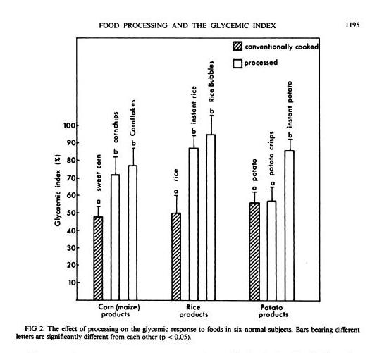
In general, industrially processed foods, which are often processed at very high temperatures to speed them through factories, have high GIs. A study in the American Journal of Clinical Nutrition [1] compared home-cooked corn, rice, and potato with processed foods based on them (instant rice, Rice Bubbles, corn chips, Cornflakes, instant potato, and potato crisps), and the processed foods had consistently higher GIs:

Another study in the British Journal of Nutrition [2] looked at 14 starchy plants prepared in different ways and found that roasting and baking raised the GI:

GI value of some of the roasted and baked foods were significantly higher than foods boiled or fried (P<0.05). The results indicate that foods processed by roasting or baking may result in higher GI. Conversely, boiling of foods may contribute to a lower GI diet.

Perhaps cooking methods that dry out the plant increase the GI.

Meals Have Lower GI

GI is calculated by eating a single food and only that food.

But what happens when you eat a meal? You’re no longer eating one food, but a mixture of foods. The baked potato may come with meat and vegetables, and with butter on top.

You might think that a weighted average of the GI of the various foods might give a good indication of the GI of the meal. Then, since fat, meat, and vegetables have a low GI, you’d expect GI of the meal to be much lower.

It turns out that the GI of meals is low – in fact, it is even lower than the average GI of the foods composing the meal.

That is the result of a new study in the American Journal of Clinical Nutrition [3]. Three meals were prepared combining a starch (potato, rice, or spaghetti) that digested to 50 g (200 calories) glucose with vegetables, sauce, and pan-fried chicken. The GIs of the meals were consistently lower than the values predicted using a weighted average of GIs of the meal components:

Meal Actual GI Predicted GI
Potato 53 63
Rice 38 51
Spaghetti 38 54

So eating a starch as part of a meal reduces GI to the range 38 to 53 – below the levels of many fruits and berries.

Fat Reduces GI

J Stanton has noted that adding a little fat to a starch is very effective in lowering its GI. In a post titled “Fat and Glycemic Index: The Myth of Complex Carbohydrates,” JS states that:

  • Flour tortillas have a GI of 30, compared to a GI of 72 for wheat bread, because tortillas are made with lard.
  • Butter reduces the glycemic index of French bread from 95 to 65.
  • A Pizza Hut Super Supreme Pizza has a GI of 30, whereas a Vegetarian Supreme has a GI of 49.

JS suggests that the reason fat does this is that it lowers the gastric emptying rate, and cites a study which showed that adding fat to starches could increase the gastric emptying time – the time for food to leave the stomach – by 50%. [4]

What’s interesting to me here is that what we really care about is not the glycemic index, but the peak blood glucose level attained after a meal. It is blood glucose levels above 140 mg/dl only that are harmful, and the harm is proportional to how high blood glucose levels rise above 140 mg/dl. So it’s the spikes we want to avoid.

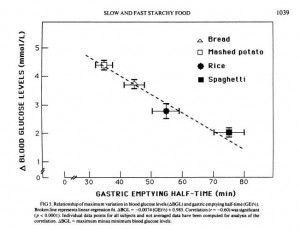
But another paper shows that gastric emptying rate is even more closely tied to peak blood glucose level than it is to glycemic index. From [5]:

So combining a starch with fat may reduce peak blood glucose levels even more than it reduces the glycemic index; which is a good thing.

Dairy reduces GI

Dairy is effective at reducing GI:

[D]airy products significantly reduced the GI of white rice when consumed together, prior to or after a carbohydrate meal. [6]

It is not likely that dairy fat alone was responsible, because whole milk worked better than butter. However, low-fat milk only reduced the GI of rice by 16%, while whole milk reduced it by 41%. So clearly dairy fats are part of the recipe, but not the whole story; whey protein may also matter.

Fiber Reduces GI

Fiber is another meal element that reduces the rise in blood sugar after eating.

Removing fiber from starchy foods increases their glycemic index [7]; adding fiber decreases it. For instance, adding a polysaccharide fiber to cornstarch reduced its GI from 83 to 58; to rice reduced its GI from 82 to 45; to yogurt from 44 to 38. [8]

So it’s good to eat starches with vegetables – the foods richest in fiber.

Acids, Especially Vinegar, Reduce GI

Traditional cuisines usually make sauces by combining a fat with an acid. Frequently used sauce acids are vinegars and citric acid from lemons, limes, or other citrus fruits.

It turns that sauce acids can substantially reduce the GI of meals. The best attested is vinegar. From a study in the European Journal of Clinical Nutrition [6]:

In the current study, the addition of vinegar and vinegared foods to white rice reduced the GI of white rice. The acetic acid in vinegar was thought to be responsible for the antihyperglycemic effect. The amount of acetic acid to be effective could be as low as that found in sushi (estimated to be about 0.2–1.5 g/100 g). The antihyperglycemic effect of vinegar is consistent with other studies performed earlier (Brighenti et al, 1995; Liljeberg & Bjorck, 1998). Although vinegar could lower GI vales, the mechanism has rarely been reported. Most studies accounted the mechanism to be due to a delay in gastric emptying. In animal studies, Fushimi (Fushimi et al, 2001) showed that acetic acid could activate gluconeogenesis and induce glycogenesis in the liver after a fasting state. It could also inhibit glycolysis in muscles. [6]

Other acids also work. Pickled foods, which are sour due to lactic acid released by bacteria, reduce the glycemic index of rice by 27% if eaten before the rice and by 25% if eaten alongside the rice [6].

Wines, especially red wines, are somewhat acidic. I haven’t seen a study of how drinking wine with a meal affects glycemic index, but it is known observationally that wine drinkers have better glycemic control and, often, long lives. [9]

So What’s the Healthiest Way to Eat “Safe Starches”?

One way to limit the likelihood of reaching dangerous blood sugar levels after a meal is by eating a relatively “low carb” diet. We recommend that sedentary people eat about 400 to 600 carb calories per day. This limits the amount eaten at any one sitting to about 200 calories / 50 g, which is the amount of a typical glucose tolerance test. It is an amount the body is well able to handle.

But the manner in which carbs are eaten may be just as important as the amount.

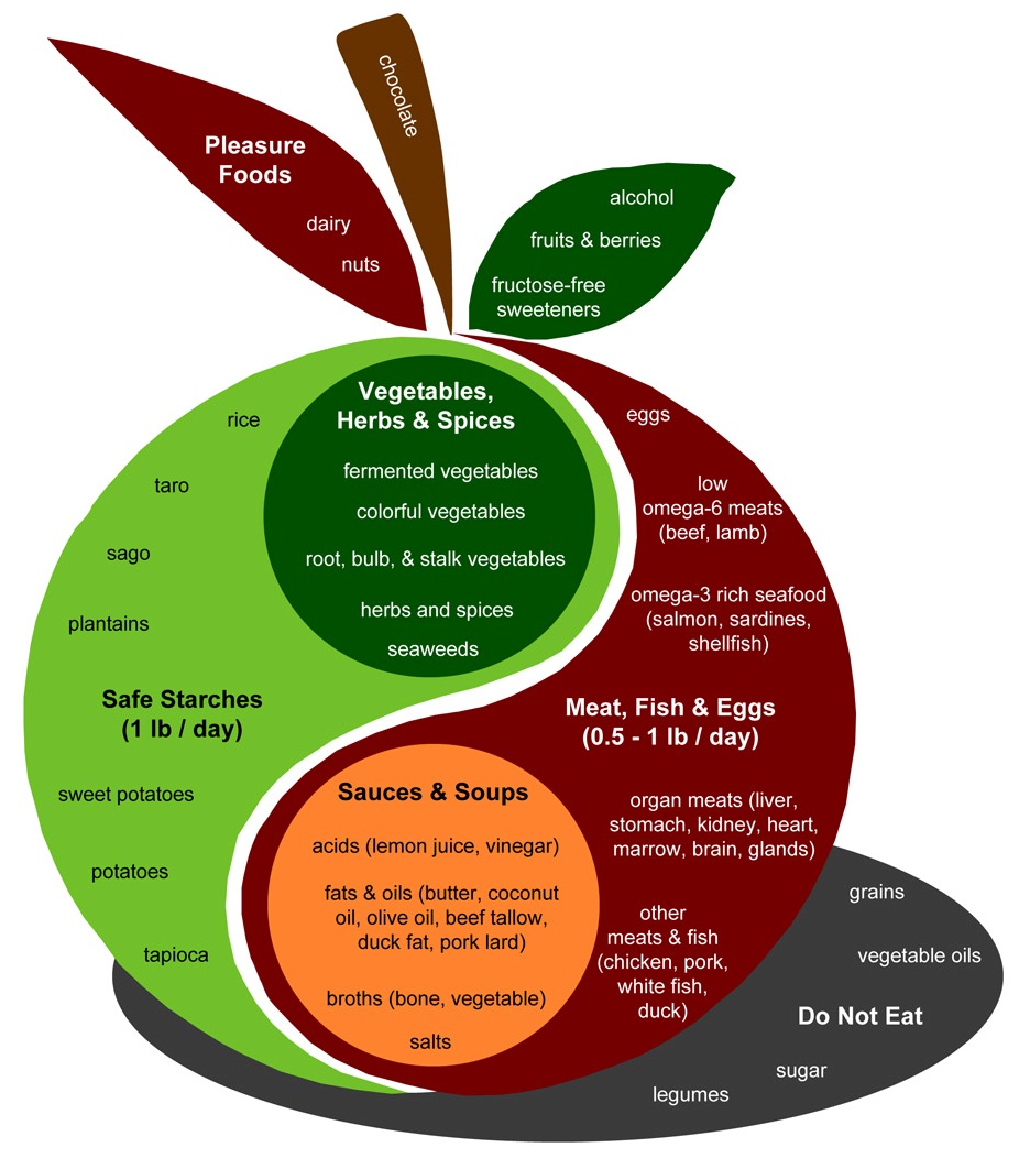
Let’s look again at the Perfect Health Diet Food Plate:

The design of a PHD meal is found in the body of the apple. Assuming two meals a day, the recipe is to combine:

  • A safe starch (roughly ½ pound, which translates to 150 to 300 carb calories);
  • A meat, fish, or egg (¼ to ½ pound);
  • A sauce made up of fats and acids such as lemon juice or vinegar;
  • Vegetables, preferably including fermented vegetables with their healthy acids;
  • (Optionally) some dairy or a glass of wine.

This is precisely the recipe which science has found minimizes the elevation of blood glucose after meals.

It seems reasonable to expect that a meal designed in this fashion will have a glycemic index around 30. The odds of 200 carb calories with a glycemic index of 30 generating blood sugar levels that are dangerous – 140 mg/dl or higher – in healthy people is very low. Even in diabetics, it may be uncommon.

So, yes, Virginia. There is a Santa Claus, and you can eat safe starches and avoid hyperglycemia too!

References

[1] Brand JC et al. Food processing and the glycemic index. Am J Clin Nutr. 1985 Dec;42(6):1192-6. http://pmid.us/4072954.

[2] Bahado-Singh PS et al. Food processing methods influence the glycaemic indices of some commonly eaten West Indian carbohydrate-rich foods. Br J Nutr. 2006 Sep;96(3):476-81. http://pmid.us/16925852.

[3] Dodd H et al. Calculating meal glycemic index by using measured and published food values compared with directly measured meal glycemic index. Am J Clin Nutr. 2011 Oct;94(4):992-6. http://pmid.us/21831990.

[4] Thouvenot P et al. Fat and starch gastric emptying rate in humans: a reproducibility study of a double-isotopic technique. Am J Clin Nutr 1994;59(suppl):781S.

[5] Mourot J et al. Relationship between the rate of gastric emptying and glucose and insulin responses to starchy foods in young healthy adults. Am J Clin Nutr. 1988 Oct;48(4):1035-40. http://pmid.us/3048076.

[6] Sugiyama M et al. Glycemic index of single and mixed meal foods among common Japanese foods with white rice as a reference food. Eur J Clin Nutr. 2003 Jun;57(6):743-52. http://pmid.us/12792658. Full text: http://www.nature.com/ejcn/journal/v57/n6/full/1601606a.html.

[7] Benini L et al. Gastric emptying of a solid meal is accelerated by the removal of dietary fibre naturally present in food. Gut. 1995 Jun;36(6):825-30. http://pmid.us/7615267.

[8] Jenkins AL et al. Effect of adding the novel fiber, PGX®, to commonly consumed foods on glycemic response, glycemic index and GRIP: a simple and effective strategy for reducing post prandial blood glucose levels–a randomized, controlled trial. Nutr J. 2010 Nov 22;9:58. http://pmid.us/21092221.

[9] Perissinotto E et al. Alcohol consumption and cardiovascular risk factors in older lifelong wine drinkers: the Italian Longitudinal Study on Aging. Nutr Metab Cardiovasc Dis. 2010 Nov;20(9):647-55. http://pmid.us/19695851.


Mobility and Health: Some Thoughts

I’d like to thank Todd Hargrove for his guest post (How to Do Joint Mobility Drills, July 26, 2011). It was thought-provoking, and I thought I’d share my reflections on it.

What Is the Goal of Exercise?

When it comes to fitness, the blogosphere tends to emphasize strength and athleticism. This is great, but there are other dimensions to health and fitness that are maybe a bit under-discussed.

As a 48-year-old recovering chronic disease patient, I am not looking to become a competitive athlete, enjoyable though that might be. Rather, I want to maximize health and longevity, and be able to freely and pleasurably move through all the challenges and opportunities life may present. I’ll be happy if I can:

  1. Be strong enough to freely manipulate my body plus a heavy load.
  2. Be fit enough to run 3-4 miles with pleasure, play an hour of tennis without getting sore, and sprint faster than common criminals.
  3. Be mobile enough to move freely and gracefully through the full natural range of motion of all joints without crackling, stiffness, or soreness.
  4. Develop good posture, circulation, and neurological function, so that my body naturally arranges itself in healthy positions.

The first three goals are not too different from Jamie Scott’s prescription for surviving a natural disaster. He asks: Could you lift yourself over a wall or up to a balcony to escape a tsunami? Sprint-jog 3-4 miles over shattered ground and obstacles to escape the liquefaction zone of an earthquake? Walk 3-4 hours over hills daily when roads are impassable? Get into a low squat to fit in a small shelter, or squeeze through a small opening?

But I have a special interest in neurological health. I had chronic ear infections as an infant, culminating in surgery, and ever since have had poor balance. My central nervous system infection made it much worse. Three years ago I had to sit down to put pants on or take them off; walked into doors; and fumbled and dropped things, as the complete loss of our former collection of wine glasses can attest. With diet and antibiotics I’ve recovered; my balance is now similar to what it was in my 20s – which is to say, poor.  I can now stand on one foot for about 20 seconds before I have to put down the other foot to balance myself; that would have been 1 or 2 seconds three years ago, but Shou-Ching can do it indefinitely. When we go hiking in the mountains, Shou-Ching clambers up or down steep rocky slopes like a mountain goat; I have to move with care.

Falls are a major cause of health impairment, broken bones, and mortality in the elderly. It would be great if I can improve nervous system function and balance before I get old and falls become dangerous.

I’m very pleased to start this blog’s discussion of fitness with Todd’s post, because mobility and neurological function are critically important to fitness at all ages – and may be crucial to good health as we age.

The Concept of Body Maps

Let me paraphrase one of the key points of Todd’s post this way:

The brain maintains “maps” of the body … These maps may become inaccurate, out of synch with the physical body … As a result the brain may believe a movement is impossible or dangerous and block its performance, even if the body is fully capable of performing the movement … With training the brain can learn the true movement capabilities of the body and revise its maps to more accurately reflect reality, thus increasing the body’s ability to move freely.

The idea that brain “maps” of the physical body, rather than the actual body, are what sets the limits to motion reminded me of a TED video I had seen by Dr. Vilayanur Ramachandran. He is a neuroscientist who investigated the problem of “phantom pain” in the lost limbs of amputation victims, and showed that the pain could be cured by “mirror box” therapies that fooled the brain into manipulating the lost arm and thereby re-drawing the brain’s body maps. Here is his fascinating TED talk:

Todd explains how improper brain maps can lead to chronic pain, and how repairing the brain maps can end the pain. This is an important idea for those suffering from chronic pain.

Use It Or Lose It

Todd observes that

While movement will clarify maps, lack of movement will tend to blur them. In a famous experiment, researchers found that sewing a monkey’s fingers together for a few weeks caused its brain to map the fingers as one unit, not as two separate parts capable of individual movements.

So if I want my brain to remember what my body is capable of, I need to regularly take my body through a diversity of movements.

This is an important reminder for someone who spends 12 hours per day at a desk. Get away from the desk, even if only for a few minutes a day, and move!

The Strategy of Slow, Mindful Movement

When I was young I wanted to do everything fast. (Shou-Ching complains that when I’m behind the wheel of a car, I think I’m still young.) But now I’m starting to appreciate the benefits of slow motion.

Todd’s list of ways to “maximize the benefit of mobility exercise” emphasizes slow, mindful movements. A few thoughts on each:

Avoid pain and threat.” Since the purpose of the brain’s body maps is to prevent dangerous movements from happening, to re-draw the maps we have to teach the brain that “dangerous” movements are actually safe. For this to be persuasive, they must actually be safe. But this corollary may be less obvious:

Make sure the movement does not … create other signs of threat such as holding the breath, grimacing, collapsing your posture, or using unnecessary tension.

I’m a fan of the mobility videos of Kelly Starrett at mobilitywod.com, and he frequently advises one never to make a “pain face” or grimace, but rather to maintain a cheerful “Zen face.” A grimace during a challenging stretch or movement may be enjoyable, but it might detract from the value of the exercise. Interesting!

Be mindful and attentive.” This one comes easily to me: I am introspective and enjoy listening to my body and paying attention to muscles, breath, and blood flow during exercise. It’s good to know that’s beneficial.

Use novel movements.” I like routine, but routine mobility drills are unproductive. Movements need to explore new capabilities.

Easy does it.” Move slowly and gently. This calls to mind the classic Chinese exercise forms, like Qi Gong and Tai Chi; they are characterized by slow, flowing, graceful movements.

Be curious, exploratory, and playful.” I like the evolutionary inference Todd makes here:

All animals engage in the most play during the times of their lives when the educational demands are the highest. This means that play is the best solution to difficult education problems that evolution has found.

I think we sometimes fall into the trap of thinking that adulthood implies seriousness and sobriety. No! Rather, good health implies lifelong playfulness.

In Boswell’s Life of Johnson, in the Dedication, Boswell writes:

It is related of the great Dr. Clarke, that when in one of his leisure hours he was unbending himself with a few friends in the most playful and frolicksome manner, he observed Beau Nash approaching; upon which he suddenly stopped. “My boys,” said he, “let us be grave – here comes a fool.”

Let us not be fools, and play!

Can Rhythmic Movement Be an Ultradian Therapy?

I’ve done several posts on the subject of circadian (day-night) rhythms, and how enhancing these rhythms with diet, light, sleep, and exercise may be therapeutic for many diseases. See, for instance, Intermittent Fasting as a Therapy for Hypothyroidism (Dec 1, 2010) and Seth Roberts and Circadian Therapy (Mar 22, 2011).

But humans have other natural biorhythms that cycle more frequently. These “ultradian rhythms” can be quite short. For instance, some hormones are released in pulses – I believe insulin and thyroid hormone may operate this way – and I believe a common interval between pulses is 6 seconds.

Many classic movement forms, like yoga or qi gong, emphasize that movement should be synchronized with breathing, and that breathing should be slow and rhythmic – often with about ten breaths per minute, or six seconds per breath.

The coincidence between these numbers intrigues me. If enhancing circadian rhythms is therapeutic for disease, might enhancing ultradian rhythms by mindful “synching” of the breath to their period be therapeutic for hormonal dysfunction?

It’s just a thought. Many people with glucose regulation issues have disrupted ultradian rhythms for insulin secretion. The ultradian clocks in their pancreatic beta cells aren’t working properly. Wouldn’t it be interesting if mindful breathing, as in yoga, could improve insulin secretion and glucose regulation?

This is not such a far out idea. Consider these quotations from recently published papers:

Mind-body modalities based on Eastern philosophy, such as yoga, tai chi, qigong, and meditation … have many reported benefits for improving symptoms and physiological measures associated with the metabolic syndrome…. Findings from the studies reviewed support the potential clinical effectiveness of mind-body practices in improving indices of the metabolic syndrome. [1]

Participation of subjects with T2DM in yoga practice for 40 days resulted in reduced BMI, improved well-being, and reduced anxiety. [2]

Yoga-nidra practiced for 30 minutes daily up to 90 days, parameters were recorded every. 30th day. Results of this study showed that most of the symptoms were subsided (P < 0.004, significant), and fall of mean blood glucose level was significant after 3-month of Yoga-nidra. This fall was 21.3 mg/dl, P < 0.0007, (from 159 +/- 12.27 to 137.7 +/- 23.15,) in fasting and 17.95 mg/dl, P = 0.02, (from 255.45 +/- 16.85 to 237.5 +/- 30.54) in post prandial glucose level. Results of this study suggest that subjects on Yoga-nidra with drug regimen had better control in their fluctuating blood glucose and symptoms associated with diabetes, compared to those were on oral hypoglycaemics alone. [3]

[F]asting plasma insulin was significantly lower in the yoga group. The yoga group was also more insulin sensitive (yoga 7.82 [2.29] v. control 4.86 [11.97] (mg/[kg.min])/(microU/ml), p < 0.001). [4]

There are fifty-six papers in Pubmed on “yoga diabetes”, and only four of them date before 2002. Most were published after 2008. This is an emerging area of research, but it would be interesting if slow, mindful movement proves to be an effective therapy for metabolic disorders. Maybe exercise doesn’t need to be vigorous to heal disorders like diabetes and obesity!

The Best Exercises for Mobility

I asked Todd what traditional movement forms he would most recommend. He replied:

In my blog I made some lists of exercises styles, traditional and modern, which are in line with what I recommend: the Feldenkrais Method, Z-Health, Alexander Technique, and tai chi are at the top of the list.

My favorite is the Feldenkrais method, but I think for purposes of your blog, some tai chi videos would be perfect, because they really provide a picture of what I’m talking about. You can’t do tai chi without observing all of the guidelines I provide at the end. And it looks cool.

You might include a point that the magic of tai chi is not so much in the specific forms they use, but in the WAY they move – smooth and slow. And the mind state while moving – mindful, relaxed, attention to small details and subtleties. You could apply this tai chi style to anything and get benefit – sitting, standing, walking, lifting weights or doing joint mobility drills.

All of these movement disciplines are extremely interesting, and I hope to get help exploring them in future blog posts. I know that a number of Z-Health Master Trainers have read our book, and hopefully one of them will teach us about Z-Health.

In closing, here are some videos of Qi Gong and Tai Chi movements. With videos available on DVD or on YouTube, there’s no need to join a class to learn mobility drills. You can play a video in your TV and practice slow, mindful, relaxed movements at home.

Perhaps the most valuable movements, in my view, are those used as “warm-up” exercises in Tai Chi or beginning movements in Qi Gong. Here is a well-made introductory video:

Here is a beautiful exhibition of Tai Chi:

Thanks, Todd. I very much appreciate the opportunity to learn about fitness from an expert!

References

[1] Anderson JG, Taylor AG. The metabolic syndrome and mind-body therapies: a systematic review. J Nutr Metab. 2011;2011:276419. http://pmid.us/21773016.

[2] Kosuri M, Sridhar GR. Yoga practice in diabetes improves physical and psychological outcomes. Metab Syndr Relat Disord. 2009 Dec;7(6):515-7. http://pmid.us/19900155.

[3] Amita S et al. Effect of yoga-nidra on blood glucose level in diabetic patients. Indian J Physiol Pharmacol. 2009 Jan-Mar;53(1):97-101. http://pmid.us/19810584.

[4] Chaya MS et al. Insulin sensitivity and cardiac autonomic function in young male practitioners of yoga. Natl Med J India. 2008 Sep-Oct;21(5):217-21. http://pmid.us/19320319.

How Does a Cell Avoid Obesity?

I am optimistic that everyone can acquire an attractive and healthy body composition, including women of a certain age. I’ll discuss what I think is the best strategy for that in future blog posts. First, some ground work.

Obesity and weight regulation is a complicated subject, and I have to confess upfront that I am not thoroughly conversant with the literature. I still learn new things every time I delve into the journals. Occasionally, I will I write posts (like today’s) that look into journal articles and molecular pathways related to obesity. This helps me explore ideas and learn. Hopefully you’ll have some fun following along.

A good starting point for an investigation into obesity would seem to be the issue of how weight regulation works in a healthy person. How does our body keep itself at its ideal weight?

Now all the problems our body has to solve, had to be solved about 2 billion years earlier by the first eukaryotic cells. Formed by the merger of fat-eating mitochondria with glucose-eating bacteria, single-celled eukaryotes had to regulate their various metabolic pathways to keep themselves from becoming too fatty or too lean (and to control the urge of mitochondria to eat their hosts!). Then, when multi-cellular organisms developed, these cellular-level mechanisms were the building blocks available for organism-level weight regulation.

So we can simplify the subject a bit by looking at individual cells. And here CarbSane gets a huge “hat tip” for finding a fascinating paper that neatly summarizes how an individual human muscle cell controls its fat and glucose levels.

Obese Cells on High-Carb Diets

The paper dates from 2004, looks at muscle cells, and has the latinate title of “Substrate cycling between de novo lipogenesis and lipid oxidation: a thermogenic mechanism against skeletal muscle lipotoxicity and glucolipotoxicity.” [1]

We can (loosely) translate “de novo lipogenesis” as “fat creation from glucose,” “lipotoxicity” as “too much fat” and “glucolipotoxicity” as “too much glucose in a cell that has too much fat.” So the paper is examining cells that are:

  1. fat;
  2. eating a high-carb diet; and
  3. disposing of excess glucose by converting it to fat.

Sounds familiar! How do these cells control their weight?

Cells Follow the Same Strategy as the Body

Glucose can be toxic and it feeds bacteria, so excess glucose is removed from the body as quickly as possible. First, it’s stored as liver and muscle glycogen; beyond that it is mostly converted to fat by de novo lipogenesis.

The main organs which do that conversion are the liver and adipose tissue, but this paper points out that muscle cells do it too:

The recent recognition that de novo lipogenesis might have relevance for lipid homeostasis in skeletal muscle stems from the realization that Sterol regulatory element binding protein-1c (SREBP-1c), a member of the family of transcription factors that regulate the expression of genes involved in lipid storage in liver and adipose tissue, is also present in skeletal muscle at a level close to that observed in the liver,41,42 … [M]ost fascinating are the very recent demonstrations that glucose alone (in the absence of insulin) can stimulate de novo lipogenesis in skeletal muscle cells….

[I]t is clear that de novo lipogenesis, although low in skeletal muscle, can be markedly stimulated in muscle cells, particularly under conditions of high glucose (and/or high insulin) concentrations. [1]

So muscle cells convert glucose to fat just as liver cells do. Indeed, they convert glucose to fat even without any insulin stimulation, just to get rid of it – but they dispose of glucose most aggressively when stimulated by insulin.

The likely reason for this is to help the body avoid glucose toxicity:

Extrapolated to conditions of postprandial elevation in blood glucose and insulin (particularly after a high-carbohydrate meal), de novo lipogenesis in skeletal muscle, like in the liver, could also contribute to blood glucose homeostasis by disposing some of the excess circulating glucose as muscle triglycerides, particularly if the glycogen stores are full. In other words, de novo lipogenesis in myocytes may provide another sink for glucose disposal through skeletal muscles. [1]

Insulin gets the muscle cells to take in more glucose and do more of this glucose-to-fat conversion. So insulin is a glucose disposal hormone. Muscle cells respond to it as an act of charity to the rest of the body.

But if muscle cells are storing fat that they manufacture from glucose, they risk becoming obese. How do they get rid of excess fat?

That gets us to the other latinate phrase in the title, “lipid oxidation,” also known as fat burning, and a key hormone, leptin. The authors write:

It is now well established that the adipocyte-derived hormone leptin, which is well known for its central role in body weight regulation in part via its control over thermogenesis, 52–55 also plays an important role in blood glucose homeostasis and in the protection of insulin-sensitive tissues against excessive ectopic lipid storage by regulating the partitioning of fatty acid away from storage towards oxidation. 56–58 [1]

Leptin is released by adipose cells in proportion to the amount of fat they are storing. High leptin levels mean “I’m obese, help me lose weight”; low leptin levels mean “I’m skinny, please don’t waste any fat, we may need it.”

Leptin helps the body regulate its weight, by two mechanisms: 

  1. Leptin promotes fatty acid oxidation, or the burning of fats.
  2. Leptin triggers thermogenesis, “creation of heat,” which warms the body and causes it to lose energy.

In short, leptin causes the body – and individual muscle cells – to transform fat into waste heat, thereby slimming down the cells and the body.

What happens if you stimulate normal muscle cells with both leptin and insulin?  This situation occurs in a healthy person with too much fat (leading to high leptin) who has eaten a high-carb meal and has lots of extra blood glucose to get rid of (leading to high insulin). The insulin triggers the glucose-to-fat conversion pathway, the leptin triggers thermogenesis – but the effect is compounded because the insulin amplifies leptin activity:

[W]e found that leptin could directly stimulate thermogenesis in skeletal muscle via ObRb,62 and that this thermogenic effect of leptin, which requires PI3K activity (since it is inhibited by wortmannin), is potentiated by insulin, a potent activator of PI3K. [1]

This makes sense: the body really wants to get rid of the excess glucose, which is toxic, but high leptin means it’s already fat and doesn’t want to get fattier. So if you’ve got too much fat and too much glucose, you really, really want to turn up the waste heat generator.

So eating some carbs, by increasing after-meal insulin, will actually tend to increase fat oxidation and waste heat generation. That is, it will lead to more calories out, at least for a few hours after a meal. Whereas a zero-carb diet, by keeping insulin low after meals, might tend to tamp down postprandial waste heat generation.

If you read CarbSane’s post, you’ll see that this is what got her excited. (Maybe it would also excite Matt Stone, advocate of the carb-overfeeding-raises-body-temperature thesis.)

We shouldn’t jump to the conclusion that eating lots of carbs is good for weight loss, since carbs may also increase appetite and calories in, or have other effects such as generating a transient glucotoxicity. But we should keep this thought in the backs of our minds: Not every response to dietary carbs works against weight loss.

The paper notes that it’s not only leptin that stimulates thermogenesis. Other hormones – including adiponectin, which is also released by adipose cells, stimulate the same thermogenic pathways.

One can also entertain the interesting possibility that, in skeletal muscle, this substrate cycling is also activated in response to other hormones and neurotransmitters (eg, adiponectin, catecholamines) particularly since adiponectin, as well as adrenergic agonists, can also stimulate AMPK activity, glucose utilization and fatty acid oxidation in skeletal muscle or adipose tissue.61,66–69 This substrate cycling between de novo lipogenesis and lipid oxidation could therefore constitute a thermogenic effector in skeletal muscle. [1]

Adiponectin is a favorite hormone of Dr. Kenneth Tourgeman of the blog Nephropal. High adiponectin levels are associated with good health; obesity is associated with low levels of adiponectin. Adiponectin, among other effects, increases insulin sensitivity – thus it would tend to promote thermogenesis not only by its own action, but also indirectly by enhancing insulin amplification of leptin-induced thermogenesis.

What If The Cell Became Hormone Resistant?

As a thought experiment, we can imagine what would happen if our healthy muscle cell became metabolically damaged – if it became resistant to some of these hormones.

If it became leptin resistant, then it would no longer dispose of excess fat via thermogenesis. The fat would collect and the cell would become obese.

If the cell remained leptin resistant but insulin sensitive, it would gradually kill itself through obesity. So leptin resistance would naturally lead to insulin resistance as the cell protects itself against lipotoxicity.

Once the cell becomes insulin resistant, then it no longer disposes of excess glucose by fat conversion. Glucose levels might become elevated. This corresponds to the condition in the body as a whole that we call metabolic syndrome or prediabetes.

High glucose levels, of course, lead to glucotoxicity or poisoning by excess glucose. The pancreatic beta cells, which produce insulin, are especially subject to glucose poisoning. Thus, prediabetes in the body, continued long enough, leads to loss of these cells and diabetes.

We can imagine two kinds of diabetes:

  • If the insulin resistance developed as a consequence of leptin resistance, then we’d have an obese diabetic.
  • If the insulin resistance developed without leptin resistance, then we’d have a skinny diabetic.

Conclusion

I like this paper a lot because it gives us a look at the key hormonal pathways involved in obesity, but in a very simple model – a single muscle cell.

It suggests that possible causes of obesity are leptin resistance and adiponectin deficiency, and that if we want to fix obesity, we may wish to look for a diet which increases leptin sensivity and/or adiponectin level.

References

[1] Dulloo AG et al. Substrate cycling between de novo lipogenesis and lipid oxidation: a thermogenic mechanism against skeletal muscle lipotoxicity and glucolipotoxicity. Int J Obes Relat Metab Disord. 2004 Dec;28 Suppl 4:S29-37.  http://pmid.us/15592483.

Choline Deficiency and Plant Oil Induced Diabetes

I’m going to deviate from my original plan for the “Dangers of a Zero-Carb Diet” series to discuss a topic that came up in the comments to the first post.

Leonie’s Diabetes and the Rose Corn Oil Trial

What prompted this diversion is Leonie’s interesting comment from Wednesday’s post:

I developed diabetes several years after being on a low carb diet. Continuing low carb to manage the diabetes did not halt its progress. It has taken about 18 months of adding more carbs (60 – 100 gr/day) to my diet to bring my fasting glucose down by a couple of mmol and eating more carbs has also lowered my Hba1c and post meal spikes significantly. I wonder if the liver is another organ that may be affected by carbohydrate deficiency.

I had not heard of such cases before, or so I thought, but Dr. Deans in the comments reminded us that Peter at Hyperlipid had noticed two similar cases in the Rose Corn Oil trial. [1] (The Rose Corn Oil trial, of course, figures prominently in our book’s discussion of PUFA toxicity.)

In the Rose Corn Oil trial, there were three arms – a normal diet arm, a high corn oil arm, and a high olive oil arm. The normal dieters were expected to eat “fried foods, fatty meat, sausages, … ice cream, cheese, … milk, eggs, and butter” while the oil arms were supposed to restrict these foods and replace them with corn or olive oil.

Here’s what happened:

Four patients were removed from the trial for other reasons. Two developed non-cardiac thromboembolism and were given anticoagulant therapy. The other two were removed because of diabetes mellitus. One of them already had mild diabetes, but glycosuria increased considerably soon after he started oil. Oil was stopped and glycosuria disappeared. Oil was restarted, but was stopped a month later because heavy glycosuria recurred. The other patient, not a previously recognized diabetic, developed glycosuria with a diabetic glucose-tolerance test a few weeks after starting oil. [1]

The patients who developed diabetes came one from the corn oil arm and one from the olive oil arm. Likewise, the patients who developed thromboembolisms came one from the corn oil arm and one from the olive oil arm. No such disasters occurred on the “fatty meat” arm.

Since all three diets were similarly fatty, it doesn’t appear to be the quantity of fat that was the issue. Rather it was the type of lipid, or some micronutrient that was present in the animal and dairy foods but lacking in the plant oils.

For insight into what the problem might be, let’s look at how scientists poison lab animals.

Insights from Diet Animal Poisoning Research

You have to pity diet researchers. It takes 60 years for bad diets to poison humans enough to significantly raise mortality rates. Yet a diet researcher is supposed to gain a Ph.D. in 4 years (or in 5 while simultaneously obtaining an MD!), do a postdoc in 2 years, win a grant in the first years of an entry-level position with PI status, and then demonstrate productive results within the term of a 2-to-5 year grant. Deadlines are pressing: A study needs to start rats or mice on two diets, and have one diet produce much better health than the other, in considerably less than a two-year time frame.

Just comparing McDonald’s fast food with a Mediterranean diet won’t do. Two years later both sets of mice will die happily of old age, with no significant differences between groups. Peer reviewers judge you to have discovered no new results. No new results means no paper, no grant, no job.

So “diet” researchers first have to become experts at quickly inducing disease in rats and mice. Find a diet that poisons animals in a few months, compare it to another diet that doesn’t, and you have a paper. Look for variations that slow or hasten the poisoning, and you have more papers. To be a highly productive scientist, one must be a skilled animal poisoner.

Various techniques have been developed for this purpose, including: knocking out some crucial gene; breeding a mutant strain that naturally develops disease; giving the animals poison with their food; or depriving them of crucial nutrients. Almost every study of diet in mice or rats uses one of these techniques.

If a missing nutrient can cause diabetes within a few years for Leonie and 12 to 18 months for the Rose Corn Oil trial volunteers, it’s likely to be pretty good at inducing disease in animals too. There’s a good chance diet animal poisoning researchers have already stumbled upon it in rats or mice.

Choline Deficiency Diseases

One of the most popular deficiency diets among researchers is the choline-deficient diet. A useful paper by Dutch scientists [2] gives a nice look at the impact of choline deficiency on rats.

Choline deficiency (CD) by itself induces metabolic syndrome (indicated by insulin resistance and elevated serum triglycerides and cholesterol) and obesity.

A combined methionine and choline deficiency (MCD) actually causes weight loss and reduces serum triglycerides and cholesterol, but induces more severe liver damage. The MCD diet prevents the body from manufacturing choline from methionine, vitamin B12, and folate, so MCD diets severely reduce choline levels; and without choline VLDL particles are not produced. Without VLDL particles, fats and cholesterol are trapped in the liver and never reach the blood and adipose cells.

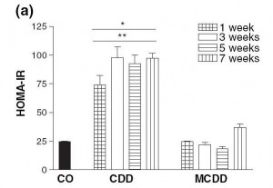
Here is a measure of insulin resistance on the two diets:

The induction of insulin resistance by the CD diet is very rapid, requiring less than a week.

Induction of insulin resistance is thought to be mediated by elevated TNF-alpha production by adipose cells and by hypertriglyceridemia. Since the MCD diet neither raised serum triglycerides nor caused obesity which induces TNF-alpha production in adipose cells, it did not cause insulin resistance.

What Does This Have to Do With Diabetes?

Insulin resistance is a key step in the development of diabetes:

  • Insulin resistance in the liver causes the liver to release more glucose into the blood (since insulin inhibits glucose release by the liver). This is discussed in a nice paper [3] found by LynMarie Daye and cited in the comments by CarbSane.
  • Peripheral insulin resistance means that the rest of the body is less sensitive to insulin. The pancreas has to produce more insulin to dispose of the excess glucose that the liver is releasing.

This elevation of insulin and glucose levels is a crucial step toward diabetes; it is “pre-diabetes.”

Persistently elevated glucose levels can then poison the beta cells of the pancreas, diminishing insulin secretion capability and causing diabetes. [4]

The Rose Corn Oil trial was not a low-carb diet, so postprandial glucose levels could easily have risen to toxic levels.

If a CD diet can cause insulin resistance in a week, it’s plausible that it might cause diabetes in 12 to 18 months, which is when the Rose Corn Oil trial patients developed it.

What About the Thromboembolism Cases?

MCD diets induce fibrinogenesis. In the blood, excess fibrin formation leads to clotting, and clots can block vessels to cause thromboembolisms. It may be that the thromboembolism cases in the Rose Corn Oil trial had methionine, folate, or B12 deficiencies to go with their choline deficiency.

Why Do Plant Oils Induce Diabetes But Not Animal Fats?

So why did diabetes develop in the corn and olive oil arms of the Rose Corn Oil trial but not the “fatty meat and dairy” arm?

Well, look at the choline content of these foods:

Choline content of one cup (~200 g) oil or fat or 227 g (1/2 lb) meat

Beef liver 968.0 mg
Cube steak (beef) 290.0 mg
Beef tallow 164.0 mg
Butter 42.7 mg
Olive oil 0.6 mg
Corn oil 0.4 mg

Source: http://nutritiondata.com.

Take away meat and dairy and replace them with plant oils, and it’s very easy to have a choline deficiency.

What Does This Have to Do With Zero-Carb Diets?

Maybe nothing … without carb consumption, postprandial glucose levels are not as high, and beta cell poisoning is less likely … but it may be that a zero-carb diet aggravates a choline deficiency in some fashion. I will leave this as a topic for further research.

UPDATE: Leonie in a new comment gives us more information: she has PCOS, goiter with nodules, and auto-antibodies. This suggests autoimmunity as a more likely explanation for her zero-carb diabetes.

Conclusion

In the book, we recommend the use of animal fats such as beef tallow for cooking, and recommend that pregnant women and vegetarians supplement with choline. We thought seriously about recommending that everyone supplement choline, but were reluctant to recommend too many supplements.

In retrospect, we should have recommended choline supplements for everyone who is overweight, has elevated blood glucose or lipids, or has elevated liver enzymes.

We have been using beef tallow as our cooking oil for several months now. It might be good practice for everyone to favor animal fats like beef tallow over plant oils for cooking.

References

[1] Rose GA et al. Corn oil in the treatment of ischaemic heart disease.  Br Med J. 1965 Jun 12;1(5449):1531-3. http://pmid.us/14288105.

[2] Veteläinen R et al. Essential pathogenic and metabolic differences in steatosis induced by choline or methione-choline deficient diets in a rat model. J Gastroenterol Hepatol. 2007 Sep;22(9):1526-33. http://pmid.us/17716355.

[3] Sonksen P, Sonksen J. Insulin: understanding its action in health and disease. Br J Anaesth. 2000 Jul;85(1):69-79. http://pmid.us/10927996.

[4] Leibowitz G et al. Glucose regulation of ?-cell stress in type 2 diabetes. Diabetes Obes Metab. 2010 Oct;12 Suppl 2:66-75. http://pmid.us/21029302.