



Bowel Disease, Part IV: Restoring Healthful Gut Flora

A healthy gut is a multi-species society: it is the cooperative product of the human body with trillions of bacterial cells from a thousand or more species.

An unhealthy gut is, more often than not, the product of a breakdown in this collaboration. Often, it is triggered by displacement of cooperative, commensal species of bacteria by pathogenic bacteria, fungi, viruses, and protozoa. This is why a long course of antibiotics, killing commensal bacteria, is often the prelude to bowel ailments.

It is difficult for the immune system to defeat gut infections without the help of commensal bacteria. Think about what the immune system has to deal with. The ulcers in ulcerative colitis are essentially the equivalent of infected skin abscesses, but in the colon. Here is a description of a bowel lesion in Crohn’s disease:

Ileal lesions in Crohn’s disease (CD) patients are colonized by pathogenic adherent-invasive Escherichia coli (AIEC) able to invade and to replicate within intestinal epithelial cells. [1]

Now imagine an infected skin abscess, but with feces spread over it three times a day, or stomach acid and digestive enzymes.  How quickly would you expect it to heal?

Commensal “probiotic” bacteria are like a mercenary army fighting on behalf of the digestive tract.  By occupying the interior lining of the digestive tract, they deprive pathogens of a “home base” that is sheltered from immune attack. If commensal bacteria dominate the gut, the immune system can usually quickly defeat infections.

This suggests that introduction of probiotic bacteria to the gut should be therapeutic for bowel disease.

Probiotic Supplements Are Inadequate

Most supermarket probiotics contain Lactobacillus or Bifidobacterium species. These species are specialized for digesting milk; they populate the guts of infants as they start breastfeeding, and are used by the dairy industry to ferment cheeses and yogurt.

These supplements are very effective at fighting acute diarrhea from most food-borne infections.  A fistful of probiotic capsules taken every hour will usually quickly supplant the pathogens and end diarrhea.

However, against more severe bowel diseases caused by chronic infections and featuring damaged intestinal mucosa, these species are usually not helpful.  One issue is that they provide only a tiny part of a healthful adult microbiome.  A recent study surveyed the bacterial species in the human gut, and found these species to be most abundant [2]:
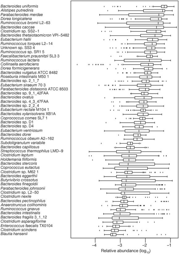
Figure: Abundant gut bacterial species

As this figure shows, Bacteroides spp. are the most common commensal bacteria, with Bacteroides uniformis alone providing almost 10% of all bacterial genes in the gut. Lactobacillus and Bifidobacterium do not appear among the 57 most abundant species.

This study showed, by the way, that patients with irritable bowel syndrome have 25% fewer types of bacterial gene in their gut than healthy people, and that the composition of bacterial genes in feces clearly distinguishes ulcerative colitis, Crohn’s disease, and healthy patients.  In other words, in the bowel diseases a few pathogenic species have colonized the gut and entirely denuded it of about 25% of the commensal species that normally populate the gut. This finding supports the idea that restoring those missing species might be therapeutic for IBS.

Bacterial Replacement Therapies Work

So if IBS patients are missing 25% of the thousand or so species that should populate the gut, or 250 species, and if common probiotics provide only 8 or so species and not the ones that are missing, how are the missing species to be restored?

The answer is simple but icky. Recall that half the dry weight of stool consists of bacteria. A healthy person daily provides a sample of billions of bacteria from every one of the thousand species in his gut. They are in his stool.

So a “fecal transplant” of a healthy person’s stool into the gut of another person will replenish the missing species.

Scientists have known for a long time that this was likely to be an effective therapy, but it is only now entering clinical practice. The New York Times recently made a stir by telling this story:

In 2008, Dr. Khoruts, a gastroenterologist at the University of Minnesota, took on a patient suffering from a vicious gut infection of Clostridium difficile. She was crippled by constant diarrhea, which had left her in a wheelchair wearing diapers. Dr. Khoruts treated her with an assortment of antibiotics, but nothing could stop the bacteria. His patient was wasting away, losing 60 pounds over the course of eight months. “She was just dwindling down the drain, and she probably would have died,” Dr. Khoruts said.

Dr. Khoruts decided his patient needed a transplant. But he didn’t give her a piece of someone else’s intestines, or a stomach, or any other organ. Instead, he gave her some of her husband’s bacteria.

Dr. Khoruts mixed a small sample of her husband’s stool with saline solution and delivered it into her colon. Writing in the Journal of Clinical Gastroenterology last month, Dr. Khoruts and his colleagues reported that her diarrhea vanished in a day. Her Clostridium difficile infection disappeared as well and has not returned since.

The procedure — known as bacteriotherapy or fecal transplantation — had been carried out a few times over the past few decades. But Dr. Khoruts and his colleagues were able to do something previous doctors could not: they took a genetic survey of the bacteria in her intestines before and after the transplant.

Before the transplant, they found, her gut flora was in a desperate state. “The normal bacteria just didn’t exist in her,” said Dr. Khoruts. “She was colonized by all sorts of misfits.”

Two weeks after the transplant, the scientists analyzed the microbes again. Her husband’s microbes had taken over. “That community was able to function and cure her disease in a matter of days,” said Janet Jansson, a microbial ecologist at Lawrence Berkeley National Laboratory and a co-author of the paper. “I didn’t expect it to work. The project blew me away.” [3]

Fecal transplants can be done without a doctor’s help:  someone else’s stool can be swallowed or inserted in the rectum. If taking feces orally, swallow a great deal of water afterward to help wash the bacteria through the stomach and its acid barrier.

Dogs and young children sometimes swallow feces.  It is unpleasant to consider, but desperate diseases call for desperate measures. Perhaps one day, healthy stools will be available in pleasant-tasting capsules, and sold on supermarket shelves.  Not yet.

Attacking Pathogenic Biofilms

Most bacterial species will build fortresses for themselves, called biofilms.  These are polysaccharide and protein meshworks that, like bone, become mineralized with calcium and other minerals. These mineralized meshworks are built on bodily surfaces, like the gut lining, and protect bacteria from the immune system, antibiotics, and other bacterial species. 

Pathogenic species known to generate biofilms include Legionella pneumophila, S. aureus, Listeria monocytogenes, Campylobacter spp., E. coli O157:H7, Salmonella typhimurium, Vibrio cholerae, and Helicobacter pylori. [4]

Biofilms favor the species that constructed them. So, once pathogens have constructed biofilms, it is hard for commensal species to displace them.

Therapies that dissolve pathogenic biofilms can improve the likelihood of success of probiotic and fecal transplant therapies. Strategies include enzyme supplements, chelation therapies, and avoidance of biofilm-promoting minerals like calcium.  Specifically:

  • Polysaccharide and protease digesting enzymes. Human digestive enzymes generally do not digest biofilm polysaccharides, but bacterial enzymes that can are available as supplements. Potentially helpful enzymes include hemicellulase, cellulase, glucoamylase, chitosanase, and beta-glucanase. Non-human protease enzymes, such as nattokinase and papain, might also help. [5]
  • Chelation therapy.  Since biofilms collect metals, compounds that “chelate” or bind metals will tend to gather in biofilms. Some chelators – notably EDTA – are toxic to bacteria.  So EDTA supplementation tends to poison the biofilm, driving bacteria out of their fortress-shelter. This prevents them from maintaining it and makes the biofilm more vulnerable to digestion by enzymes and commensal bacteria. It also tends to reduce the population of pathogenic bacteria.
  • Mineral avoidance. The supply of minerals, especially calcium, iron, and magnesium, can be a rate-limiting factor in biofilm formation. Removal of calcium can cause destruction of biofilms. [6] We recommend limiting calcium intake while bowel disease is being fought, since the body can meet its own calcium needs for an extended period by pulling from the reservoir in bone. Upon recovery, bone calcium can be replenished with supplements. Iron is another mineral which promotes biofilms and might be beneficially restricted. We do not recommend restricting magnesium.

Some commercial products are available which can help implement these strategies. For instance, Klaire Labs’ InterFase (http://www.klaire.com/images/InterFase_Update_Article.pdf) is a popular enzyme supplement which helps digest biofilms, and a version containing EDTA is available (InterFase Plus).

Attacking Biofilms With Berries, Herbs, Spices, Vinegar, and Whey

Plants manufacture a rich array of anti-microbial compounds for defense against bacteria.

There is reason to believe that traditional herbs and spices, which entered the human diet during the Paleolithic and have been passed down through the generations for tens of thousands of years, were selected by our hunter-gatherer ancestors as much for their ability to promote gut health as for their taste. Dr. Art Ayers notes that:

Plants are adept at producing a wide array of chemicals with refined abilities to block bacterial functions. So when researchers sought chemicals to solve the problem of pathogens forming biofilms, it was natural to test plant extracts for inhibiting compounds. In a recent article [7], D.A. Vattem et al. added extracts from dietary berries, herbs and spices to bacterial pathogens, including the toxin producing Escherichia coli (EC) O157:H7, and checked for the ability to produce a chemical that signals the formation of a biofilm. The effective phytochemicals inhibited the bacteria from recognizing a critical density of bacteria, i.e. quorum sensing, and responding with the production of the biofilm-triggering chemical.

Blueberry, raspberry, cranberry, blackberry and strawberry extracts were effective as quorum sensing inhibitors (QSIs). Common herbs such as oregano, basil, rosemary and thyme were also effective. Turmeric, ginger and kale were also tested and found to contain QSIs. [8]

A few other remedies can weaken biofilms:

  • Acetic acid in vinegar can solubilize the calcium, iron, and magnesium in biofilms, removing these minerals and weakening the biofilm; citric acid binds calcium and can disrupt biofilms. [9]
  • Lactoferrin, a molecule in milk whey, binds iron and inhibits biofilm formation and growth. [10]
  • N-acetylcysteine can destroy or inhibit biofilms. [11]

Conclusion

Fecal transplants are the best probiotic. Tactics to disrupt pathogenic biofilms can assist probiotics in bringing about re-colonization of the digestive tract by commensal bacteria.

Along with a non-toxic diet (discussed in Part II) and nutritional support for the immune system and gut (discussed in Part III), these steps to improve gut flora make up a natural program for recovery from bowel disease.

UPDATE: Please read the cautions by two health professionals, annie and Jesse, about potential dangers of self-treatment with fecal transplants and EDTA. It is always better to pursue these therapies with a doctor’s assistance and monitoring.

Related Posts

Other posts in this series:

  1. Bowel Disorders, Part I: About Gut Disease July 14, 2010
  2. Bowel Disease, Part II: Healing the Gut By Eliminating Food Toxins m July 19, 2010
  3. Bowel Disease, Part III: Healing Through Nutrition July 22, 2010

References

[1] Lapaquette P, Darfeuille-Michaud A. Abnormalities in the Handling of Intracellular Bacteria in Crohn’s Disease. J Clin Gastroenterol. 2010 Jul 7. [Epub ahead of print]. http://pmid.us/20616747.

[2] Qin J et al. A human gut microbial gene catalogue established by metagenomic sequencing. Nature. 2010 Mar 4;464(7285):59-65. http://pmid.us/20203603.

[3] Carl Zimmer, “How Microbes Defend and Define Us,” New York Times, July 12, 2010,  http://www.nytimes.com/2010/07/13/science/13micro.html.

[4] Donlan RM. Biofilms: microbial life on surfaces. Emerg Infect Dis. 2002 Sep;8(9):881-90. http://pmid.us/12194761.

[5] Tets VV et al. [Impact of exogenic proteolytic enzymes on bacteria]. Antibiot Khimioter. 2004;49(12):9-13. http://pmid.us/16050494.

[6] Kierek K, Watnick PI. The Vibrio cholerae O139 O-antigen polysaccharide is essential for Ca2+-dependent biofilm development in sea water. Proc Natl Acad Sci U S A. 2003 Nov 25;100(24):14357-62. http://pmid.us/14614140.  Geesey GG et al. Influence of calcium and other cations on surface adhesion of bacteria and diatoms: a review. Biofouling 2000; 15:195–205.

[7] Vattem DA et al. Dietary phytochemicals as quorum sensing inhibitors. Fitoterapia. 2007 Jun;78(4):302-10. http://pmid.us/17499938.

[8] Art Ayers, “Spices are Antimicrobial and Inhibit Biofilms,” Dec. 7, 2008, http://herbal-properties.suite101.com/article.cfm/spices_are_antimicrobial_and_inhibit_biofilms.

[9] Art Ayers, “Cure for Inflammatory Diseases,” Sept. 2, 2009, http://coolinginflammation.blogspot.com/2009/09/cure-for-inflammatory-diseases.html. Desrosiers M et al. Methods for removing bacterial biofilms: in vitro study using clinical chronic rhinosinusitis specimens. Am J Rhinol. 2007 Sep-Oct;21(5):527-32. http://pmid.us/17883887.

[10] O’May CY et al. Iron-binding compounds impair Pseudomonas aeruginosa biofilm formation, especially under anaerobic conditions. J Med Microbiol. 2009 Jun;58(Pt 6):765-73. http://pmid.us/19429753.

[11] Cammarota G et al. Biofilm demolition and antibiotic treatment to eradicate resistant Helicobacter pylori: A clinical trial. Clin Gastroenterol Hepatol. 2010 May 14. [Epub ahead of print] http://pmid.us/20478402. Zhao T, Liu Y. N-acetylcysteine inhibit biofilms produced by Pseudomonas aeruginosa. BMC Microbiol. 2010 May 12;10:140. http://pmid.us/20462423.
Lactose Intolerance: Often A Result of ‘Silent’ Wheat-Derived Bowel Disease

In the comments to last week’s post on food toxins and bowel disease, I stated that, “People with bowel problems will usually do well to avoid dairy proteins, but my belief is that people with healthy bowels can usually manage dairy.”

Today, Peter links to some evidence for that proposition.

In 2005 a group led by Antonio Gasbarrini at Catholic University in Rome studied patients who experienced diarrhea and bloating after dairy consumption. They gave all patients an H2-lactose breath test, which tests the ability of the small intestine to digest the milk sugar lactose, and tests for celiac disease antibodies.

The found that 24% of those with lactose malabsorption, but only 2% of those who could digest lactose, tested positive for celiac disease antibodies. [1] It appears that destruction of the small intestine by wheat is a leading cause of inability to digest lactose – the prerequisite for lactose intolerance.

A 2008 follow-up study showed that when these patients gave up wheat and other gluten-containing grains, they recovered the ability to digest lactose:

The present study shows that a large proportion of CD patients experience a regression of lactose malabsorption after receiving a gluten-free diet. This may be related to normalization of the brush border with an improvement of lactase enzyme activity. LBT should be performed after 12 months in CD patients on a gluten-free diet in order to assess the persistence/disappearance of lactose malabsorption, thus avoiding an unnecessary lactose-free diet. [2]

Now these papers deal only with lactose malabsorption, not problems with partially digested cow casein peptides, which are the most important cause of difficulties from dairy.  So they don’t prove that dairy is safe for those who don’t eat grains and have healthy bowels.

However, they do give a bit of encouragement to those of us who are loathe to follow Loren Cordain’s advice to give up dairy.  

[1] Ojetti V et al. High prevalence of celiac disease in patients with lactose intolerance. Digestion. 2005;71(2):106-10. http://pmid.us/15775678.

[2] Ojetti V et al. Regression of lactose malabsorption in coeliac patients after receiving a gluten-free diet. Scand J Gastroenterol. 2008;43(2):174-7. http://pmid.us/17917999.

Bowel Disease, Part III: Healing Through Nutrition

[UPDATED August 2015 with updates in italic . – Paul]

Bowel diseases are characterized by chronic infection of the gut lining (and sometimes immune cells), wounded and inflamed gut tissue, and autoimmune attacks on the gut.

Malnutrition contributes to bowel disease by impairing immunity, impairing gut motility, and slowing intestinal healing.

Conversely, bowel diseases impair nutrient absorption along with the rest of digestion, exacerbating malnutrition.  To avoid a vicious spiral, bowel disease patients should be especially attentive to their nutritional needs.

The first step toward good nutrition is to eat the Perfect Health Diet, including all of our supplemental foods. For gut health, egg yolks are especially important. Also important are extracellular matrix components from bones and joints; vegetables, herbs, and spices; and healthy fats (which trigger bile production, bile being beneficial for the gut). See our Recommended Supplements page for more on the supplemental foods.

We no longer recommend taking a multivitamin. For various reasons multivitamin formulas are incomplete:

  • Some nutrients, such as magnesium and vitamin C, are too bulky to fit in a single pill.
  • Some, such as vitamin D and iodine, have no “one size fits all” dose that manufacturers can safely include.  They therefore include a low dose that is safe for all, meaning that most receive an insufficiency.
  • Others, like melatonin, may be unnecessary for the general population but are likely to benefit bowel disease patients.

Here, then, are a few supplements that bowel disease patients may find to be helpful additions to their multivitamin.

Vitamin D3 and Partners

Vitamin D has been called the “antibiotic vitamin” [1] because it triggers the body’s production of natural antibiotic compounds.

Vitamin D is needed for the production of the antimicrobial peptides cathelicidin and beta-defensin 2, which are produced mainly in immune cells and in epithelial cells lining the gut. [2, 3] These antimicrobial peptides normally saturate the mucosal barrier, where they kill most bacteria, enveloped viruses, fungi, and protozoa.

Evidence has accumulated that deficiencies in antimicrobial peptides are causal factors in bowel diseases:

  • In Crohn’s disease, a deficiency of antimicrobial peptides allows pathogens to invade. [4, 5, 6]
  • Reduced expression of intestinal defensins predicts diarrhea two months in advance. [7]
  • When antimicrobial peptides are induced therapeutically, intestinal infections are relieved. [8]
  • Mice with no vitamin D function due to knockout of the vitamin D receptor experience bacterial overgrowth of the intestine, and even mild injury to the colon results in the death of the mouse. [9]

There is increasing awareness that vitamin D is needed for defense against infections generally. [10]

Vitamin D has other benefits besides strengthening immunity. It also suppresses autoimmunity.  For instance, there is evidence for an inverse relationship between vitamin D levels and auto-antibody levels [11]. Some autoimmune patients have experienced a disappearance of auto-antibodies upon supplementation with vitamin D. [12]

Since bowel diseases are the result of infections and autoimmunity, normalization of vitamin D levels is probably extremely helpful.

Vitamin D is also associated with reduced risk of colorectal cancer. [13] Bowel disease patients are at elevated risk for colorectal cancer.

Sunshine should be sought regularly, and supplements added to bring serum 25-hydroxyvitamin D levels to at least 40 ng/ml. In addition, vitamin D should be accompanied by supplementation of two key partners:

  • Vitamin K2 is needed for proper vitamin D function.  Most inflammatory bowel disease patients are severely deficient in vitamin K2. [14] A good daily supplement should include 100 mcg of the MK-7 form, perhaps combined with some synthetic MK-4 and plant-derived vitamin K1.
  • Magnesium is needed for proper vitamin D function and many people are deficient.  200 mg/day magnesium citrate (which is better absorbed than magnesium oxide) is appropriate.

Melatonin

Melatonin is a crucial hormone which is evolutionarily conserved across all nearly all animals, indicating that it is essential to health. Most know that it is produced in the pineal gland of the brain during sleep, but it is less well known that it is abundantly produced by the gut. Much of the body’s melatonin gathers in the gut, where melatonin concentrations are 100-fold greater than in blood and 400-fold greater than in the pineal gland. [15]

In the gut melatonin reduces inflammation, stimulates immune function, fosters tissue repair and helps regenerate the epithelium. [15] Melatonin also has antimicrobial effects. [16]

Clinical trials have found that melatonin can be beneficial in treating bowel conditions. [17, 18, 19] Melatonin seems to be especially effective at reducing abdominal pain. [20, 21]

To maximize night-time melatonin levels, it is best to sleep in a totally darkened room; avoid eating food at night; and avoid exercising at night. Melatonin can also be supplemented.  Supplemental melatonin should be taken immediately before bed. Time-release tablets are best, otherwise fluctuating melatonin levels may cause waking in the middle of the night. If early waking does occur, reduce the dose.

Thyroid and Immune Minerals:  Selenium and Iodine

Selenium and iodine are critical for thyroid and immune function. Adequate thyroid hormone and a well-functioning immune system, in turn, are essential for gut health.

The thyroid hormone T4 is 65% iodine by weight, and the active thyroid hormone T3 is 59% iodine by weight.  Selenium-containing deiodinase enzymes are required to convert inactive thyroid hormone to its active form. Either iodine or selenium deficiency can cause hypothyroidism, or a deficiency of thyroid hormone.

Gut problems, especially constipation, are among the primary symptoms of hypothyroidism. Thyroid hormone is important for proper wound healing – and therefore for recovery from bowel disease.

Selenium and iodine are also essential for immune function.  Iodine along with the enzyme myeloperoxidase is needed to produce respiratory bursts – the burst of reactive oxygen species (ROS) that white blood cells use to kill pathogens.  Selenium is necessary both to strip iodine from thyroid hormone in the white blood cells, and to maintain (via the enzyme glutathione peroxidase) the function of the antioxidant glutathione which protects both white blood cells and gut cells from ROS.  Deficiency of either selenium or iodine leads to an immediate reduction in the killing activity of white blood cells.

Iodine was widely prescribed for infectious diseases in the 19th century. The Nobel laureate Dr. Albert Szent Györgyi, the discoverer of vitamin C, recounted this anecdote:

When I was a medical student, iodine in the form of KI was the universal medicine. Nobody knew what it did, but it did something and did something good. We students used to sum up the situation in this little rhyme:

If ye don’t know where, what, and why

Prescribe ye then K and I. [22]

Doses as large as 1 gram potassium iodide, containing 770 mg of iodine, were given. In practice, however, it’s highly desirable to start with a low dose of iodine, around 1 mg/day, and allow the thyroid to adapt before gradually increasing the dose.

The great danger of high doses of iodine is that it will make autoimmune attacks, as well as attacks on pathogens, more powerful. Therefore large supplemental doses of iodine should be taken only after grains and legumes have been eliminated from the diet for at least 3 months. Bowel disease patients should also be tested for the presence of thyroid auto-antibodies before beginning high-dose iodine.

Related minerals: 

  • Myeloperoxidase requires iron (heme), and unfortunately anemia due to iron deficiency is common in bowel disease patients, especially among menstruating women. [23] A good way to judge the need for iron is to measure blood ferritin levels, which should be 50 ng/ml or higher.

Thyroid hormone

If auto-antibodies are present, then hypothyroidism cannot be repaired by iodine supplementation. Yet thyroid hormone is necessary for gut healing.  In such cases, prescription thyroid hormone should be taken.

Hypothyroidism is widely undiagnosed, because the “normal” range of thyroid stimulating hormone (TSH) is far too wide. TSH levels over 1.5 mIU/L may indicate a subclinical hypothyroidism that is sufficient to measurably raise mortality. [24] Anyone with a TSH over 1.5 mIU/L and a basal body temperature below 98 F should consider obtaining prescription thyroid hormone to test whether it helps relieves hypothyroidism-associated symptoms such as constipation and improves general health. Generally, a good dose of thyroid hormone will eliminate symptoms of hypothyroidism and reduce TSH to 2.0 or so – still elevated, to stimulate thyroid healing.

Antioxidants and Bile Supports: Vitamin C, Glutathione, N-Acetylcysteine, Taurine, Glycine

Since the main immune defense (and autoimmune) mechanisms in the gut involve around ROS-producing respiratory bursts, the gut of any bowel disease patient is a ROS-rich environment.

It is therefore desirable to maximize the ability of both gut and immune cells to protect themselves against ROS with native antioxidants.

Foremost among the native antioxidants is glutathione, the primary immune and gut antioxidant. Glutathione may be supplemented directly, or its levels may be raised by supplementing with vitamin C and N-acetylcysteine.

Vitamin C has other important functions:  it is needed for wound healing and to maintain the collagen-based extracellular matrix which backs the gut and gives it integrity. One of the symptoms of scurvy (extreme vitamin C deficiency) is bleeding from the mucus membranes, including the gut lining.

A Japanese study found that vitamin C was highly protective against ulcerative colitis, reducing incidence by 55%. [25]

In rats, glutathione deficiency leads to elevated infection-induced bowel inflammation. [26] Glycine (the most abundant amino acid in extracellular matrix) and taurine both support glutathione synthesis.

Related minerals: 

  • Zinc and copper are both required for the function of another antioxidant, zinc-copper superoxide dismutase.  We recommend supplementing dietary intake with another 15 mg zinc and 2 mg copper. This can be achieved by taking a daily multivitamin plus eating occasional beef or lamb liver.
  • Magnesium is needed for glutathione synthesis. As noted before, 200 mg/day magnesium citrate is a highly desirable supplement for bowel disease patients.

Magnesium and copper deficiencies contribute to necrotizing enterocolitis [27], and probably worsen all bowel diseases.

Bile is an important aid to gut health, in part because it helps to clear the small intestine of bacteria. Bile needs vitamin C for its manufacture and needs to be conjugated with glycine or taurine. Glycine can be obtained from food as extracellular matrix material, or as a powder which you can sprinkle on food. Taurine is an excellent supplement for patients with gut disorders.

Summary

Although not a complete list of the vitamins and minerals which may be helpful to bowel disease patients, these are among the most important – and most often overlooked:

  • Vitamin D3 sufficient to raise serum 25-hydroxyvitamin D above 40 ng/ml.
  • Vitamin K2, at least 100 mcg/day.
  • Magnesium citrate or bis-glycinate, 200 mg/day.
  • Melatonin, if needed for deep restful sleep.
  • Selenium, 200 mcg/week.
  • Iodine, 225 mcg/day.
  • Thyroid hormone sufficient to bring TSH below 2.0.
  • Vitamin C, 1 g/day.
  • Glutathione, 500 mg/day, preferably in the reduced form, taken between meals on an empty stomach with a full glass of water (since it is destroyed by stomach acid).
  • N-acetylcysteine, 500 mg/day.
  • Iron, zinc, and copper sufficient to relieve deficiencies.
  • Taurine, 1 g/day.
  • Glycine (if insufficient extracellular matrix is eaten), up to 5 g/day.

Related Posts

Other posts in this series:

  1. Bowel Disorders, Part I: About Gut Disease July 14, 2010
  2. Bowel Disease, Part II: Healing the Gut By Eliminating Food Toxins m July 19, 2010
  3. Bowel Disease, Part IV: Restoring Healthful Gut Flora July 27, 2010

References

[1] “The antibiotic vitamin: deficiency in vitamin D may predispose people to infection,” Science News, Nov 11, 2006, http://findarticles.com/p/articles/mi_m1200/is_20_170/ai_n16865477/.

[2] Liu PT et al. Cutting edge: vitamin D-mediated human antimicrobial activity against Mycobacterium tuberculosis is dependent on the induction of cathelicidin. J Immunol. 2007 Aug 15;179(4):2060-3. http://pmid.us/17675463.

[3] Lehrer RI, Ganz T. Defensins of vertebrate animals. Curr Opin Immunol. 2002 Feb;14(1):96-102. http://pmid.us/11790538.

[4] Rivas-Santiago B et al. Susceptibility to infectious diseases based on antimicrobial peptide production. Infect Immun. 2009 Nov;77(11):4690-5. http://pmid.us/19703980.

[5] Wehkamp J et al. Inducible and constitutive beta-defensins are differentially expressed in Crohn’s disease and ulcerative colitis. Inflamm Bowel Dis. 2003 Jul;9(4):215-23. http://pmid.us/12902844.

[6] Barrier dysfunction due to distinct defensin deficiencies in small intestinal and colonic Crohn’s disease. Mucosal Immunol. 2008 Nov;1 Suppl 1:S67-74. http://pmid.us/19079235

[7] Kelly P et al. Reduced gene expression of intestinal alpha-defensins predicts diarrhea in a cohort of African adults. J Infect Dis. 2006 May 15;193(10):1464-70. http://pmid.us/16619196.

[8] Wehkamp J et al. Defensins and cathelicidins in gastrointestinal infections. Curr Opin Gastroenterol. 2007 Jan;23(1):32-8. http://pmid.us/17133082.

[9] Froicu M, Cantorna MT. Vitamin D and the vitamin D receptor are critical for control of the innate immune response to colonic injury. BMC Immunol. 2007 Mar 30;8:5. http://pmid.us/17397543.

[10] Yamshchikov AV et al. Vitamin D for treatment and prevention of infectious diseases: a systematic review of randomized controlled trials. Endocr Pract. 2009 Jul-Aug;15(5):438-49. http://pmid.us/19491064.

[11] Goswami R et al. Prevalence of vitamin D deficiency and its relationship with thyroid autoimmunity in Asian Indians: a community-based survey. Br J Nutr. 2009 Aug;102(3):382-6. http://pmid.us/19203420.

[12] Dr. John Cannell, The Vitamin D Newsletter, March 9, 2009.

[13] Woolcott CG et al. Plasma 25-hydroxyvitamin D levels and the risk of colorectal cancer: the multiethnic cohort study. Cancer Epidemiol Biomarkers Prev. 2010 Jan;19(1):130-4. http://pmid.us/20056631.

[14] Kuwabara A et al. High prevalence of vitamin K and D deficiency and decreased BMD in inflammatory bowel disease. Osteoporos Int. 2009 Jun;20(6):935-42. http://pmid.us/18825300.

[15] Bubenik GA. Gastrointestinal melatonin: localization, function, and clinical relevance. Dig Dis Sci. 2002 Oct;47(10):2336-48. http://pmid.us/12395907.

[16] Tekbas OF et al. Melatonin as an antibiotic: new insights into the actions of this ubiquitous molecule. J Pineal Res. 2008 Mar;44(2):222-6. http://pmid.us/18289175.

[17] Sánchez-Barceló EJ et al. Clinical uses of melatonin: evaluation of human trials. Curr Med Chem. 2010;17(19):2070-95. http://pmid.us/20423309.

[18] Terry PD et al. Melatonin and ulcerative colitis: evidence, biological mechanisms, and future research. Inflamm Bowel Dis. 2009 Jan;15(1):134-40. http://pmid.us/18626968.

[19] Chang FY, Lu CL.Treatment of irritable bowel syndrome using complementary and alternative medicine. J Chin Med Assoc. 2009 Jun;72(6):294-300. http://pmid.us/19541564.

[20] Lu WZ et al. Melatonin improves bowel symptoms in female patients with irritable bowel syndrome: a double-blind placebo-controlled study. Aliment Pharmacol Ther. 2005 Nov 15;22(10):927-34. http://pmid.us/16268966.

[21] Song GH et al. Melatonin improves abdominal pain in irritable bowel syndrome patients who have sleep disturbances: a randomised, double blind, placebo controlled study.  Gut. 2005 Oct;54(10):1402-7. http://pmid.us/15914575.

[22] Szent-Györgyi, A. (1957) Bioenergetics. New York: Academic Press, p. 112.

[23] Gomollón F, Gisbert JP. Anemia and inflammatory bowel diseases. World J Gastroenterol. 2009 Oct 7;15(37):4659-65. http://pmid.us/19787829.

[24] Asvold BO et al. Thyrotropin levels and risk of fatal coronary heart disease: the HUNT study. Arch Intern Med. 2008 Apr 28;168(8):855-60. http://pmid.us/18443261.

[25] Sakamoto N et al. Dietary risk factors for inflammatory bowel disease: a multicenter case-control study in Japan. Inflamm Bowel Dis. 2005 Feb;11(2):154-63. http://pmid.us/15677909.

[26] van Ampting MT et al. Intestinal barrier function in response to abundant or depleted mucosal glutathione in Salmonella-infected rats. BMC Physiol. 2009 Apr 17;9:6. http://pmid.us/19374741.

[27] Caddell JL. A review of evidence for a role of magnesium and possibly copper deficiency in necrotizing enterocolitis. Magnes Res.1996 Mar;9(1):55-66. http://pmid.us/8819095.

Bowel Disease, Part II: Healing the Gut By Eliminating Food Toxins

The gut is the front line of health.  The human gut houses 100 trillion bacteria from a thousand different species [1]; they weigh several pounds and make up about half the dry weight of stool. To control these bacteria 70% to 80% of the body’s immune cells are normally found in and around the gut.

A healthy gut is protected by a mucosal layer that is designed to promote commensal (friendly) bacteria, while providing a barrier to pathogenic bacteria.  Humans have evolved ways to “feed” commensal species of bacteria.  For instance:

  • Human mucus is made of glycoproteins, or compounds made of protein and sugar. Certain probiotic bacteria, such as Bifidobacterium bifidum, are able to digest human mucus. [2] Thus, the human intestine has evolved to produce “food” for beneficial gut bacteria, assuring that they are maintained even during long fasts.
  • Mother’s milk contains special sugars, called human milk oligosaccharides, which specifically feed Bifidobacterium bifidum and assure that this species successfully colonizes the baby’s intestine and wards off infection.  [3]

The absence of this protective barrier of mucus and friendly bacteria makes the intestine extremely vulnerable to infectious disease.  Premature babies who are fed formula, not human breast milk, often contract a dangerous intestinal infection, necrotizing enterocolitis. [3]

In addition to pathogenic bacteria, the gut is confronted by a heavy load of toxins. Bruce Ames and Lois Gold have estimated that the average person eats 5,000 to 10,000 different plant toxins, amassing to 1500 mg per day, plus 2000 mg of burnt toxins generated during cooking. [4]

Today’s post will focus on how those 1500 mg of natural plant toxins damage the intestinal wall and its mucosal barrier, thereby bringing about infectious bowel diseases.

Cereal Grain Toxicity

Grasses became the staple foods of agriculture because of their rich yields:  a single plant may generate tens of thousands of seeds annually. 

Yet this prolific seed production has always made grasses attractive to herbivores, and caused seeds to evolve high levels of toxins designed to poison mammalian digestive tracts, thus enabling their seeds to pass through herbivore guts undigested.  It is these toxins that make the cereal grains so dangerous to human health.

The effectiveness of grain toxins at sabotaging human digestion is illustrated by the increase in fecal mass they produce:

For every gram of wheat bran eaten, fecal weight increases by 5.7 grams. [5]

By inhibiting human digestion, wheat toxins dramatically increase the amount of undigested starch reaching the colon. This increased food supply substantially increases the bacterial population – and the presence of starch, which is ordinarily unavailable in the colon, favors the growth of pathogenic species.

Unfortunately wheat toxins do much more than inhibit digestion of food.  They also damage the gut itself.

Wheat contains an ingenious cocktail of toxins:

  • Gluten, a complex of proteins, inspires on immune response which inflames the intestine in at least 83% of people [6], and makes the intestine permeable, allowing gut bacteria and their toxins to enter the body. [7] Gluten triggers anti-wheat antibodies in at least 30% of the population, and auto-antibodies – that is, antibodies that attack human cells – in at least 0.4% of the population. [8] These unlucky folks suffer celiac disease, which devastates the intestine, as well as autoimmune thyroiditis. [9]
  • Opioid peptides produce effects similar to morphine and heroin.  Wheat opioids have been implicated as causes of schizophrenia. [10]
  • Wheat germ agglutinin is a lectin, or protein that binds sugars. At extremely low doses, a few parts per billion, WGA causes gut inflammation and leakiness. At typical dietary doses, WGA causes shedding of the intestinal brush border and shrinkage in the surface area of the intestine. [11] WGA alone can induce celiac disease in rats. [12]

By unknown mechanisms, grains can induce vitamin deficiency diseases. Wheat and oats induce rickets [13] while corn induces pellagra. [14]

Since as little as 1 milligram of gluten per day can prevent recovery from bowel disease [15], it is essential that grains be eliminated entirely from the diet.

Legume Toxicity

Legumes also contain an array of toxins which suspend digestion and damage the gut. Some examples:

  • Phytohaemagglutinin, a kidney bean lectin, makes the gut leaky; blocks stomach acid production, promoting bacterial overgrowth of the small intestine; overpopulates the gut with immature cells that are easily colonized by E. coli and other pathogens; disturbs the mucus and shortens villi. [16]
  • Alpha-amylase inhibitors in legumes prevent starch digestion and leads to gut bloating and multiplication of pathogenic gut bacteria. [17]
  • Antibodies to soy proteins have been identified in duodenitis, Crohn’s disease, ulcerative colitis, and coeliac disease, and these diseases are sometimes cured when soy is removed from the diet. [18]

It should be noted that peanut and soybean allergies are among the most common allergies. This testifies to the significant immune response legume toxins can generate.

Omega-6 Toxicity

Most people are familiar with the evidence linking the omega-6 to omega-3 ratio to cardiovascular disease.  Most Americans have an omega-6 to omega-3 ratio in tissue that is ten-fold too high; cultures with a better omega-6 to omega-3 ratio, such as Greenland Inuit and Japanese, have much lower rates of heart disease.

Since the effect of an excessive omega-6 to omega-3 is to greatly increase inflammation while impairing immune function, it’s logical that an elevated ratio would worsen inflammatory bowel disease.

And it does. The EPIC (European Prospective Investigation into Cancer and Nutrition) study took food diaries from 203,193 people and followed them for four years [19]: 

  • Being in the upper quartile of intake of omega-6 fatty acids raised the risk of ulcerative colitis by 149%.
  • Being in the upper quartile of intake of the omega-3 DHA, which is abundant in salmon and sardines, reduced the risk of ulcerative colitis by 36% – 77% after adjustment for omega-6 intake. (Apparently those who ate more omega-3s also ate more omega-6s.)

Another study found that being in the upper third of DHA intake reduced the risk of ulcerative colitis by about 50%. [20]

The upshot:  anyone with inflammatory bowel diseases should strictly limit omega-6 consumption and strive to eat a pound per week of salmon or sardines.

Fructose Toxicity

Fructose is a sugar that is toxic and useless to humans – but it is a rich source of energy to bacteria. Fructose consumption strongly promotes bacterial growth in the intestine and increases levels of bacterial endotoxins in the body. [21]

Of Fiber

Most people think that fiber is indigestible, and that it comes out in their stool. This is not true. Fiber is indigestible to humans, but not to bacteria.  Fiber is bacterial food that enables gut bacteria to multiply. Bacteria, not undigested food, make up most of the dry weight of stool. [22]

Doctors often recommend fiber to bowel disease patients. While not wholly without merit, this advice usually backfires.

There are three problems: helping bacteria feed and multiply may be undesirable; fiber, such as the brans of cereal grains, often contains toxic proteins; and, finally, whole grain fibers and other “roughage” scrape and injure the intestinal wall.  Dr. Paul L. McNeil explains that:

When you eat high-fiber foods, they bang up against the cells lining the gastrointestinal tract, rupturing their outer covering. [23]

That can’t be a good thing.

And it isn’t. In the Diet and Reinfarction Trial (DART), published in 1989, 2,033 British men were divided into a high-fiber group and a control group.  The high-fiber group ate whole grains and doubled their grain fiber intake from 9 to 17 grams per day. The result?  Deaths in the high fiber group were 22% higher over the course of the study – 9.9% of the control group died versus 12.1% of the high-fiber group. [24]

Softer soluble fibers from fruits and some vegetables are much more likely to help than wheat bran, but even they may be a good thing only in moderation, or only in a healthy bowel. Fiber feeds pathogenic bacteria as well as probiotic bacteria, and increases the populations of both. When the gut is damaged and leaky, more bacteria mean more bacterial toxins and more pathogens infiltrating the body. A low-fiber diet, leading to reduced bacterial populations in the gut, may be desirable for bowel disease patients.

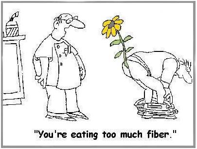
Yes, it is possible to get too much fiber!

(via Peter at Hyperlipid)

Other Toxic Foods

Nightshade plants and seeds of all species can also contain toxins and may also need to be restricted in people with damaged intestines.

Dairy proteins, especially pasteurized cow casein, are also often problematic for people with damaged intestines.

In general, any foods that commonly produce allergies are likely to give trouble to people with damaged intestines.

All plant proteins are risky, as are dairy and egg proteins. It is possible to minimize risk by:

  • Eating clarified butter, but refraining from protein-rich dairy foods.
  • Eating cooked egg yolks, but avoiding the protein-rich egg whites.

Summary of Toxic Foods to Eliminate or Avoid

In short, bowel disease patients should eliminate toxic foods from their diet:

  • Eliminate all grains except rice. Wheat, oats, and corn, and their products such as wheat flour, cornstarch, bread, and pasta, must be eliminated.
  • Eliminate all legumes, especially soy, beans, and peanuts.
  • Eliminate omega-6 rich oils, such as soybean oil, safflower oil, corn oil, peanut oil, and canola oil.
  • Eliminate fructose sugars, except from fruits and berries.  Drink no sugar-containing beverages.
  • Minimize fiber to keep down gut bacterial populations and avoid mechanical injuries to the intestinal wall.
  • Minimize other potentially toxic protein sources. In general, protein should be obtained from animal and fish meats, not eggs, dairy, or plants. However, fats from dairy and eggs are highly desirable.  

When gut health is restored, dairy and fiber may be restored to the diet. However, the major toxic foods – grains, legumes, omega-6-rich oils, and most fructose – should be eliminated for life.

Filling the Gaps in the Diet

For most people, eliminating grains will leave a large gap in the diet.  Foods that should be used to fill that gap include:

  • Healthful plant foods are the safe starches, such as white rice, taro, sweet potatoes and yams, and fruits and berries.
  • Saturated-fat rich oils and fats, like beef tallow, clarified butter, coconut oil, palm oil, cocoa butter (yes, chocolate desserts are healthy!), along with modest quantities of olive oil and lard. Make homemade salad dressings with these oils, rather than buying supermarket dressings made with soybean or canola oil.
  • Further balance the omega-6 to omega-3 ratio by eating low-omega-6 meats, such as the red meats (beef and lamb) and seafood, and eating 1 lb per week salmon or sardines for fresh omega-3 fats.

When the small intestine is damaged, fatty foods may be difficult to tolerate, since the enzymes that digest dietary fats and proteins may also digest human cells.  In such cases the diet must focus on starchy foods like rice until bowel health is restored. Be sure to supplement with vitamins and minerals in such cases.

Conclusion

Eliminating food toxins may cure bowel diseases and always improves prognosis.  Pedro Bastos of The Paleo Diet Update writes:

From the feedback we have received over the years, IBS patients respond dramatically and rapidly to the Paleo Diet. Their experience indicates that milk, grains and legumes seem to be the main culprits in this condition via a number of mechanisms. [The Paleo Diet Update v.4, #18]

Auto-antibodies generated by wheat against the thyroid, pancreas, and gut typically disappear within 3 to 6 months after removal of wheat from the diet. [25, 26] This is probably a reasonable estimate for the time frame in which bowel disease patients can expect significant improvements from the elimination of dietary food toxins.

Related Posts

Other posts in this series:

  1. Bowel Disorders, Part I: About Gut Disease July 14, 2010
  2. Bowel Disease, Part III: Healing Through Nutrition July 22, 2010
  3. Bowel Disease, Part IV: Restoring Healthful Gut Flora July 27, 2010

References

[1] Fujimura KE et al. Role of the gut microbiota in defining human health. Expert Rev Anti Infect Ther. 2010 Apr;8(4):435-54. http://pmid.us/20377338.

[2] Ruas-Madiedo P et al. Mucin degradation by Bifidobacterium strains isolated from the human intestinal microbiota. Appl Environ Microbiol. 2008 Mar;74(6):1936-40. http://pmid.us/18223105.

[3] Bode L. Human milk oligosaccharides: prebiotics and beyond. Nutr Rev. 2009 Nov;67 Suppl 2:S183-91. http://pmid.us/19906222. Hat tip Dr. Art Ayers.

[4] Ames BN, Gold LS. Paracelsus to parascience: the environmental cancer distraction. Mutation Research 2000 Jan 17; 447(1):3-13. http://pmid.us/10686303

[5] Cummings JH. The effect of dietary fibre on fecal weight and composition. Pp 547–73 in: Spiller GA, ed. Handbook of dietary fibre in human nutrition. 2nd ed. Boca Raton, FL: CRC Press, 1993.

[6] Bernardo D et al. Is gliadin really safe for non-coeliac individuals? Production of interleukin 15 in biopsy culture from non-coeliac individuals challenged with gliadin peptides. Gut 2007 Jun;56(6):889-90. http://pmid.us/17519496.

[7] Drago S et al. Gliadin, zonulin and gut permeability: Effects on celiac and non-celiac intestinal mucosa and intestinal cell lines.  Scand J Gastroenterol. 2006 Apr;41(4):408-19. http://pmid.us/16635908.

[8] Not T et al. Celiac disease risk in the USA: high prevalence of antiendomysium antibodies in healthy blood donors. Scand J Gastroenterol. 1998 May;33(5):494-8. http://pmid.us/9648988.

[9] Sollid LM, Jabri B. Is celiac disease an autoimmune disorder? Curr Opin Immunol. 2005 Dec;17(6):595-600. http://pmid.us/16214317.

[10] Singh MM et al Wheat gluten as a pathogenic factor in schizophrenia. Science. 1976 Jan 30;191(4225):401-2. http://pmid.us/1246624. Dohan FC et al 1984 Is schizophrenia rare if grain is rare? Biol Psychiatry. 1984 Mar;19(3):385-99. http://pmid.us/6609726.

[11] Lorenzsonn V, Olsen WA. In vivo responses of rat intestinal epithelium to intraluminal dietary lectins. Gastroenterology. 1982 May;82(5 Pt 1):838-48. http://pmid.us/6895878.

[12] Sjölander A et al. Morphological changes of rat small intestine after short-time exposure to concanavalin A or wheat germ agglutinin. Cell Struct Funct. 1986 Sep;11(3):285-93. http://pmid.us/3768964.

[13] Mellanby E. (March 15 1919) An experimental investigation on rickets. The Lancet 193(4985):407-412.

[14] Carpenter KJ, Lewin WJ. A critical review: A reexamination of the composition of diets associated with pellagra. J Nutr 1985 May;115(5):543–552. http://pmid.us/3998856.

[15] Biagi F et al. A milligram of gluten a day keeps the mucosal recovery away: a case report. Nutr Rev. 2004 Sep;62(9):360-3. http://pmid.us/15497770.

[16] Kordás K et al. Phytohaemagglutinin inhibits gastric acid but not pepsin secretion in conscious rats. J Physiol Paris. 2001 Jan-Dec;95(1-6):309-14. http://pmid.us/11595455. Pusztai A et al. Kidney bean lectin-induced Escherichia coli overgrowth in the small intestine is blocked by GNA, a mannose-specific lectin. J Appl Bacteriol. 1993 Oct;75(4):360-8.  http://pmid.us/8226393. Prykhod’ko O et al. Precocious gut maturation and immune cell expansion by single dose feeding the lectin phytohaemagglutinin to suckling rats. Br J Nutr. 2009 Mar;101(5):735-42. http://pmid.us/18644165.

[17] Pusztai A et al. Inhibition of starch digestion by alpha-amylase inhibitor reduces the efficiency of utilization of dietary proteins and lipids and retards the growth of rats. J Nutr. 1995 Jun;125(6):1554-62. http://pmid.us/7782910.

[18] Haeney MR et al. Soya protein antibodies in man: their occurrence and possible relevance in coeliac disease.  J Clin Pathol. 1982 Mar; 35(3):319-22.  http://pmid.us/7040491.

[19] IBD in EPIC Study Investigators. Linoleic acid, a dietary n-6 polyunsaturated fatty acid, and the aetiology of ulcerative colitis: a nested case-control study within a European prospective cohort study. Gut. 2009 Dec;58(12):1606-11. http://pmid.us/19628674.

[20] John S et al. Dietary n-3 polyunsaturated fatty acids and the aetiology of ulcerative colitis: a UK prospective cohort study. Eur J Gastroenterol Hepatol. 2010 May;22(5):602-6. http://pmid.us/20216220.

[21] Bergheim I et al. Antibiotics protect against fructose-induced hepatic lipid accumulation in mice: role of endotoxin. J Hepatol. 2008 Jun; 48(6): 983-92. http://pmid.us/18395289.

[22] Stephen AM et al. Effect of changing transit time on colonic microbial metabolism in man. Gut. 1987 May;28(5):601-9. http://pmid.us/3596341.

[23] Quoted in Science Daily, http://www.sciencedaily.com/releases/2006/08/060823093156.htm. Hat tip Dr. Michael Eades.

[24] Burr ML et al. Effects of changes in fat, fish, and fibre intakes on death and myocardial reinfarction: diet and reinfarction trial (DART). Lancet. 1989 Sep 30;2(8666):757-61. http://pmid.us/2571009. Hat tip Stephan Guyenet.

[25] Berti I et al. Usefulness of screening program for celiac disease in autoimmune thyroiditis. Dig Dis Sci. 2000 Feb;45(2):403-6. http://pmid.us/10711459.

[26] Mainardi E et al. Thyroid-related autoantibodies and celiac disease: a role for a gluten-free diet? J Clin Gastroenterol. 2002 Sep;35(3):245-8. http://pmid.us/12192201.