Search Results for: selenium - Page 5

DHA and Angiogenesis: The Bottom Line

So I thought I’d finish up the series on DHA and angiogenesis by discussing 2 issues:

1.      First, an assertion: The pathway by which oxidized DHA drives angiogenesis may be really important for human health.

2.      Second, the $64,000 question: Is there evidence that high levels of dietary DHA promotes diseases of pathological angiogenesis? What about other dietary factors bearing on DHA oxidation?

Significance of the Oxidized DHA Link to Angiogenesis

The papers discussed in Friday’s post about a major angiogenesis pathway stimulated by oxidized DHA (Omega-3s, Angiogenesis and Cancer: Part II, April 29, 2011) may not seem important to many readers. But to cancer researchers and pharmaceutical companies, this is blockbuster work.

A tumor is, in the words of Hal Dvorak, “a wound that never heals.” [1] To support growth, cancers invoke the wound healing process – especially, creation of new blood vessels, or angiogenesis. But the tumor prevents the wound healing process from completing. If it ever did complete, then the tumor itself would be healed. It would cease to grow and become benign.

It’s been recognized for decades that an ability to block angiogenesis would effectively constitute a cure for cancer. The William Li video explains why: nearly everyone gets microscopic tumors that never develop the ability to induce angiogenesis. Life-threatening cancer is the result of tumors that can induce angiogenesis. No angiogenesis, and no one would die of cancer.

But existing anti-angiogenic cancer therapies have produced disappointing results. Avastin, an anti-angiogenic drug targeting VEGF (vascular endothelial growth factor), has been estimated to extend colon cancer patient lifespan by only 6 weeks.  (Nevertheless, Avastin generated $7.3 billion in revenue last year. Imagine how much money there would be in an anti-angiogenic therapy that worked!)

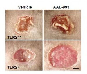
The work I discussed last Friday suggests a reason for that failure. Recall these pictures:

If only the VEGF pathway is blocked (upper right), there is almost as much angiogenesis and wound healing as in a normal wound (upper left). But when both the VEGF and TLR-2 angiogenic pathways are blocked (lower right), there is no wound healing.

If these are the operative pathways in cancer also, then blocking the TLR-2 angiogenesis pathway might be the key to cancer therapy.

But cancer is not the only disease of pathological angiogenesis. Others include:

  • Age-related macular degeneration, diabetic retinopathy, and retinopathy of prematurity – three common causes of blindness.
  • Atherosclerosis, which often features angiogenic vessels in thickened arterial walls.
  • Vascular malformations and tumors.
  • Obesity. Adipose tissue utilizes angiogenic pathways, and angiogenesis inhibition prevents the deposition of fat.
  • Rosacea, psoriasis, and some other skin conditions.
  • Endometriosis, uterine fibroids, and some other causes of female infertility.
  • Rheumatoid arthritis.
  • Crohn’s disease.
  • Preeclampsia.

It may be that the TLR-2 pathway is key to these diseases as well, and that a treatment that inhibits this pathway can cure or improve all of these diseases.

Add up the size of these markets and a pharmaceutical company executive would swoon.

Luckily, we’re not pharmaceutical company executives. But we can still get excited over possibilities to improve these diseases through diet and anti-microbial medicine.

Infections as Contributing Causes of These Diseases

TLR-2 is stimulated by other things besides oxidized DHA. In particular, TLR-2 is an immune molecule which is stimulated by pathogen proteins. As Wikipedia notes:

TLR-2 recognizes many bacterial, fungal, viral, and certain endogenous substances.

This tells us that many pathogens may stimulate angiogenesis through the TLR-2 pathway. As a result, anti-microbial medicines might help treat some diseases of pathological angiogenesis.

Some antibiotics, including doxycycline and minocycline, are known to exercise anti-angiogenic effects independent of the antibiotic effects. [2]

Diet-Induced Angiogenesis

Many foods affect angiogenesis. In fact, cancer studies have identified dozens of plant foods, from garlic to tomatoes to leeks, that possess anti-angiogenic properties.

However, foods can also promote angiogenesis. Let’s stick to the oxidized DHA pathway and see if there’s evidence that foods drive it.

You’ll recall the recipe was:

DHA + oxidative stress + retinyl protein = TLR-2 driven angiogenesis

If this pathway is important in human disease, then we should expect diseases of angiogenesis to be worsened by adding the ingredients on the left.

Specifically, cancer, AMD, rosacea, and so forth should be worsened by high doses of DHA, high doses of vitamin A, and low doses of antioxidant minerals like zinc or selenium.

Is there any evidence for that pattern?

Cancer Studies

First, let me give my bottom line on the Brasky study that kicked off this series. High tissue levels of DHA were associated with increased risk of high-grade prostate cancer, and the oxidized DHA angiogenesis pathway provides a mechanism for this association. What’s not clear is why tissue DHA levels were high. EPA levels were also elevated in the high-grade prostate cancers, but not by nearly as much as DHA levels. EPA and DHA appear together in fish and fish oil, so this suggests that fish consumption contributed to but was not the primary cause of the elevated tissue DHA. The drug finasteride greatly raised risk of high-grade prostate cancer, but the paper did not break down the DHA-cancer association between the finasteride and placebo arms. The most likely explanation, in my view, is that finasteride increases conversion of EPA to DHA and creates artificially high tissue DHA levels. The high DHA levels combined with oxidative stress drive cancer through the TLR-2 angiogenesis pathway.

A clever but unlikely alternative explanation was suggested by Peter at Hyperlipid: perhaps extra dietary fish oil raises testosterone levels. Prostate cancer is a hormone-dependent cancer and can be promoted by testosterone, just as breast cancer is promoted by estrogen. Possible supporting evidence comes from a paper showing an inverse association between metabolic syndrome / diabetes and prostate cancer. The trouble with this idea is that (a) this effect should have been strongest in the low-grade cancers, since diabetes reduced the incidence of low-grade cancers, but in the Brasky study DHA had no association with low-grade cancers, (b) fish oil lowers testosterone levels in rats, (c) in the Brasky study high-grade prostate cancers were strongly associated with obesity and the obese generally have low testosterone levels, and (d) surprisingly, high-grade prostate cancers are associated with low testosterone, not high. So one could argue that fish oil might promote high-grade prostate cancer by lowering testosterone!

A unified explanation along this line would be: Finasteride raises DHA levels, and DHA lowers testosterone. Low testosterone reduces incidence of low-grade prostate cancers but makes it much more likely they will progress to high-grade. Thus, finasteride reduces prostate cancer incidence but increases high-grade prostate cancer incidence and overall prostate cancer mortality. Fits all the facts. Could be.

My bottom line: the Brasky study is weak evidence for anything, but it does raise a whiff of evidence that high dietary fish oil intake might encourage a transition from low-grade to high-grade cancer.

What about other ingredients in the recipe? Does increasing retinyl levels raise cancer risk?

Retinyl palmitate (vitamin A) has been tested in clinical trials for its effect on cancer risk. The trials had to be cut short when it was found that vitamin A increased cancer mortality:

The Carotene and Retinol Efficacy Trial (CARET) was a multicenter randomized, double-blind placebo-controlled chemoprevention trial testing whether daily supplementation with 30 mg β-carotene + 25,000 IU retinyl palmitate would reduce lung cancer risk among 18,314 heavy smokers, former heavy smokers and asbestos-exposed workers. The intervention ended 21 months early in January, 1996 when interim analysis found evidence that the supplements increased the risk of lung cancer and total mortality in this high-risk population by 28% and 17%, respectively (10). [3]

After the study ended participants were tracked for years afterward. Those who had received vitamin A during the trial, but especially those in the vitamin A arm who took additional supplements (mainly multivitamins which are rich in A, but possibly also fish oil), had more high-grade prostate cancers:

As a proportion of the total prostate cancer cases, more men who were randomized to the active arm developed high-grade prostate cancer (Gleason 7-10) than in the placebo arm (44.6% vs. 40.1%, respectively)….

For aggressive prostate cancer, men in the CARET intervention arm who used additional supplements had a relative risk for aggressive prostate cancer (Gleason >or=7 or stage III/IV) of 1.52 (95% CI, 1.03-2.24; P < 0.05), relative to all others. [3]

Interestingly, in the placebo arm taking multivitamins and other supplements reduced cancer risk.

Other studies have found similar results.

Men with higher retinol concentrations at baseline were more likely to develop prostate cancer (quintile 5 vs. quintile 1 hazard ratio = 1.19, 95% confidence interval: 1.03, 1.36; P(trend) = 0.009). The results were similar for aggressive disease. Joint categorization based on baseline and 3-year retinol levels showed that men who were in the highest quintile at both time points had the greatest increased risk (baseline/3-year quintile 5/quintile 5 vs. quintile 1/quintile 1 hazard ratio = 1.31, 95% confidence interval: 1.08, 1.59). In this largest study to date of vitamin A status and subsequent risk of prostate cancer, higher serum retinol was associated with elevated risk, with sustained high exposure conferring the greatest risk. [4]

Carotenoids, which can generally be converted to vitamin A, are also associated with higher cancer risk. There is one exception – lycopene:

Lycopene was inversely associated with prostate cancer risk (comparing highest with lowest quartiles, odds ratio (OR) = 0.65, 95% confidence interval (CI): 0.36, 1.15; test for trend, p = 0.09), particularly for aggressive disease (comparing extreme quartiles, OR = 0.37, 95% CI: 0.15, 0.94; test for trend, p = 0.04). Other carotenoids were positively associated with risk. [5]

What’s special about lycopene? Wikipedia explains:

Lycopene may be the most powerful carotenoid quencher of singlet oxygen,[18] being 100 times more efficient in test tube studies of singlet-oxygen quenching action than vitamin E … The absence of the beta-ionone ring structure for lycopene increases its antioxidant action….

Lycopene is not modified to vitamin A in the body

So lycopene does not increase retinyl levels, but does act as an extraordinarily powerful antioxidant, thus reducing oxidative stress! If you wanted a good food for stopping the DHA – angiogenesis pathway, you’ve found it: tomatoes.

Hmmm, tomatoes go well with salmon …

That gets us to the third part of the recipe, oxidative stress. If oxidized DHA drives angiogenesis, then antioxidants should be preventative for these diseases.

The evidence here is rather mixed, because with the exception of the negative effects of vitamin A, most antioxidants seem to have little effect on cancer. Nevertheless, I’ll give some studies. Selenium is a antioxidant mineral due to its role in glutathione peroxidase:

Serum selenium was inversely associated with risk of prostate cancer (comparing highest to lowest quartiles, OR = 0.71, 95% CI 0.39-1.28; p for trend = 0.11), with similar patterns seen in both blacks and whites. [6]

Zinc is an antioxidant due to its role in zinc-copper superoxide dismutase. Prostate cancer is associated with low tissue levels of zinc. [7, 8] High dietary intake of zinc is associated with lower rates of prostate cancer. [9]

N-acetylcysteine is an antioxidant supplement that is a precursor to glutathione. N-acetylcysteine has been shown to prevent angiogenesis and has been proposed as a likely cancer preventative, but this is as yet untested. [10]

Other Diseases of Angiogenesis

I’ll skip those for now, other than to note that fish oil is a well-known trigger of rosacea. Is it possible that the mechanism is via TLR-2 activation by oxidized DHA?

Conclusion

At the moment there’s some puffs of smoke but no fire. Observational studies weakly link high DHA, high vitamin A, and low antioxidant status to diseases of angiogenesis such as cancer.

This pattern would be consistent with the idea that the natural pathway used in wound healing to trigger angiogenesis – DHA oxidation and combination with retinyl protein to trigger TLR-2 pathways – is also important for cancer progression.

It suggests a strategy of reduced fish oil and vitamin A consumption and increased intake of certain antioxidants (such as lycopene, zinc, selenium, or NAC) may be helpful against cancer.

However, this idea needs testing. No study in animal cancer models has tested this dietary combination.

Given the many proven benefits of moderate amounts of fish oil, I don’t see a reason yet to alter our recommendation that healthy people should eat a pound of fish per week. That said, I do think very high intakes of fish or fish oil are ill advised. And I’m intrigued by the idea that dietary changes may have the potential to play a powerful role in recovery from diseases of angiogenesis such as cancer.

References

[1] Dvorak HF. Tumors: wounds that do not heal. Similarities between tumor stroma generation and wound healing. N Engl J Med. 1986 Dec 25;315(26):1650-9. http://pmid.us/3537791.

[2] Yao JS et al. Comparison of doxycycline and minocycline in the inhibition of VEGF-induced smooth muscle cell migration. Neurochem Int. 2007 Feb;50(3):524-30. http://pmid.us/17145119.

[3] Neuhouser ML et al. Dietary supplement use and prostate cancer risk in the Carotene and Retinol Efficacy Trial. Cancer Epidemiol Biomarkers Prev. 2009 Aug;18(8):2202-6. http://pmid.us/19661078.

[4] Mondul AM et al. Serum retinol and risk of prostate cancer. Am J Epidemiol. 2011 Apr 1;173(7):813-21. http://pmid.us/21389041.

[5] Vogt TM et al. Serum lycopene, other serum carotenoids, and risk of prostate cancer in US Blacks and Whites. Am J Epidemiol. 2002 Jun 1;155(11):1023-32. http://pmid.us/12034581.

[6] Vogt TM et al. Serum selenium and risk of prostate cancer in U.S. blacks and whites. Int J Cancer. 2003 Feb 20;103(5):664-70. http://pmid.us/12494476.

[7] Sarafanov AG et al. Prostate cancer outcome and tissue levels of metal ions. Prostate. 2011 Jan 26. doi: 10.1002/pros.21339. [Epub ahead of print] http://pmid.us/21271612.

[8] Costello LC, Franklin RB. Zinc is decreased in prostate cancer: an established relationship of prostate cancer! J Biol Inorg Chem. 2011 Jan;16(1):3-8. http://pmid.us/21140181.

[9] Epstein MM et al. Dietary zinc and prostate cancer survival in a Swedish cohort. Am J Clin Nutr. 2011 Mar;93(3):586-93. http://pmid.us/21228268.

[10] Noonan DM et al. Angiogenesis and cancer prevention: a vision. Recent Results Cancer Res. 2007;174:219-24. http://pmid.us/17302199.

Omega-3s, Angiogenesis and Cancer: Part II

On Tuesday (Omega-3 Fats, Angiogenesis, and Cancer: Part I, April 26, 2011) I introduced the issue of possible relationships between omega-3 fatty acids, their lipid peroxidation products, and diseases of angiogenesis such as cancer, and promised to discuss a possible mechanism today.

It may be well, however, to start by saying a little bit more about the Brasky paper [1] linking prostate cancer to DHA.

Denise Minger’s Commentary on the Brasky Paper

Denise Minger wrote a commentary on this paper for Mark’s Daily Apple, which is excellent. Her conclusion – “given the oxidation-prone nature of all polyunsaturated fats, a massive intake of omega-3’s – despite their brilliance in moderation – could potentially do more harm than good” – is the proper one.

A few of Denise’s observations, however, could stand elaboration.

The study measured the fraction of serum phospholipid fatty acids in various polyunsaturated and trans-fat species, not dietary intake. This is the right parameter to measure, as fatty acid profiles can be measured precisely while dietary intakes assessed through questionnaires are notoriously unreliable. Also, phospholipids are the fats in cell membranes, and these are the ones involved in the inflammatory signaling pathways long thought to drive cancer risk. So cell membrane lipid measurements have the best chance to demonstrate a link to cancer risk.

Denise makes the important point, however, that the connection between dietary fish oil intake and serum fatty acid profile is not simple. Higher DHA intake raises phospholipid DHA levels, but lower intake of non-omega-3 fats also raises the DHA fraction. She points to a study [2] comparing a low-fat diet (20% fat, 6.7% PUFA, n-6:n-3 ratio 11.1) to a high-fat diet (45% fat, 15% PUFA, n-6:n-3 ratio 12.3).  The low-fat diet had more of its fat in the form of long-chain omega-3s, but the specific DHA intake on the diets was not reported. Membrane DHA ended up 28% higher on the low-fat diet.

So if DHA is dangerous, low-fat dieters will be in the most trouble. Another reason to eat a high-fat diet!

Does this affect our interpretation of the Brasky study? I don’t think it affects it much, because study participants were healthy at the start of the study with no history of cancer and macronutrient intakes don’t vary a lot among the general public. Americans vary surprisingly little from the median of about 50% carbs, 15% protein, and 35% fat – so it’s likely that the quartile with high tissue DHA levels were also high fish oil consumers.

However, study participants were followed for 7 years, at which point their prostate cancer status was assessed. Incidence of low-grade prostate cancers had no association with start-of-the-study DHA intake, but incidence of high-grade prostate cancers was strongly associated.

Here are a couple of possible explanations for this pattern:

  1. DHA is bad: DHA doesn’t drive early cancer development but does drive late-stage cancer growth – i.e. the transition from low-grade to high-grade cancer. So the DHA consumers got the high-grade cancers. Angiogenesis does, in fact, drive the shift from low-grade to high-grade cancer, so a DHA-angiogenesis association would be consistent with this explanation.
  2. Hospital diet advice is bad: DHA was a marker at the start of the study for conscientious, educated, disciplined persons who followed health advice and ate fish oil. When these people were diagnosed with low-grade cancer, they followed the dietary advice of their cancer dietitian. The dietitian’s advice?  Eat lots of wheat, whole grains, legumes, and vegetable oils. It could be the conscientious folks who followed bad diet advice from the hospital dietitian who got the high-grade cancers.

So there is a possible confounding effect.

Another of Denise’s assertions is that there is an “otherwise consistent train of research showing that DHA seems protective at best (and neutral at worst).” Now it is true that there are more studies showing DHA to have benefits against cancer than harm. But this trend is hardly consistent, and the vast majority of studies have failed to detect a relationship.

In the comments to Tuesday’s post, eric linked to a 2005 meta-review of studies on omega-3 fats and cancer. [3] The reviewers looked at 1,210 journal articles and found a mixed bag of mostly insignificant evidence:

Significant associations between omega-3 consumption and cancer risk were reported for lung cancer in two studies; for breast cancer in one; for prostate cancer in one; and for skin cancer in one. However, for lung cancer, one of the significant associations was for increased cancer risk and the other was for decreased risk (four other risk ratios were not significant for lung cancer). For breast cancer, five other estimates did not show a significant association. Only one study assessed skin cancer risk. No effects were reported for cancers of the aerodigestive tract, bladder cancer, colorectal cancer, lymphoma, ovarian cancer, pancreatic cancer, or stomach cancer. Thus, omega-3 fatty acids do not appear to decrease overall cancer risk.

Data were insufficient to permit assessment of a temporal or dose-response relationship. [3]

So the score was 4 studies finding that DHA is associated with less cancer, 1 that it is associated with more, and a boatload that it had no association.

Now there are two ways of interpreting this general insignificance of DHA against cancer. One is to note that there are slightly more studies showing DHA to have benefits than harm, and therefore to judge that DHA might be helpful against cancer.

But another, equally plausible, interpretation is this. Most Americans eat far too much omega-6, and their omega-6 to omega-3 tissue ratio is too high, which is pro-inflammatory via the COX-2 pathway. Eating omega-3s including DHA reduces inflammation by downregulating the COX-2 pathway. This accounts for the well-attested benefits of DHA against cardiovascular disease. Now, cancer is promoted by COX-2 pathway inflammation, which is why COX-2 inhibitors such as aspirin and ibuprofen are protective against cancer. [4] DHA’s action to downregulate this pathway must generate an anti-cancer effect. But, unlike aspirin and ibuprofen, DHA has no observable effect on overall cancer risk. This suggests that DHA has other effects, unrelated to its anti-inflammatory activity, that are cancer promoting. These counterbalance the benefits from its anti-inflammatory effect. If DHA has pro-angiogenic effects that are independent of COX-2 mediated inflammation, then this could account for the observations.

One reason an association of DHA with high-grade cancer may have been missed is that it would be detected only in large studies able to segregate cancers by grade. Brasky et al note:

In the European Prospective Investigation into Cancer and Nutrition (EPIC) (12), the highest quintile of percent DHA was associated with elevated risks of both low-grade (relative risk (RR) = 1.53, 95% CI: 0.96, 2.44) and high-grade (RR = 1.41, 95% CI: 0.76, 2.62) prostate cancer. They also reported significant positive associations of the percent EPA with high-grade prostate cancer (RR = 2.00, 95% CI: 1.07, 3.76). Given that the Prostate Cancer Prevention Trial and the European Prospective Investigation into Cancer and Nutrition, the 2 largest studies of blood levels of phospholipid fatty acids, reported increased risks of high-grade prostate cancer with high levels of ω-3 fatty acids, it remains a possibility that these fatty acids promote tumorigenesis. [1]

If there were no other evidence linking DHA to angiogenesis, the Brasky and EPIC study associations would be interesting, but unlikely to change anyone’s mind. Denise points out the need for other evidence – especially, mechanistic evidence – to make the connection more plausible:

We haven’t sleuthed out any mechanism that could explain why DHA (but not other polyunsaturated fats) promotes rapid tumor growth.

And this is where today’s post comes in. In fact, there is a known mechanism by which DHA but not other polyunsaturated fats can promote rapid tumor growth. Shou-Ching told me about it a few months ago.

DHA and Angiogenesis in Macular Degeneration

Let’s start by going back to 2003 and a paper on the role of a compound called carboxyethylpyrrole (CEP) in age-related macular degeneration (AMD). [5] AMD is an eye disease caused by improper angiogenesis. Basically, malformed blood vessels overgrow the eye, causing retinal detachment and blindness. It afflicts 35% of those over age 75, and is the leading cause of blindness in developed countries. CEP? Well, the paper explains:

Free radical-induced oxidation of docosahexaenoate (DHA)-containing lipids generates ω-(2-carboxyethyl)pyrrole (CEP) protein adducts that are more abundant in ocular tissues from AMD than normal human donors…. The CEP adduct uniquely indicates oxidative modification from DHA derivatives because CEP protein modifications cannot arise from any other common polyunsaturated fatty acid. [5]

CEP is uniquely produced by oxidation of DHA, not other PUFAs. Its abundance depends on DHA abundance, availability of retinyl proteins, and the level of oxidative stress.

CEP is elevated in AMD. The correlation is strong: a person in whom the immune system is trying but failing to clear elevated CEP levels almost invariably has macular degeneration (AMD):

Of individuals (n = 13) exhibiting both antigen and autoantibody levels above the mean for non-AMD controls, 92% had AMD. [5]

So CEP is a great marker for AMD. Is it causal?

Well, first it’s worth noting that the retina is uniquely vulnerable to DHA oxidation:

Although rare in most human tissues, DHA is present in ~80 mol % of the polyunsaturated lipids in photoreceptor outer segments (13). The abundance of DHA in photoreceptors, the high photooxidative stress in retina, and the fact that DHA is the most oxidizable fatty acid in humans (13) suggests that DHA oxidation products may have possible utility as biomarkers for AMD susceptibility. [5]

Oxidation is linked to AMD, and antioxidants slow AMD progression:

Oxidative damage appears to contribute to the pathogenesis of AMD (4) based on epidemiological studies showing that smoking significantly increases the risk of AMD (1, 24). The molecular mechanism for how smoking enhances the risk for AMD is not known. We speculate that reactive oxygen and nitrogen species derived from tobacco smoke in the lungs leads to oxidative protein modifications in the blood that contribute to drusen formation and choroidal neovascularization. Results from a recent clinical trial (5) also demonstrate that the progression of AMD can be slowed in some individuals by high daily doses of antioxidant vitamins and zinc. Direct evidence of oxidative damage in AMD donor eye tissues include elevated levels of CEP adducts uniquely derived from the oxidative fragmentation of DHA (6). [5]

This is where things stood in 2003. By 2010 this group, led by Case Western Reserve University chemist Robert G. Salomon, had established that administering CEP to mice can cause AMD:

To test the hypothesis that this hapten is causally involved in initiating an inflammatory response in AMD, we immunized C57BL/6J mice with mouse serum albumin (MSA) adducted with CEP. Immunized mice develop antibodies to CEP, fix complement component-3 in Bruch’s membrane, accumulate drusen below the retinal pigment epithelium during aging, show decreased a- and b-wave amplitudes in response to light, and develop lesions in the retinal pigment epithelium mimicking geographic atrophy, the blinding end-stage condition characteristic of the dry form of AMD. Inflammatory cells are present in the region of lesions and may be actively involved in the pathology observed. [6]

This constitutes the first really good animal model for AMD. [6]

How does this relate to cancer? That leads us to a Nature paper from October 2010 [7], from the group of Tatiana Byzova at the Cleveland Clinic.

DHA, Immunity, and Angiogenesis

This is a rich paper. Briefly, CEP has a physiological function: it is transiently elevated in wounds and recruits immune cells from bone marrow to the site of the wound. These immune cells further increase oxidative stress and promote angiogenesis; CEP levels are highest at the time of peak angiogenesis. CEP is highly elevated in cancers. Unlike in wounds, where CEP is elevated for a few days, in cancers CEP elevation is chronic.

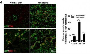
Here’s a staining comparing CEP in normal skin and in melanoma:

The CEP is co-localized with CD68, a glycoprotein which binds to LDL and is found on macrophages, and with CD31, a membrane marker of neutrophils, macrophages, and endothelial cells. CEP is marking endothelial cells and white blood cells in angiogenic vessels, and possibly LDL.

It turns out that CEP drives angiogenesis by attaching to an immune receptor, Toll-like receptor 2 (TLR2). There are two major pathways for angiogenesis: one driven by vascular endothelial growth factor (VEGF), which is dominant in conditions of hypoxia (oxygen starvation), and one by TLR2. Of these, the TLR2 pathway may in some contexts be more important. Here are pictures of wound healing in mice:

On the upper left is a normal mouse. On the upper right is a similar wound treated with the VEGF inhibitor AAL-993. This wound is rather like a cancer treated with the VEGF inhibitor Avastin. Wound healing is slightly impaired, but still works.

On the lower left is a similar wound with no VEGF inhibition, but the TLR2 pathway blocked by TLR2 knockout. The wound can’t scab and doesn’t heal successfully. If TLR2 is knocked out and VEGF inhibited, there is no wound healing at all (lower right).

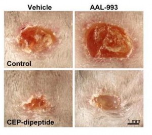
You can accelerate angiogenesis and wound healing by adding CEP to the wound.

In the bottom row, CEP has been added. Left is without VEGF inhibition, right with.

If you administer CEP-neutralizing antibodies to a normal wound, wound healing takes more than twice as long. This confirms that angiogenesis driven specifically by CEP (and therefore by DHA oxidation) is part of healthy wound healing.

Tumors use these same pathways to generate vessels and feed their growth. As the paper notes:

[T]umors implanted in TLR2-/- mice exhibited dramatically decreased vascularization and increased areas of necrosis. [7]

Here’s the paper’s conclusion:

Altogether our results establish a novel mechanism of angiogenesis that is independent of hypoxia-triggered VEGF expression. The products of lipid oxidation are generated as a consequence of oxidative stress and are recognized by TLR2, possibly in a complex with TLR1 on ECs, and promote angiogenesis in vivo, thereby contributing to accelerated wound healing and tissue recovery. If high levels of CEP and its analogs accumulate in tissues, it may lead to excessive vascularization, e.g. in tumors. Contribution of the CEP/TLR2 axis to angiogenesis varies in different physiological settings possibly depending on the extent of oxidative stress. CEP-driven angiogenesis may be an attractive therapeutic target, especially in cancers resistant to anti-VEGF therapy. Inflammation and oxidation-driven angiogenesis may occur in other pathologies, for example atherosclerosis, where arterial thickening can depend on its microvasculature. In these settings, there is an extensive generation of oxidative products which might promote atherogenesis via TLR2. Indeed, it was shown that TLR2?/? mice are protected from atherosclerosis, and this effect could be mediated by cells other than bone marrow-derived29. Thus, along with pathogen- and danger-associated molecular patterns, TLR2 recognizes an oxidation-associated molecular pattern. This new function of TLR2 as a sensor of oxidative stress reveals the shortcut link between innate immunity, oxidation and angiogenesis. [7]

Connection to Vitamin A

DHA is oxidized to a compound called HOHA which then combines with a protein, generally a retinyl (vitamin A-derived) protein to form CEP.

Cancers generate lots of CEP from DHA, and perhaps one way they do that is by generating lots of retinyl proteins. Cancers are known to have disturbed vitamin A biology with lots of retinyl:

Disturbance in vitamin A metabolism seems to be an important attribute of cancer cells. Retinoids, particularly retinoic acid, have critical regulatory functions and appear to modulate tumor development and progression. The key step of vitamin A metabolism is the esterification of all-trans retinol, catalyzed by lecithin/retinol acyltransferase. In this work we show that malignant melanoma cells are able to esterify all-trans retinol and subsequently isomerise all-trans retinyl esters into 11-cis retinol, whereas their benign counterparts – melanocytes are not able to catalyze these reactions. Besides, melanoma cell lines express lecithin/retinol acyltranseferase both at the mRNA and protein levels. In contrast, melanocytes do not express this enzyme … [8]

I haven’t looked much into this literature but it may speak to higher cancer risk with excessive vitamin A intake. Thus high-vitamin A cod liver oil may be a double risk for cancer patients.

Conclusion

It looks like we have a recipe for angiogenesis:

DHA + retinyl + oxidative stress = angiogenesis

This recipe is invoked normally and properly during wound healing. But it is also invoked excessively in pathological contexts – notably in cancers and age-related macular degeneration, probably also in other angiogenesis-associated diseases such as arthritis, rosacea, obesity, psoriasis, endometriosis, dementia, and multiple sclerosis.

In the case of cancer, DHA oxidation to CEP might transform miniscule, harmless cancers to high-grade, life-threatening cancers.

Should this possibility affect our dietary omega-3 recommendations? Well, we need to know the relative importance of the three ingredients on the left side of the above equation in producing angiogenesis. Chris Kresser wondered in the comments Tuesday whether oxidation may be the key factor:

I question whether DHA supplementation would truly play a causative role in the absence of a *pro-oxidative environment*.

In other words, perhaps in someone eating a SAD, not exercising, under a lot of stress, etc. DHA is more easily oxidized and thus potentially carcinogenic.

But in someone that is keeping all other oxidative risk factors low (i.e. they’re avoiding n-6, exercising, managing stress, reducing exposure to chemical toxins, etc.) I tend to doubt that supplementing with DHA could cause significant harm.

That’s the last piece of the puzzle: how do we minimize the level of oxidized DHA?

As I replied to Chris in the comments, low-carb Paleo dieters are not out of the woods in regard to oxidative stress. Oxidative stress is generated normally during metabolism, immune function – and by cancers. If anti-oxidant minerals like zinc, copper, and selenium and vitamins like vitamin C are deficient, then oxidative stress can be very high on a low-carb Paleo diet.

At the moment, I think it’s prudent to eat no more than 1 pound of salmon or similar cold-water fish per week, to avoid further EPA/DHA supplements, and to avoid low-fat diets which tend to elevate membrane DHA levels. Moderate omega-3 consumption is especially important for those suffering from diseases of pathological angiogenesis – especially cancer. DHA is essential for good health – but in excess, it is probably dangerous.

References

[1] Brasky TM et al. Serum Phospholipid Fatty Acids and Prostate Cancer Risk: Results From the Prostate Cancer Prevention Trial. Am. J. Epidemiol. April 24, 2011 DOI: 10.1093/aje/kwr027 (Will be at http://pmid.us/21518693.)

[2] Raatz SK et al. Total fat intake modifies plasma fatty acid composition in humans. J Nutr. 2001 Feb;131(2):231-4. http://pmid.us/11160538.

[3] MacLean CH, Newberry SJ, Mojica WA, et al. Effects of Omega-3 Fatty Acids on Cancer. Summary, Evidence Report/Technology Assessment: Number 113. AHRQ Publication Number 05-E010-1, February 2005. Agency for Healthcare Research and Quality, Rockville, MD. http://www.ahrq.gov/clinic/epcsums/o3cansum.htm.

[4] Harris RE. Cyclooxygenase-2 (cox-2) and the inflammogenesis of cancer. Subcell Biochem. 2007;42:93-126. http://pmid.us/17612047.

[5] Gu X et al. Carboxyethylpyrrole protein adducts and autoantibodies, biomarkers for age-related macular degeneration. J Biol Chem. 2003 Oct 24;278(43):42027-35. http://pmid.us/12923198.

[6] Hollyfield JG et al. A hapten generated from an oxidation fragment of docosahexaenoic acid is sufficient to initiate age-related macular degeneration. Mol Neurobiol. 2010 Jun;41(2-3):290-8. http://pmid.us/20221855.

[7] West XZ et al. Oxidative stress induces angiogenesis by activating TLR2 with novel endogenous ligands. Nature. 2010 Oct 21;467(7318):972-6. http://pmid.us/20927103.

[8] Amann PM et al. Vitamin A metabolism in benign and malignant melanocytic skin cells: Importance of lecithin/retinol acyltransferase and RPE65. J Cell Physiol. 2011 Apr 4. doi: 10.1002/jcp.22779. [Epub ahead of print] http://pmid.us/21465477.

Causes and Cures for Constipation

Constipation seems to be very common; we’re frequently asked about it. Constipation can afflict low-carb dieters: It was widely reported among Atkins dieters, and is the most common side effect reported on clinical ketogenic diets for epilepsy. [1]

Fortunately constipation is usually easily cured. There are a few common causes, and most of them have simple fixes.

Antioxidant Deficiency

Among the most common causes of constipation among low-carbers is deficient antioxidant capacity.

The gut is a challenging environment, full of oxidizing compounds. If glutathione and other antioxidants become deficient in intestinal cells, then the gut becomes leaky [2], leading to inflammation and the potential for constipation. Oxidative stress can also lead to loss of neurons or neuronal axons from the colon and consequently a loss of motility. Constipation is a common symptom of irritable bowel syndrome.

The cure is simple: supplement. Specifically:

  • Selenium, 200 mcg/day (perhaps 400 mcg during a starting period). Selenium is needed for glutathione peroxidase, a glutathione recycling enzyme.
  • Vitamin C, 1 g/day (perhaps 4 g/day during a starting period). Vitamin C and glutathione recycle one another.
  • Zinc and copper. Total zinc intake should be 30-50 mg/day including food and multivitamin; copper intake should be 2-5 mg/day. Food typically provides 15 mg zinc and 1 mg copper.
  • Cysteine and glycine. These are the component amino acids for glutathione. For constipation I would suggest eating some beef gelatin for glycine, and taking one 500 mg capsule of N-acetylcysteine per day.

Egg yolks also increase glutathione production [3], and eating a dozen per week is highly recommended. Lipoic acid may also help. [4]

Glutamine, a supplement frequently recommended for gut ailments, can also enhance glutathione production [5]. However, I would generally avoid this, because it can promote proliferation of pathogenic bacteria.

Hypothyroidism

Hypothyroidism may be the most common cause of constipation.

A vicious circle often develops: Gut problems lead to autoimmune hypothyroidism, and constipation is only one symptom of the damage hypothyroidism does to the gut. The vicious circle can be broken by treating the hypothyroidism.

The cure:

  1. Eliminate wheat, soy, and other autoimmune-promoting or goitrogenic toxins from the diet.
  2. Supplement with thyroid supporting nutrients like:
    • Selenium (as above).
    • Magnesium (citrate or chelate) 200 mg/day.
    • Zinc and copper (as above).
    • Iodine.
  3. Improve circadian rhythm. Circadian rhythm therapies for hypothyroidism were discussed in two posts, Intermittent Fasting as a Therapy for Hypothyroidism (Dec 1, 2010) and Seth Roberts and Circadian Therapy (Mar 22, 2011).
  4. See your doctor to consider replacing thyroid hormone and investigating related problems. Your doctor can prescribe thyroid hormone and can explore related problems that may contribute to hypothyroidism, such as adrenal fatigue or iron deficiency.

As always, a few cautions about iodine supplementation. Before starting iodine, eliminate wheat from the diet and supplement with selenium, copper, and magnesium. If you have thyroid-related effects from copper supplementation, before proceeding further supplement copper until your thyroid no longer reacts. Start iodine at a low dose, say 500 mcg (0.5 mg) per day. Increase the iodine dose no faster than one doubling per month. When you increase the dose, if you have a reaction to the higher dose, phase it in: say, alternate between 0.5 mg and 1 mg for a week before trying 1 mg/day every day. Go slowly, plan on spending 4 months to reach 3 mg/day, which is a quarter Iodoral tablet. Tablets can be cut into pieces with razor blades and liquid solutions can be diluted; don’t hesitate to reduce doses.

Insufficient Dietary Fat and/or Bile

Not long ago Jamie Scott (That Paleo Guy), a very smart nutritional advisor, discussed his solution for constipation. There were several gems in there, for instance, that foods that stimulate opioid receptors can cause constipation [5b]:

For example, both gluten grains (and I suspect grains in general) plus dairy contain opiate-like compounds that serve to reduce the motility (movement) of your gut.  This effect is commonly seen in those who have to take the likes of codeine (also an opiate) for any length of time.

Chocolate also has opioid peptides. But don’t worry, chocolate isn’t usually the cause of constipation!

But Jamie’s main advice was:

[M]any of the people I work with just are not eating enough fat….

People are a bit skeptical when I suggest increasing fat intake to overcome constipation.  Thankfully I can now point to a clinical study in which a high fat diet, in very short time, increased gastrointestinal transit time, reducing the likelihood of constipation occurring;

Gastrointestinal transit, post-prandial lipaemia and satiety following 3 days high-fat diet in men

High-fat diets promote stool passage for several reasons.

  1. Just a tiny bit of undigested fat can help grease the passage of stool through the colon. Even modest amounts of fat in the stool make it difficult to retain feces and cause fecal incontinence. This is why indigestible oils, like jojoba oil or Olestra, cause diarrhea.
  2. A second factor is that bile is released into the intestine when fats are eaten, and bile acids have a laxative effect. Both the primary bile acid chenodeoxycholic acid (CDCA) and the secondary bile acid deoxycholic acid (DCA) act as laxatives by inducing water secretion in the colon. An excess of bile therefore causes diarrhea and an insufficiency can cause constipation. [6] This is why people who have had their gallbladders removed typically develop diarrhea: bile is constantly leaking into the digestive tract.

Just be sure that the high fat you eat comes from healthy natural sources, not high-omega-6 industrially processed seed oils.

Also, bile acid metabolism has a diurnal rhythm. [7] Improved bile acid metabolism may be another pathway by which circadian rhythm therapies could help constipation.

Insufficient Gut Flora

Fiber is often recommended for constipation, and though I believe insufficient fiber is rarely the primary factor in constipation, it can help by several mechanisms.

First, bacteria, dead and alive, form a large fraction of the stool, and the more fiber you eat, the more bacteria you will have. Bacteria are surrounded by fatty cell membranes which are a little bit slippery.

Second, and probably more important, gut bacteria tend to increase the laxative effect of bile.

This is because the body controls water secretion in the colon in part by sulfation of bile acids; sulfation eliminates the induction of water secretion.

Gut bacteria usually want more water in the colon than the body does, so they’ve evolved sulfatase enzymes that desulfate human bile acids. [8, 9] The desulfated bile causes water release and the stool becomes soft and loose.

Some bacterial species do this more than others – especially virulent strains that spread by inducing diarrhea – and I’m sure everyone’s experienced at some time in their lives what happens when one of these species overpopulates the gut. We often call this “food poisoning” and it can be treated by large doses of probiotics, to displace the bad species with bacteria that release fewer sulfatase enzymes.

At the opposite extreme, if gut bacteria are lacking then bile acids will be excessively sulfated and won’t induce water secretion. Constipation may develop.

Thus, antibiotics can induce constipation. Presumably a zero-carb, zero-fiber diet would also make constipation more likely.

Adding some fiber to your high-fat diet, therefore, can relieve constipation.

Choline

Even though choline deficiency is not a proven cause of constipation, I’ll put this in because choline is so important for health. Choline is needed to produce the neurotransmitter acetylcholine. A deficiency of acetylcholine is associated with loss of colonic motility and constipation. [10]

Egg yolks and liver are good sources of choline. There’s nothing wrong with choline supplementation either.

Hypercalcemia

Some people over-do vitamin D supplementation and/or calcium supplementation. Elevated blood calcium levels, which can be brought about by too much vitamin D, will cause constipation. If you supplement either vitamin D or calcium and have constipation, ask your doctor to check serum 25OHD, 1,25D, and calcium levels.

Gut Infections

Some pathogenic bacteria are able to paralyze the gut and induce constipation. Some bacterial species that induce diarrhea can also induce constipation when they choose – for instance, C. difficile. This is one reason why patients with bowel disorders such as Irritable Bowel Syndrome or ulcerative colitis often experience both constipation and diarrhea.

Antibiotics like vancomycin that work well against Clostridium have successfully reversed cases of constipation. [11]

Infection-induced constipation may also cause bloating, gas, acid reflux, and gastroparesis, so if your constipation is accompanied by these symptoms you should see a doctor.

Don’t over-use laxatives

It can be risky to over-use laxatives. The riskiest is probably senna, which promotes bowel movements by (a) inhibiting water absorption by the colon and (b) promoting muscle spasms in the colon wall. Unfortunately, senna is a toxin that can cause lasting damage, notably to nerves.

If you must use laxatives, magnesium citrate liquids are the best. This will restore your body’s magnesium status as well as promote clearing of the bowels.

Conclusion

With good diet and nutrition, and normalization of thyroid function, constipation is usually easily cured. The exceptional cases are those of chronic gut infections; these may require some detective work on the part of a doctor.

I’m sure this reads like a laundry list, but it should give those with constipation some ideas!

References

[1] Neal EG et al. The ketogenic diet for the treatment of childhood epilepsy: a randomised controlled trial. Lancet Neurol. 2008 Jun;7(6):500-6. http://pmid.us/18456557.

[2] Rao R. Oxidative stress-induced disruption of epithelial and endothelial tight junctions. Front Biosci. 2008 May 1;13:7210-26. http://pmid.us/18508729.

[3] Young D et al. Egg yolk peptides up-regulate glutathione synthesis and antioxidant enzyme activities in a porcine model of intestinal oxidative stress. J Agric Food Chem. 2010 Jul 14;58(13):7624-33. http://pmid.us/20540508.

[4] Chandrasekharan B et al. Colonic motor dysfunction in human diabetes is associated with enteric neuronal loss and increased oxidative stress. Neurogastroenterol Motil. 2011 Feb;23(2):131-8, e26. http://pmid.us/20939847.

[5] Cao Y et al. Glutamine enhances gut glutathione production. JPEN J Parenter Enteral Nutr. 1998 Jul-Aug;22(4):224-7. http://pmid.us/9661123.

[5b] Tuteja AK et al. Opioid-induced bowel disorders and narcotic bowel syndrome in patients with chronic non-cancer pain. Neurogastroenterol Motil. 2010 Apr;22(4):424-30, e96. http://pmid.us/20100280.

[6] Hofmann AF et al. Altered bile acid metabolism in childhood functional constipation: inactivation of secretory bile acids by sulfation in a subset of patients. J Pediatr Gastroenterol Nutr. 2008 Nov;47(5):598-606. http://pmid.us/18955863.

[7] Abrahamsson H et al. Altered bile acid metabolism in patients with constipation-predominant irritable bowel syndrome and functional constipation. Scand J Gastroenterol. 2008;43(12):1483-8. http://pmid.us/18788050.

[8] Huijghebaert SM, Eyssen HJ. Specificity of bile salt sulfatase activity from Clostridium sp. strains S1. Appl Environ Microbiol. 1982 Nov;44(5):1030-4. http://pmid.us/7181500.

[9] Ridlon JM et al. Bile salt biotransformations by human intestinal bacteria. J Lipid Res. 2006 Feb;47(2):241-59. http://pmid.us/16299351.

[10] Burleigh DE. Evidence for a functional cholinergic deficit in human colonic tissue resected for constipation. J Pharm Pharmacol. 1988 Jan;40(1):55-7. http://pmid.us/2896776.

[11] Celik AF et al. The effect of oral vancomycin on chronic idiopathic constipation. Aliment Pharmacol Ther. 1995 Feb;9(1):63-8. http://pmid.us/7766746.

Answer Day: What Causes High LDL on Low-Carb Paleo?

First, thank you to everyone who commented on the quiz. I enjoyed reading your thoughts.

Is High LDL Something to Worry About?

Perhaps this ought to be the first question. Jack Kronk says “I don’t believe that high LDL is necessarily a problem” and Poisonguy writes “Treat the symptoms, Larry, not the numbers.” Poisonguy’s comment assumes that the LDL number is not a symptom of trouble. Is it?

I think so. It helps to know a little about the biology of cholesterol and of blood vessels.

When cells in culture plates are separated from their neighbors and need to move, they make a lot of cholesterol and transport it to their membranes. When cells find good neighbors and settle down, they stop producing cholesterol.

The same thing happens in the body. Any time there is a wound or injury that needs to be healed, cholesterol production gets jacked up.

When people have widespread vascular injuries, cholesterol is produced in large quantities by cells lining blood vessels. Now, to repair injuries cells have to coordinate their functions. Endothelial cells are the coordinators of vascular repair: they direct other cell types, like smooth muscle cells and fibroblasts, in the healing of vascular injuries.

To heal vascular injuries, these cells not only need more cholesterol for movement; they also need to multiply. It turns out that LDL, which carries cholesterol, also causes vascular cells to reproduce (“mitogenesis”):

The best-characterized function of LDLs is to deliver cholesterol to cells. They may, however, have functions in addition to transporting cholesterol. For example, they seem to produce a mitogenic effect on endothelial cells, smooth muscle cells, and fibroblasts, and induce growth-factor production, chemotaxis, cell proliferation, and cytotoxicity (3). Moreover, an increase of LDL plasma concentration, which is observed during the development of atherosclerosis, can activate various mitogen-activated protein kinase (MAPK) pathways …

We also show … LDL-induced fibroblast spreading … [1]

If endothelial cells are the coordinators of vascular repair, and LDL particles their messengers to fibroblasts and smooth muscle cells, then ECs should be able to generate LDL particles locally. Guess what:  ECs make a lipase whose main effect is to decrease HDL levels but can also convert VLDL and IDL particles into LDL particles and remove fat from LDL particles to make them into small, dense LDL:

Endothelial lipase (EL) has recently been identified as a new member of the triglyceride lipase gene family. EL shares a relatively high degree of homology with lipoprotein lipase and hepatic lipase …

In vitro, EL has hydrolyzed phospholipids in chylomicrons, very low density lipoprotein (VLDL), intermediate density lipoprotein and LDL. [2]

Immune cells, of course, are essential for wound healing and they should be attracted to any site of vascular injury. It turns out that immune cells have LDL receptors and these receptors may help them congregate at sites of vascular injury. [3]

I don’t want to exaggerate the state of the literature here:  this is a surprisingly poorly investigated area. But I believe these things:

1.      Cholesterol and LDL particles are part of the vascular wound repair process.

2.      Very high LDL levels are a marker of widespread vascular injury.

Now this is not the “lipid hypothesis.” Compare the two views:

  • The lipid hypothesis:  LDL cholesterol causes vascular injury.
  • My view:  LDL cholesterol is the ambulance crew that arrives at the scene of the crime to help the victims. The lipid hypothesis is the view that ambulance drivers should be arrested for homicide because they are commonly found at murder scenes.

So, to Poisonguy, on my view high LDL numbers are a symptom of vascular injury and are a cause for concern.

Big-Picture View of the Cause of High LDL

So, on a micro-level, I think vascular damage causes high LDL. But what causes vascular damage?

Here I notice a striking difference in commenters’ perspectives and mine. I tend to take a big-picture, top-down view of biology. There are three basic causes of nearly all pathologies:

1.      Toxins, usually food toxins.

2.      Malnutrition.

3.      Pathogens.

The whole organization of our book is dictated by this view. It is organized in four Steps. Step One is about re-orienting people’s views of macronutrients away from high-grain, fat-phobic, vegetable-oil-rich diets toward diets rich in animal fats. The other steps are about removing the causes of disease:

1.      Step Two is “Eat Paleo, Not Toxic” – remove food toxins.

2.      Step Three is “Be Well Nourished” – eliminate malnutrition.

3.      Step Four is “Heal and Prevent Disease” – address pathogens by enhancing immunity and, where appropriate, taking advantage of antibiotic therapies.

So when someone offers a pathology, any pathology, my first question is: Which cause is behind this, and which step do they need to focus on?

In Larry’s case, he had been eating low-carb Paleo for years. So toxins were not a problem.

Pathogens might be a problem – after all, he’s 64, and everybody collects chronic infections which tend to grow increasingly severe with age – but Larry hadn’t reported any other symptoms. More to the point, low-carb Paleo diets typically enhance immunity, yet Larry had fine LDL numbers before adopting low-carb Paleo and then his LDL got worse. So it wouldn’t be infectious in origin unless his diet had suppressed immunity through malnutrition – in which case the first step would be to address the malnutrition.

Step Three, malnutrition, was the only logical answer. The conversion to Paleo removes a lot of foods from the diet and could easily have removed the primary sources of some micronutrients.

So I was immediately convinced, just from the time-course of the pathology, that the cause was malnutrition.

Micronutrient Deficiencies are Very Common

In the book (Step Three) we explain why nearly everyone is deficient in micronutrients. The problems are most severe for minerals:  water treatment removes minerals from water, and mineral depletion of soil by industrial agriculture leads to mineral deficiencies in farmed plants and grain-fed animals.

This is why our “essential supplements” include a multimineral supplement plus additional quantities of five minerals – magnesium, copper, chromium, iodine, and selenium. Vitamins get a lot of attention, but minerals are where the big health gains are.

Copper Deficiency and LDL

Some micronutrient deficiencies are known to cause elevated LDL.

Readers of our book know that copper causes vascular disease; blog readers may be more familiar with an excellent post by Stephan, “Copper and Cardiovascular Disease”, discussing evidence that copper deficiency causes cardiovascular disease. As I’ve just argued that cardiovascular disease causes high LDL, it shouldn’t be a surprise that copper deficiency also causes hypercholesterolemia:

Copper and iron are essential nutrients in human physiology as their importance is linked to their role as cofactors of many redox enzymes involved in a wide range of biological processes, as well as in oxygen and electron transport. Mild dietary deficiencies of both metals … may cause long-term deleterious effects in cardiovascular system and alterations in lipid metabolism (3)….

Several studies showed a clear correlation among copper deficiency and dyslipidemia. The main alterations concern higher plasma CL and triglyceride (TG) concentrations, increased VLDL-LDL to HDL lipoproteins ratio, and the shape alteration of HDL lipoproteins.  [4]

The essentiality of copper (Cu) in humans is demonstrated by various clinical features associated with deficiency, such as anaemia, hypercholesterolaemia and bone malformations. [5]

Over the last couple of decades, dietary copper deficiency has been shown to cause a variety of metabolic changes, including hypercholesterolemia, hypertriglyceridemia, hypertension, and glucose intolerance. [6]

Copper deficiency is, I believe, the single most likely cause of elevated LDL on low-carb Paleo diets. The solution is to eat beef liver or supplement.

So, was my advice to Larry to supplement copper?  Yes, but that was not my only advice.

Other Micronutrient Deficiencies and Elevated LDL

Another common micronutrient deficiency that causes elevated LDL cholesterol is choline deficiency that is NOT accompanied by methionine deficiency. That is discussed in my post “Choline Deficiency and Plant Oil Induced Diabetes”:

Choline deficiency (CD) by itself induces metabolic syndrome (indicated by insulin resistance and elevated serum triglycerides and cholesterol) and obesity.

A combined methionine and choline deficiency (MCD) actually causes weight loss and reduces serum triglycerides and cholesterol …

I quote both these effects because it illustrates the complexity of nutrition. A deficiency of a micronutrient can present with totally different symptoms depending on the status of other micronutrients.

Julianne had a really nice comment, unfortunately caught in the spam filter for a while, with a number of links. She mentions vitamin C deficiency and, with other commenters, noted the link between hypothyroidism and elevated LDL. As one cause of hypothyroidism is iodine or selenium deficiency, this is another pathway by which mineral deficiencies can elevate LDL.

UPDATE: Mike Gruber reduced his LDL by 200 mg/dl by supplementing iodine. Clearly iodine can have big effects!

Other commenters brought up fish oil. They may be interested to know that fish oil not only balances omega-6 to modulate inflammatory pathways, it also suppresses endothelial lipase and thus moderates the LDL-raising and HDL-lowering effect of vascular damage:

On the other hand, physical exercise and fish oil (a rich source of eicosapentaenoic acid and docosahexaenoic acid) suppress the activity of EL and this, in turn, enhances the plasma concentrations of HDL cholesterol. [7]

Whether this effect is always desirable is a topic for another day.

My December Advice to Larry

So what was my December advice to Larry?

It was simple. In adopting a low-carb Paleo diet, he had implemented Steps One and Two of our book. My advice was to implement Step Three (“Be well nourished”) by taking our recommended supplements. Eating egg yolks and beef liver for copper and choline is a good idea too.

Just to cover all bases, I advised to include most of our “therapeutic supplements” as well as all the “essential supplements.”

Since December, Larry has been taking all the recommended supplements and eating 5 ounces per week of beef liver. As I noted yesterday, Larry’s LDL decreased from 295 mg/dl to 213 mg/dl, HDL rose from 74 mg/dl to 92 mg/dl, and triglycerides fell from 102 to 76 mg/dl since he started Step Three. This is all consistent with a healthier vasculature and reduced production of endothelial lipase.

Conclusion

Some people think there is something wrong with a diet if supplements are recommended. They believe that a well-designed diet should provide sufficient nutrition from food alone, and that if supplements are advised then the diet must be flawed.

I think this is quite mistaken. The reality is that Paleolithic man was often mildly malnourished, and modern man – due to the absence of minerals from treated water and agriculturally produced food, and the reduced diversity and higher caloric density of our foods – is severely malnourished compared to Paleolithic man.

We recommend eating a micronutrient-rich diet, including nourishing foods like egg yolks, liver, bone broth soups, seaweed, fermented vegetables, and so forth. But I think it’s only prudent to acknowledge and compensate for the widespread nutrient depletion that is so prevalent today. Even when nutrient-rich food is regularly eaten, micronutrient deficiencies are still possible.

Eating Paleo-style is not enough to guarantee perfect health. Luckily, supplementation of the key nutrients that we need for health and that are often missing from foods will often get us the rest of the way.

References

[1] Dobreva I et al. LDLs induce fibroblast spreading independently of the LDL receptor via activation of the p38 MAPK pathway. J Lipid Res. 2003 Dec;44(12):2382-90. http://pmid.us/12951358.

[2] Paradis ME, Lamarche B. Endothelial lipase: its role in cardiovascular disease. Can J Cardiol. 2006 Feb;22 Suppl B:31B-34B. http://pmid.us/16498510.

[3] Giulian D et al. The role of mononuclear phagocytes in wound healing after traumatic injury to adult mammalian brain. J Neurosci. 1989 Dec;9(12):4416-29. http://pmid.us/2480402.

[4] Tosco A et al. Molecular bases of copper and iron deficiency-associated dyslipidemia: a microarray analysis of the rat intestinal transcriptome. Genes Nutr. 2010 Mar;5(1):1-8. http://pmid.us/19821111.

[5] Harvey LJ, McArdle HJ. Biomarkers of copper status: a brief update. Br J Nutr. 2008 Jun;99 Suppl 3:S10-3. http://pmid.us/18598583.

[6] Aliabadi H. A deleterious interaction between copper deficiency and sugar ingestion may be the missing link in heart disease. Med Hypotheses. 2008;70(6):1163-6. http://pmid.us/18178013.

[7] Das UN. Long-chain polyunsaturated fatty acids, endothelial lipase and atherosclerosis. Prostaglandins Leukot Essent Fatty Acids. 2005 Mar;72(3):173-9. http://pmid.us/15664301.