Monthly Archives: October 2010 - Page 4

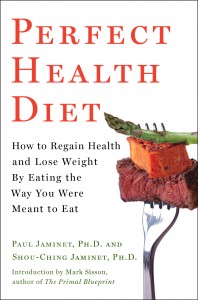
Guess the Book Cover

I’ve been told that, barring last-minute surprises, the proof copy of the book will be shipped to us today. It should arrive tomorrow. Assuming we approve, we should be able to begin shipping copies shortly.

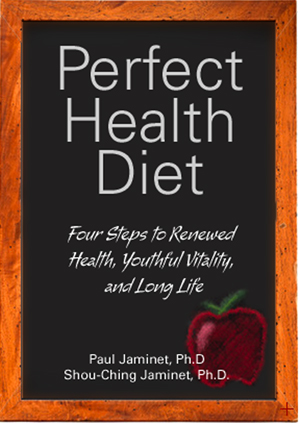
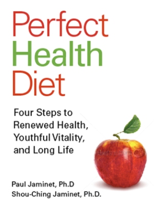
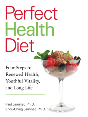
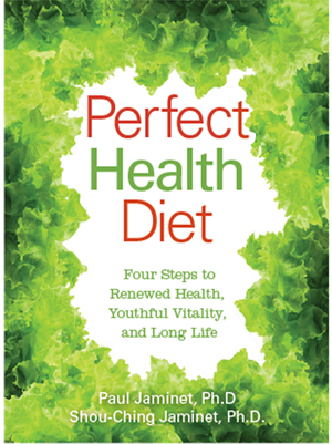
I thought it might be fun to show some of the book cover designs we considered. Can you guess which design we used?  Answer tomorrow.

(A)

(B)

(C)

(D)

By the way, if any of you are looking for a graphic designer, we strongly recommend Monika Chas, who designed our book cover and logo. Thanks Monika!

The Red Sox (USDA) Diet, Lymphoma, and Vascular Malformations

I want to say a bit more about the “Red Sox diet” – which basically consists of a rigorous adherence to the Dietary Guidelines put out by the US Department of Agriculture – and the diseases it causes.

My first post discussed two of the main problems with this diet:

  • Grain toxins are abundant in the diet.
  • Vitamin K2 is almost absent.

My second post discussed why vitamin D deficiency is also a concern. Insufficient vitamin D is especially dangerous for people who eat a lot of grains, since wheat, oats, and other grains interfere with vitamin D action and can induce rickets.

These three factors alone have tremendous influence on disease and mortality rates:

  • In the China Study, wheat consumption is more strongly associated with mortality and cardiovascular disease than any other dietary factor.
  • Vitamin D status has a huge impact on health. Those in the highest quartile of serum vitamin D have 35% lower mortality than those in the third quartile, and 52% lower mortality than those in the bottom quartile. [1]
  • Vitamin K2 status is nearly as important for health. In the Rotterdam Study, those ingesting a mere 41 mcg of vitamin K2 a day had 29% lower mortality (57% lower heart disease mortality) than those in the bottom third of K2 consumption. [2]

So it pays to remove grains (except rice) from the diet, get plenty of sunshine, and plenty of vitamin K2 from animal and dairy fats.

Today’s Diseases: Lymphoma and Cavernous Malformation

Last week we were mainly concerned with the Red Sox broken bone epidemic. Today I want to look at two other diseases that have struck Red Sox players in recent years – lymphoma, and cerebral cavernous malformation – to see if they might be caused by grain toxins and by vitamin D and K2 deficiencies.

In recent years among Red Sox players and prospects:

  • Pitcher Jon Lester developed anaplastic large cell lymphoma in 2006. He was diagnosed shortly after making his major league debut.
  • First baseman Anthony Rizzo, a top minor league prospect, developed Hodgkin’s lymphoma in 2008.
  • Top outfielder Ryan Westmoreland developed a cavernous malformation in his brain in March 2010. It began to cause headaches and numbness, and required surgery which has left him with a difficult recovery, comparable to that from a severe stroke.

These diseases are rare:

  • Lymphoma strikes about 1 person in 4,500 in the US each year.
  • Cerebral cavernous malformations develop in about 1 person in 200, but most are asymptomatic; only 5% of those (i.e., 1 person in 4,000) develop headaches, and fewer still need brain surgery to remedy neurological defects. A cavernous malformation of the severity of Mr. Westmoreland’s is an extremely rare occurrence.

Let’s compare Red Sox lymphoma incidence with its expected value. (Since we have two cases of lymphoma, statistics are better than for cavernous malformations.)

The Red Sox have a major league team and 6 minor league teams, with about 175 players total. So the Red Sox should expect 0.039 cases of lymphoma per year, or less than 0.2 cases in five years. They’ve experienced two, ten-fold higher than expected.

One might expect elite athletes to be healthier than the public, and thus to have lower lymphoma rates than the general population. Indeed, if professional baseball players developed lymphoma as frequently as the public, major league baseball players would experience 1.2 cases per year. A Google search turns up a couple of baseball players who developed lymphoma while playing – former first baseman Andres Galarraga, diagnosed 1999, and Scott Hodges, who played briefly for the Expos in 2004 – and several others who developed it after retirement – former Yankee star Roger Maris, diagnosed in 1983, and former Red Sox speedster Dave Roberts, diagnosed in 2010. Overall, however, the MLB rate seems to be much lower than 1.2 cases per year. The Red Sox lymphoma rate may be as much as 50-fold higher than their MLB peers.

Is this epidemic of lymphoma just bad luck? Or is it bad diet?

Grain Consumption and Lymphoma

Wheat and other grains contain toxic proteins. One kind is called gluten. About 99.6% of people are genetically capable of developing antibodies to wheat gluten; about 11% of the population develops systemic IgG antibodies against gluten. In some of these, auto-antibodies, or antibodies that target both gluten and self tissue, develop. These auto-antibodies primarily attack the thyroid and gut, but they can also destroy the heart and other organs. Severe auto-immune disease is called celiac disease.

Celiac disease patients tend to have leaky guts that let wheat toxins into the body, and (unless they quit eating wheat) the dysfunction and inflammation caused by these toxins generates other diseases. Up to 20% of celiac disease patients develop cancers, and 50% of those cancers are lymphomas. [3] The likelihood of intestinal lymphoma is 77 times higher in celiac disease patients than in the general population.

Going in the other direction, about 35% of Hodgkin lymphoma patients and 25% of non-Hodgkin lymphoma patients have anti-wheat IgG antibodies [4], compared to 11% in the general population.

It certainly looks like grain toxicity, or autoimmunity caused by grain consumption, may contribute to lymphoma development. The more wheat is consumed, the more likely that anti-wheat antibodies – and the various diseases caused by wheat toxins – will develop.

Vitamin D and Lymphoma

Vitamin D protects against many cancers; the protective effect is strongest for solid tumor cancers like breast cancer, ovarian cancer, and colon cancer. But it may also protect against lymphoma.

An Australian study found that “risk of non-Hodgkin lymphoma fell with increasing reported sun exposure hours” [5]. A recent review reported “an inverse association between sun exposure and NHL risk.” Too little data was available for Hodgkin lymphoma to form a conclusion. [6]

Vitamin K and Lymphoma

Vitamin K exhibits strong anti-cancer effects, especially against blood cancers. [7] Vitamin K can block proliferation of T-lymphoma cells [8] and induce cell death in B-lymphoma cells and myeloma cells [9]. Vitamin K has been tested in clinical trials against leukemias but not lymphomas, but has shown positive results. In AML specifically, a dose of 45 mg/day K2 led to regression of AML in 71% of patients in Japanese pilot studies. [10] In the ECKO trial, vitamin K supplementation reduced cancer incidence by 75%. [11]

Cavernous Malformations

If you’re not tired – and I apologize for stuffing so much into one post, but for the benefit of the Red Sox and their players I want to get it out – let’s look at Ryan Westmoreland’s cavernous malformation.

A cavernous malformation is a degeneration of blood vessels consisting of:

  • Missing matrix. Capillaries are dilated and thin-walled: they have the usual monolayer of endothelial cells but crucially, elastic fibers generated by smooth muscle cells are absent from the vessel walls. A fibrotic wall of variable thickness forms.
  • Hemorrhage. The capillaries usually show visible bleeding. Clotting may be present

Other pathological features commonly but not always seen in cavernous malformations are “haemosiderin deposits, gliosis, thrombosis, fibrotic changes, hyalinised vessel walls, calcification and cholesterol crystals.” [12]

Let’s work from this body of defects and see how they may be induced by grain toxins and vitamin D and K2 deficiencies.

Vitamin K and Vascular Malformations

In infants, vitamin K deficiency produces vascular malformations. [13]

Vitamin K deficiency is also known to produce hemorrhage. In fact, a leading cause of hemorrhagic stroke is vitamin K deficiency:

  • In one study of the causes of hemorrhagic stroke, 20 of 24 victims had vascular malformations, and 5 cases had clinical vitamin K deficiency [14]. (All 24 were probably subclinically vitamin K deficient.)
  • A Chinese study of hemorrhagic strokes in children concluded, “Vitamin K deficiency was a major etiology in 72 of 94 hemorrhagic strokes (76.6%).” [15]

What about the calcification observed in many vascular malformations?  Vascular calcification is the universal signal of vitamin K2 deficiency. Interestingly, vitamin K2 deficiency specifically leads to damage to the elastin matrix:

[M]ineralization induced by warfarin was observed limited to elastic fibers … In a recent paper, dermal elastic fiber calcification has been extensively described in patients affected by coagulation disorders due to a genetic defect in vitamin K recycling … [16]

Warfarin is a drug that interferes with vitamin K2. It seems clear that a loss of K2 function, due to deficiency, genetic defects, or warfarin, leads to specific damage to the elastin matrix.

Gluten and Cavernous Malformations

Another reason the elastin matrix might be missing from the vessels in a vascular malformation is autoimmune attack.

Both human elastin and wheat gluten are elastic proteins with similar structures. Intriguingly, it has been reported that anti-wheat antibodies may trigger autoimmune attacks on elastin. [17]

Could Vitamin D Play a Role?

While vitamin D deficiency has not been directly tied to cavernous malformations, it has been linked to hemorrhagic stroke. [18]

If Vitamin D deficiency contributed to his vascular malformation and cerebral bleeding, then grain consumption would have exacerbated the problem.

In Edward Mellanby’s pioneering experiments, he induced the disease by feeding dogs a diet of oats or wheat bread, and then cured it by adding cod liver oil (which contains vitamin D). Either dietary fats or sunlight cured rickets; a cereal-based diet combined with confinement indoors caused rickets. [19]

Grain consumption remains the leading risk factor for rickets in the world today. Today, rickets is mainly found in sunny countries such as Nigeria, South Africa, and Bangladesh, where it is the result of “cereal-based diets with limited variety.” [20]

In recent decades, more progress has been made in understanding how wheat and other grains induce rickets. First, wheat consumption leads to rapid loss of vitamin D. Eating just 20 g (0.7 ounces) per day of wheat bran causes vitamin D to be depleted 43% faster. [21] Second, wheat germ agglutinin, a wheat toxin, can block activation of the Vitamin D Receptor. [22]

Ryan Westmoreland was at risk for vitamin D deficiency. He lives in Rhode Island; the low sun during the northern winter, combined with the tendency to cover the skin when outdoors in winter, tends to induce a vitamin D deficiency.

Vitamin D levels are lowest in March, at the end of winter. Mr. Westmoreland experienced his cavernous malformation in March.

Conclusion

As with most diseases, we don’t yet know precisely what dietary factors contribute to lymphomas and cavernous malformations. There is plenty of room to disagree about the degree to which diet contributed to the Red Sox players’ lymphomas and cerebral hemorrhaging.

Nevertheless, we believe that:

  • Diseases don’t happen by accident. On a nourishing and toxin-free diet, the body should be able to maintain great health for a century or more.
  • Diseases are caused by food toxins, malnourishment, and pathogens. Eating a toxin-rich, nutrient-poor diet undermines the immune system and creates vulnerability to pathogens. So food toxins and malnourishment may be considered the “root cause” of disease.

Unfortunately, a toxin-rich, nutrient-poor diet is what the Red Sox have embraced. It seems quite possible that their high-grain, high-soy, high-vegetable oil, low vitamin K2 diet, combined with possibly insufficient vitamin D in some players, has raised players’ risk for many diseases 10-fold or more.

Ten-fold or more seems to be roughly the disease incidence rate the Red Sox are actually experiencing.

I believe that the Red Sox are paying the price for their diet in injuries, disease, and – very likely – weaker athletic performance. It behooves them to adopt a healthier diet.

References

[1] Dobnig H et al. Independent association of low serum 25-hydroxyvitamin d and 1,25-dihydroxyvitamin d levels with all-cause and cardiovascular mortality. Arch Intern Med. 2008 Jun 23;168(12):1340-9. http://pmid.us/18574092.

[2] Geleijnse JM et al. Dietary Intake of Menaquinone Is Associated with a Reduced Risk of Coronary Heart Disease: The Rotterdam Study. J Nutr. 2004 Nov;134(11):3100-5. http://pmid.us/15514282.

[3] Volta U et al. High prevalence of celiac disease in the Italian general population. Dig Dis Sci. 2001 Jul;46(7):1500-5. http://pmid.us/11478502. Freeman HJ. Adult celiac disease and its malignant complications. Gut Liver. 2009 Dec;3(4):237-46. http://pmid.us/20431755.

[4] Cil T et al. Screening for Celiac disease in Hodgkin and non-Hodgkin lymphoma patients. Turk J Gastroenterol. 2009 Jun;20(2):87-92. http://pmid.us/19530040.

[5] Hughes AM et al. Sun exposure may protect against non-Hodgkin lymphoma: a case-control study. Int J Cancer. 2004 Dec 10;112(5):865-71. http://pmid.us/15386383.

[6] Negri E. Sun exposure, vitamin D, and risk of Hodgkin and non-Hodgkin lymphoma. Nutr Cancer. 2010 Oct;62(7):878-82. http://pmid.us/20924963.

[7] Lamson DW, Plaza SM. The anticancer effects of vitamin K. Altern Med Rev. 2003 Aug;8(3):303-18. http://pmid.us/12946240.

[8] Kovalenko DV, Zelenin AV. Molecular mechanisms of the antiproliferative effect of vitamin K3 on Jurkat cells. Biochemistry (Mosc). 1999 Apr;64(4):468-72. http://pmid.us/10231603.

[9] Tsujioka T et al. The mechanisms of vitamin K2-induced apoptosis of myeloma cells. Haematologica. 2006 May;91(5):613-9. http://pmid.us/16670066.

[10] Miyazawa K et al. Vitamin K2 therapy for myelodysplastic syndromes (MDS) and post-MDS acute myeloid leukemia: information through a questionnaire survey of multi-center pilot studies in Japan. Leukemia. 2000 Jun;14(6):1156-7. http://pmid.us/10865985.

[11] Cheung AM et al. Vitamin K supplementation in postmenopausal women with osteopenia (ECKO trial): a randomized controlled trial. PLoS Med. 2008 Oct 14;5(10):e196. http://pmid.us/18922041.

[12] Frischer JM et al. Cerebral cavernous malformations: congruency of histopathological features with the current clinical definition. J Neurol Neurosurg Psychiatry. 2008 Jul;79(7):783-8. http://pmid.us/17986498.

[13] Berger TM et al. Imaging diagnosis and follow-up of infantile hepatic haemangioendothelioma: a case report. Eur J Pediatr. 1994 Feb;153(2):100-2. http://pmid.us/8157013.

[14] Takeshita M et al. Hemorrhagic stroke in infancy, childhood, and adolescence. Surg Neurol. 1986 Nov;26(5):496-500. http://pmid.us/3764653.

[15] Wang JJ et al. Risk factors for arterial ischemic and hemorrhagic stroke in childhood. Pediatr Neurol. 2009 Apr;40(4):277-81. http://pmid.us/19302940.

[16] Gheduzzi D et al. Matrix Gla protein is involved in elastic fiber calcification in the dermis of pseudoxanthoma elasticum patients. Lab Invest. 2007 Oct;87(10):998-1008.  http://pmid.us/17724449.

[17] Bödvarsson S et al. Dermatitis herpetiformis–an autoimmune disease due to cross-reaction between dietary glutenin and dermal elastin? Scand J Immunol. 1993 Dec;38(6):546-50. http://pmid.us/8256113.

[18] Pilz S et al. Low vitamin d levels predict stroke in patients referred to coronary angiography. Stroke. 2008 Sep;39(9):2611-3. http://pmid.us/18635847.

[19] Mellanby E. (March 15 1919) An experimental investigation on rickets. The Lancet 193(4985):407-412. Reprinted in Nutrition. 1989 Mar-Apr; 5(2): 81-6; discussion 87. http://pmid.us/2520279.

[20] Pettifor JM. Nutritional rickets: deficiency of vitamin D, calcium, or both? Am J Clin Nutr. 2004 Dec;80(6 Suppl):1725S-9S. http://pmid.us/15585795.

[21] Batchelor AJ, Compston JE. Reduced plasma half-life of radio-labelled 25-hydroxyvitamin D3 in subjects receiving a high-fibre diet. Br J Nutr. 1983 Mar;49(2):213-6. http://pmid.us/6299329.  

[22] Miyauchi Y et al. Importin 4 Is Responsible for Ligand-independent Nuclear Translocation of Vitamin D Receptor. J Biol Chem. 2005 Dec 9;280(49):40901-8. http://pmid.us/16207705.

Move Over, Jillian Michaels!

Frostie the Cockatoo has a workout video:

Red Sox Players Should Manage Their Vitamin D and Melatonin Status

Everyone should routinely measure their 25-hydroxyvitamin D status and use sun exposure or vitamin D3 supplementation to attain a level around 40 ng/ml.

This is especially important for those whose occupations keep them out of the sun. Night workers, especially, may have difficulty manufacturing sufficient vitamin D.

In my last post on the amazing and unnatural fragility of Red Sox players’ bones, I didn’t discuss vitamin D – a crucial nutrient for bone health, and health generally – because I thought that baseball players must get a great deal of sun exposure.

But on second thought, that may not be the case.

The Red Sox Are Night Workers

Most games start in the evening, around 7:05 pm. Games typically last between three and four hours. Players often eat a post-game meal at midnight, and may not sleep until 2 or 3 am. On road stands, every third game is followed by travel, so that players arrive in the new city early in the morning. As it is often difficult to sleep on planes, it’s reasonable to guess that many players sleep during daylight hours.

Night Workers Are At High Risk for Fractures

The Nurses’ Health Study found that night workers generally had a 37% higher risk of bone fractures. But slender night workers – those with a BMI below 24 – had a 136% higher risk of fractures. [1]

Jacoby Ellsbury, with the most fragile bones on the Red Sox, is one of the more slender players, and might be particularly vulnerable to the night work effect. At 6’1”, 185 pounds, his BMI is 24.4.

Vitamin D Is Crucial for Bone Health

Bone mineralization is optimized at serum 25-hydroxyvitamin D levels near 40 ng/ml. [2] Randomized controlled trials have found substantial reductions in fracture rates with vitamin D supplementation, for instance a 58% reduction in non-vertebral fractures and a 37% reduction in hip fractures. [2]

Even slight deficiencies can weaken bones. In mild vitamin D deficiency, serum PTH becomes elevated in order to increase conversion of 25(OH)D to the more active form of 1,25(OH)D to compensate for the insufficiency of 25(OH)D; however, elevation in PTH increases bone resorption, leading to additional bone loss. [3]

Vitamin D Optimization Improves Athletic Performance

In addition to its effects on bones, Vitamin D is also critical for muscle function and coordination. [3] Prolonged vitamin D deficiency is associated with severe muscle weakness which improves within several weeks of vitamin D supplementation. [4] In another study, quickness of movement was proportional to serum 25-hydroxyvitamin D levels. As the 25(OH)D level rose from 9.0 to 37.6 ng/mL, time to perform an 8-foot mobility test decreased by 0.67 seconds. [5] In another study, vitamin D supplementation reduced the rate of falls by 49%. [2]

For elite athletes, optimizing vitamin D can make a significant difference. After noticing that athletic performance was consistently better in summer than winter, the East German and Soviet athletic machines began programs of vitamin D supplementation. These programs coincided with the rise of these nations to the top of the Olympic medal lists. [6]

Melatonin Is Also Important

Melatonin, the “hormone of darkness,” is released during sleep, but only under conditions of quiet and darkness. Even a small amount of light can disrupt melatonin production.

Melatonin has both direct and indirect effects on bone. [7] It directly affects bone mineralization and activity of osteoclasts and osteoblasts – the two cell types responsible for bone remodeling and healing – and indirectly affects bone through its effects on other hormones such as cortisol. Melatonin levels tend to decline with age, and this may be responsible in part for the higher rates of osteoporosis in the elderly. In rats, melatonin co-participates in bone loss in a model of osteoporosis. [8]

Melatonin may also help athletic performance. In rats, 4 weeks of melatonin supplementation just before sleep led to reduced lactate levels during exercise, delayed exhaustion, and increased glycogen reserves. [9, 10]

Conclusions

Everyone, but especially professional athletes who work at night, should monitor serum 25-hydroxyvitamin D levels and get sun exposure or D3 supplements at mid-day to achieve 40 ng/ml.

Everyone, but especially professional athletes who work at night, should sleep in rooms with totally opaque drapes, so that the room remains completely dark after the sun rises, until natural waking. Artificial light sources should be eliminated, for instance by turning LCD clocks face down. Melatonin supplementation may also be worth consideration, especially in the elderly or those suffering from chronic infections; time-release melatonin at bed-time is optimal.

These steps will help optimize status of two hormones crucial for bone health and, possibly, athletic performance.

Maintaining optimal vitamin D and melatonin status is tricky for night workers. It should be a priority for the Red Sox. Have they done it?

References

[1] Feskanich D et al. Nightshift work and fracture risk: the Nurses’ Health Study. Osteoporos Int. 2009 Apr;20(4):537-42. http://pmid.us/18766292.

[2] Bischoff-Ferrari HA et al. Positive association between 25-hydroxy vitamin D levels and bone mineral density: a population-based study of younger and older adults. Am J Med. 2004 May 1;116(9):634-9. http://pmid.us/15093761.

[3] Lane NE. Vitamin D and systemic lupus erythematosus: bones, muscles, and joints. Curr Rheumatol Rep. 2010 Aug;12(4):259-63. http://pmid.us/20429045.

[4] Prabhala A et al. Severe myopathy associated with vitamin D deficiency in western New York. Arch Intern Med. 2000 Apr 24;160(8):1199-203. http://pmid.us/10789615.

[5] Bischoff-Ferrari HA et al. Higher 25-hydroxyvitamin D concentrations are associated with better lower-extremity function in both active and inactive persons aged > or =60 y. Am J Clin Nutr. 2004 Sep;80(3):752-8. http://pmid.us/15321818.

[6] Cannell JJ et al. Athletic performance and vitamin D. Med Sci Sports Exerc. 2009 May;41(5):1102-10. http://pmid.us/19346976.

[7] Cardinali DP et al. Melatonin effects on bone: experimental facts and clinical perspectives. J Pineal Res. 2003 Mar;34(2):81-7. http://pmid.us/12562498.

[8] Ostrowska Z et al. Assessment of the relationship between dynamic pattern of nighttime levels of melatonin and chosen biochemical markers of bone metabolism in a rat model of postmenopausal osteoporosis. Neuro Endocrinol Lett. 2001;22:129–136. http://pmid.us/11335889.

[9] Kaya O et al. Melatonin supplementation to rats subjected to acute swimming exercise: Its effect on plasma lactate levels and relation with zinc. Neuro Endocrinol Lett. 2006 Feb-Apr;27(1-2):263-6. http://pmid.us/16648794.

[10] Kaya O et al. Effect of melatonin supplementation on plasma glucose and liver glycogen levels in rats subjected to acute swimming exercise. Pak J Pharm Sci. 2010 Jul;23(3):241-4. http://pmid.us/20566433.