Category Archives: Pork

Neu5gc, Red Meat, and Human Disease: Part I

A number of people asked for comments on the most recent red meat scare, including Nicole, Ryan, and Mishkin on the blog, JT Olds on Twitter, and others on Facebook. You probably saw some of the headlines:

The article Nicole linked is a bit more scientifically inclined: “Possible Link Between Red Meat Consumption And Increased Cancer Risk Identified” (IFL Science). Here’s the press release version from UCSD: “Sugar Molecule Links Red Meat Consumption and Elevated Cancer Risk in Mice”. In the blogosphere, Stephan has summarized the issue in the context of a post on red meat and cancer.

The headlines are based on a paper [01] that reported that, in mice genetically altered to lack a sugar (Neu5gc) that humans also lack, feeding Neu5gc and injecting anti-Neu5gc antibodies generates inflammation which can promote the growth of cancers.

Significance of Neu5gc

The paper itself is a rather artificial scenario whose significance will be determined by future work. So analyzing this single paper would not be interesting. But I think it’s worthwhile to look into the broader idea that eating Neu5gc-bearing meats might be inflammatory or a source of autoimmunity.

In terms of PHD recommendations, this could affect the relative emphasis we place on different meats. If Neu5Gc is a true health risk, then we would want to emphasize seafood more and red meat less.

Another benefit to thinking about Neu5gc is that it may give us some insight into what a PHD “autoimmune protocol” should look like.

Background: Evolutionary History of Neu5gc

All cells in multicellular organisms are coated in carbohydrates, and the carbohydrates terminate in one of 43 sialic acids. In mammals, two forms of predominate: Neu5Ac and Neu5Gc. Each mammalian cell has tens or hundreds of millions of molecules of Neu5Ac and Neu5Gc on its surface. [02]

Neu5Gc is made from Neu5Ac, but the gene for making Neu5Gc was inactivated in the human lineage shortly before the emergence of Homo. The mutation occurred 3.2 million years ago and reached fixation – that is, all ancestral hominids had come to have the mutated gene – 2.9 million years ago. [03] This very rapid fixation indicates there was strong selection in support of the mutation.

In fact, this mutation by itself may have led to a speciation event, after which our ancestors could no longer mate with other apes. From that point on, Neu5gc-less females had difficulty producing children with males who retained the Neu5gc gene, because they would form antibodies against Neu5gc-coated sperm, making fertilization unlikely. [04]

Why was losing Neu5gc selected in our ancestors? Two possibilities are likely:

  • Loss of Neu5gc improved brain function.
  • Loss of Neu5gc (temporarily) reduced vulnerability to (ancestral) pathogens.

It should be noted that Neu5Gc has been lost independently in some other mammals as well – ferrets and new world monkeys. New world monkeys such as capuchins and spider monkeys also experienced a brain expansion, and ferrets are notably smart, so either explanation might be relevant to these cases of “convergent evolution.”

Neu5gc and Brain Function

Carbohydrates are extremely important for intercellular interactions. Indeed, the incorporation of carbohydrates into cell membranes and extracellular matrix is what made possible the rise of multicellular life.

In no organ are intercellular interactions as complex or consequential as in the brain. Not surprisingly, then, carbohydrates including the sialic acids are important to brain function.

The human brain is extraordinarly rich in sialic acids: neural membranes have 20 times more sialic acids than membranes of other human cell types. Animal brains are also enriched in sialic acids relative to their other tissues, but not as much as in humans; the human brain has 2-4 times more sialic acids the brains of other mammals. [05]

Curiously, though, Neu5gc is rare in the brains of all animals. Neu5gc is strongly suppressed, by about 98%, in the brains of all vertebrates, suggesting that its presence inhibits brain function. [06] It appears that Neu5gc is somehow toxic to brain function.

Loss of the gene for Neu5gc completely eliminated Neu5gc from the hominid brain. If Neu5gc does impair brain function, mutational inactivation of Neu5gc would have improved brain function. If so, the mutational inactivation of Neu5gc could have been driven by the same evolutionary forces that, soon after, selected for the tremendous expansion of the hominid brain.

Incidentally, dietary sialic acids — except for Neu5Gc – appear to be nutritious for humans, and especially for the developing infant brain. Breast milk is exceptionally rich in sialic acids, almost all of it Neu5Ac. Formula, by contrast, has much lower levels of sialic acids (0.21 mmol/L compared to 3.72 mmol/L in breast milk). Breast fed infants have nearly twice as many sialic acids in saliva than formula fed infants, confirming that milk sialic acids are taken up by the body and utilized.

Animal studies show that sialic acids in breast milk nourish the brain. Sialic acids facilitate neurotransmission between neurons. When piglet milk is supplemented with sialic acids, brain sialic acid levels are increased, and the piglets learn faster and make fewer mistakes in maze tests. [05] Rodents also perform better on tests of learning and memory after sialic acid supplementation. [07]

Not only does formula have fewer sialic acids than breast milk, cow milk based formulas have some Neu5Gc. [05] It has been observed that formula-fed infants have lower IQs than breast-fed infants. Sialic acids might help explain that. The lack of nourishing Neu5Ac and the presence of toxic Neu5Gc in formula might lastingly impair brain function in formula-fed infants.

Neu5Gc and Infection Risk

As the outermost molecules in the carbohydrate coat surrounding cells, sialic acids are the first contact point for pathogens seeking entry to the cell, and for immune cells seeking to detect whether the cell is native or foreign.

There has been a “Red Queen” evolutionary arms race in which pathogens evolved ways to utilize sialic acids for cell entry, or to hide from the immune system; and animals evolved changes to their sialic acids to frustrate the pathogens. [08]

Many pathogens interact with sialic acids in order to adhere to and gain entry into the cell. Pathogens generally rely on a single specific endocytic route for cell entry. This often requires binding to a specific sialic acid as the initial point of attachment.

Pathogens that specifically utilize Neu5Gc to enter cells include canine and feline parvoviruses [09]; pathogens that specifically utilize Neu5Ac include adeno-associated viruses and the minute virus of mice (MVM) [10].

A human pathogen that uses sialic acids to enter cells is the malaria protozoan. Plasmodium falciparum causes severe disease in humans and enters cells via Neu5Ac; Plasmodium reichenowi causes milder disease in chimpanzees and gorillas and enters cells via Neu5Gc. Plasmodium falciparum appears to have evolved recently – possibly reaching its current form only 10,000 years ago when the rise of agriculture and animal husbandry brought humans and mosquitos into closer proximity – while Plasmodium reichenowi is thought to resemble the ancestral form that would have afflicted hominids and apes 3.5 million years ago.

Possibly, the gene for Neu5Gc was inactivated to protect ancestral hominids from malaria. With the loss of Neu5Gc, hominids would have become immune to P. reichenowi. [11] [12]

Unfortunately, after P. falciparum’s adaptation to Neu5Ac which is overabundant in humans, we now suffer from more severe malaria than chimpanzees or gorillas (the “malignant malaria” mystery). [13]

In addition to entry points for microbes, sialic acids can be entry points for microbial toxins. For example, Shiga toxin from shigatoxigenic E. coli binds to Neu5Gc. [14]

Sialic Acid Concealment and the Gut Microbiome

The immune system is sensitive to the composition of the carbohydrate coat on a cell. White blood cells have a number of sialic acid detectors on their surfaces (called Siglecs, for sialic acid Ig-superfamily lectins). Some, which bind to human sialic acids, inhibit immune responses. Others, which bind to non-human sialic acids, activate immune responses.

Thus, when white blood cells contact a cell bearing human sialic acids, the immune system interprets it as “self” and tamps down immunity. When it detects foreign sialic acids, the immune system treats the cell as “foreign” and is more likely to attack it.

Some microbes – including commensal gut microbes – have been living in humans long enough that they have learned to take up sialic acids, chiefly Neu5Ac, and incorporate them into lipopolysaccharides on their cell membranes. This suppresses immunity toward them. [15]

A number of human pathogens have learned the same trick. Pathogens that incorporate sialic Neu5Ac into their cell membranes for the purpose of mimicking human cells and evading human immune defenses include Escherichia coli K1, Haemophilus influenzae, Pasteurella multocida, Neisseria spp., Campylobacter jejuni and Streptococcus agalactiae. [16]

Due to this “molecular mimickry” of human molecules, it has been suggested that these bacteria – especially Haemophilus influenzae and Neisseria spp. – may be sources of autoimmunity. [17]

While some bacteria can synthesize sialic acids themselves, most obtain it from their environment. These bacteria release enzymes called sialidases to cleave the sialic acids from food in the digestive tract, from surrounding cells, or from mucus. [15] Bacteria can obtain Neu5Ac from human tissue and mucus as well as food, but Neu5Gc only from food, chiefly beef and pork.

Neu5Gc in Human Tissue

Although humans can no longer synthesize Neu5Gc, we still have all the cellular machinery for utilizing it. When dietary Neu5Gc is absorbed into the body and enters cells, it can be incorporated into glycoproteins bound for the cell surface glycocalyx, just as Neu5Ac is.

As a result, Neu5Gc of dietary origin appears at low levels on the surface of human cells.

Neu5gc is found at high levels in all mammals except humans, ferrets, and new world monkeys; birds and reptiles do not produce Neu5Gc at all, and fish and shellfish produce only low levels. So, of the four major meat groups – beef, pork, chicken, and fish – Neu5gc is obtained predominantly from the red meats, beef and pork.

Among human cells, Neu5Gc appears at highest levels on tumor cells, especially metastatic cells. [21] This makes Neu5Gc a potential target for cancer therapy.

Neu5Gc as an Immunogen

Neu5gc expressed on cell walls is a potential immunogen. When pig organs are transplanted to humans, Neu5Gc is the second most important cause of rejection, after the α1,3-galactose (αGal) epitope. [20]

Anti-Neu5gc antibodies have been found in 85% of humans. [18] It is thought that antibodies form in early childhood after dietary Neu5Gc is incorporated by certain gut bacteria into lipooligosaccharides that can generate antibodies. Some of these antibodies may cross-react with compounds human cells form from dietary Neu5Gc; these human molecules are then known as “xeno-autoantigens.” [21]

Summary

Neu5Gc from mammalian meats, such as beef and pork, is incorporated into the cell surface coats and walls of gut microbes and some human cells, mainly in the gut and in tumors. Neu5gc in bacterial walls is immunogenic and 85% of people have detectable antibodies to Neu5Gc. Eating beef and pork supplies antigens for these antibodies, potentially triggering inflammation. There are concerns that this inflammation may have negative health effects.

Next up: Neu5Gc and autoimmunity.

Note: May Perfect Health Retreat

Interested in a luxury vacation combined with an education in how to be healthy for the rest of your life? Science classes, cooking classes, movement and relaxation classes, personal health coaching, gourmet food, all on a magnificent beach, with hot tubs and salt water pools? Come to the Perfect Health Retreat! Spaces are currently 2/3 filled and the retreat is expected to sell out early. Visit here for more info or email me at paul@perfecthealthretreat.com.

PHRetreat_img7_300x200px

References

[01] Samraj AN et al. A red meat-derived glycan promotes inflammation and cancer progression. Proc Natl Acad Sci U S A. 2014 Dec 29. pii: 201417508. [Epub ahead of print]. http://pmid.us/25548184.

[02] Kraemer PM. Sialic acid of mammalian cell lines. J Cell Physiol. 1966 Feb;67(1):23-34. http://pmid.us/5327858. Was 21

[03] Hayakawa T, Aki I, Varki A, Satta Y, Takahata N. Fixation of the human-specific CMP-N-acetylneuraminic acid hydroxylase pseudogene and implications of haplotype diversity for human evolution. Genetics. 2006 Feb;172(2):1139-46. http://pmid.us/16272417. Full text: http://www.ncbi.nlm.nih.gov/pmc/articles/PMC1456212/. Was 22

[04] Ghaderi D et al. Sexual selection by female immunity against paternal antigens can fix loss of function alleles. Proc Natl Acad Sci U S A. 2011 Oct 25;108(43):17743-8. http://pmid.us/21987817. was 2

[05] Wang B. Molecular mechanism underlying sialic acid as an essential nutrient for brain development and cognition. Adv Nutr. 2012 May 1;3(3):465S-72S. http://pmid.us/22585926. Full text: http://www.ncbi.nlm.nih.gov/pmc/articles/PMC3649484/. Was 31

[06] Davies LR, Varki A. Why Is N-Glycolylneuraminic Acid Rare in the Vertebrate Brain? Top Curr Chem. 2013 Mar 8. [Epub ahead of print] http://pmid.us/23471785. was 8

[07] Wang B. Sialic acid is an essential nutrient for brain development and cognition. Annu Rev Nutr. 2009;29:177-222. http://pmid.us/19575597. was 32

[08] Varki A. Colloquium paper: uniquely human evolution of sialic acid genetics and biology. Proc Natl Acad Sci U S A. 2010 May 11;107 Suppl 2:8939-46. http://pmid.us/20445087. was 51

[09] Löfling J et al. Canine and feline parvoviruses preferentially recognize the non-human cell surface sialic acid N-glycolylneuraminic acid. Virology. 2013 May 25;440(1):89-96. http://pmid.us/23497940. was 54

[10] Wu Z et al. Alpha2,3 and alpha2,6 N-linked sialic acids facilitate efficient binding and transduction by adeno-associated virus types 1 and 6. J Virol. 2006 Sep;80(18):9093-103. http://pmid.us/16940521. was 55

[11] Varki A, Gagneux P. Human-specific evolution of sialic acid targets: explaining the malignant malaria mystery? Proc Natl Acad Sci U S A. 2009 Sep 1;106(35):14739-40. http://pmid.us/19717444. was 57

[12] Martin MJ et al. Evolution of human-chimpanzee differences in malaria susceptibility: relationship to human genetic loss of N-glycolylneuraminic acid. Proc Natl Acad Sci U S A. 2005 Sep 6;102(36):12819-24. http://pmid.us/16126901. was 58

[13] Rich SM et al. The origin of malignant malaria. Proc Natl Acad Sci U S A. 2009 Sep 1;106(35):14902-7. http://pmid.us/19666593/.

[14] Byres E et al. Incorporation of a non-human glycan mediates human susceptibility to a bacterial toxin. Nature. 2008 Dec 4;456(7222):648-52. http://pmid.us/18971931.

[15] Varki A, Gagneux P. Multifarious roles of sialic acids in immunity. Ann N Y Acad Sci. 2012 Apr;1253:16-36. http://pmid.us/22524423. Full text: http://www.ncbi.nlm.nih.gov/pmc/articles/PMC3357316/

[16] Severi E, Hood DW, Thomas GH. Sialic acid utilization by bacterial pathogens. Microbiology. 2007 Sep;153(Pt 9):2817-22. http://pmid.us/17768226. Full text: http://mic.sgmjournals.org/content/153/9/2817.long.

[17] Harvey HA, Swords WE, Apicella MA. The mimicry of human glycolipids and glycosphingolipids by the lipooligosaccharides of pathogenic neisseria and haemophilus. J Autoimmun. 2001 May;16(3):257-62. http://pmid.us/11334490.

[18] Zhu A, Hurst R. Anti-N-glycolylneuraminic acid antibodies identified in healthy human serum. Xenotransplantation. 2002 Nov;9(6):376-81. http://pmid.us/12371933.

[19] Takahashi T et al. N-glycolylneuraminic acid on human epithelial cells prevents entry of influenza A viruses that possess N-glycolylneuraminic acid binding ability. J Virol. 2014 Aug;88(15):8445-56. http://pmid.us/24829344.

[20] Park JY et al. α1,3-galactosyltransferase deficiency in germ-free miniature pigs increases N-glycolylneuraminic acids as the xenoantigenic determinant in pig-human xenotransplantation. Cell Reprogram. 2012 Aug;14(4):353-63. http://pmid.us/22775484.

[21] Samraj AN, Läubli H, Varki N, Varki A. Involvement of a non-human sialic Acid in human cancer. Front Oncol. 2014 Feb 19;4:33. http://pmid.us/24600589.

The Trouble With Pork, Part 3: Pathogens

We started this series with a look at remarkably strong correlations between pork consumption and liver cirrhosis mortality, liver cancer, and multiple sclerosis (Pork: Did Leviticus 11:7 Have It Right?, Feb 8, 2012). In Part 2, we looked at omega-6 fats in industrial pork meat and toxins in processed pork products as possible causes (The Trouble with Pork, Part 2, Feb 15, 2012).

That second post left us with several clues that some pathogen (or pathogens) that (a) infects both pigs and humans and (b) can be transmitted from pigs to humans via the eating of pork, is responsible for the disease associations. These clues include:

  1. The risk is higher for fresh pork than processed pork. Processed pork is generally cured or smoked, both steps that are anti-microbial.
  2. Eating fiber, which increases gut bacterial populations and enhances immune vigilance of the gut, is protective.
  3. The disease risk is specifically associated with two organs – the central nervous system (multiple sclerosis) and the liver (cirrhosis, hepatocellular carcinoma). Pathogens are more likely than other pork components to have tissue specificity.

Our mission today is to try to track down the pathogen(s), and figure out how to minimize risk of infection.

Pigs And Zoonotic Infections

Scientists studying xenotransplantation – the transplantation of animal organs into a person to replace a failing organ – have had the best luck with pig organs. Pigs are easier to work with than primates, not dramatically different in size than humans, and their organs are less likely to provoke rejection than those of other mammals. This suggests a similarity of biology between pigs and humans.

But biological similarity has its downsides. A large number of pathogens can infect both pigs and humans. More than any other animal, pigs pass pathogens to humans.

Indeed, investigators have been surprised at how frequently pathogens pass back and forth. According to a new study (discussed at Aetiology) of the evolutionary history of methicillin-resistant Staphylococcus aureus (MRSA), S. aureus was passed to pigs by their human caretakers. In pigs, which are routinely given antibiotics by industrial food producers, S. aureus picked up resistance genes to tetracyclines and methicillins. The resulting antibiotic-resistant ST398 strain was passed back to humans.

Wikipedia lists some of the pathogens that flourish in both pigs and humans and can infect humans who eat infected pork, usually undercooked pork:

Although all of these pathogens are potential concerns, I do not see strong specific links between the above pathogens and our three pork-associated diseases – liver cirrhosis, liver cancer, and multiple sclerosis.

However, there is another pathogen capable of infecting humans from pork that is a strong candidate: hepatitis E virus (HEV).

Hepatitis E

Hepatitis E was first observed in a 1955 outbreak in New Delhi, India. It generally produces an acute disease that lasts for several weeks; most victims recover with few symptoms, but in a few this acute illness progresses into a severe liver disease that can be fatal. About 2% of all infections lead to death from this acute liver disease; death rates are higher in pregnant women.

Hepatitis E seems to have evolved in the last millennium: There are four known genotypes, all of which infect humans and two of which infect pigs, and their common ancestor dates to 536 to 1344 years ago. [1] However, the pig-infecting genotypes 3 and 4 of Hepatitis E underwent a notable population expansion in the twentieth century, during which there has been “an extensive genetic divergence of HEV strains and high prevalence of HEV infections in many parts of the world.” [2]

The human-only genotypes of Hepatitis E are transmitted by fecal contamination of drinking water and are prevalent only in developing countries with poor sanitation; but the pig-and-human genotypes are transmitted primarily through pork consumption:

[G]enotypes 3 and 4 are associated with sporadic disease attributable to exposure to body fluids of infected swine [8] and ingestion of food products from pigs, boars and deer [11], [16], [18]. [1]

Hepatitis E seems to be most prevalent in Asia, the Middle East, and Africa:

Hepatitis E is the most important or the second most important cause of acute clinical hepatitis in adults throughout Asia, the Middle East and Africa. [8]

However, it has been spreading to Europe and the Americas:

HEV was rarely identified in industrialized countries, and the few reported cases of infection were usually in someone who had recently traveled to an endemic region. In the past few years this pattern has changed, as cases of endemic or autochthonous hepatitis E have been diagnosed with increasing frequency in individuals who have not traveled abroad….

Cases have been reported with increasing regularity throughout Western Europe, as well as in some Eastern European countries. [7]

The genotypes that coinfect humans and pigs may have originated in East Asia:

All but one genotype 4 sequence originated either from China or Japan…. [T]he genotype 3 sequences were divided into 3.1 and 3.2 clades … [A]lthough 87.5% of the clade 3.1 variants were from Asia and 60% of the clade 3.2 variants were from Europe (Table S1), these clades were found to have similar histories (Fig. 6). [1]

Historically, China and Japan did not raise cattle for food and pigs have been the major source of meat. Even today in southern China, pigs are often kept in the yards of homes, and close contact between pigs and humans facilitates zoonotic transmission.

At pig farms, Hepatitis E virus seems to spread readily. A Japanese study reported:

[O]ur estimates imply that more than 95% of pigs are infected before the age of 150 days. [3]

Presumably this is due to fecal-oral transmission among pigs in close quarters. At French farms, 65% of pigs were found to be hepatitis E infected at age 90 days. [4]

Transmission to Humans Via Pork

Can humans get infected by eating pork products? It now seems clear that the answer is yes.

A French study found that the genotype distribution of hepatitis E infecting humans is identical to the genotype distribution in pigs at slaughterhouses:

Frequent zoonotic transmission of hepatitis E virus (HEV) has been suspected, but data supporting the animal origin of autochthonous cases are still sparse. We assessed the genetic identity of HEV strains found in humans and swine during an 18-month period in France. HEV sequences identified in patients with autochthonous hepatitis E infection (n = 106) were compared with sequences amplified from swine livers collected in slaughterhouses (n = 43). Phylogenetic analysis showed the same proportions of subtypes 3f (73.8%), 3c (13.4%), and 3e (4.7%) in human and swine populations. Furthermore, similarity of >99% was found between HEV sequences of human and swine origins. These results indicate that consumption of some pork products, such as raw liver, is a major source of exposure for autochthonous HEV infection. [5]

As hepatitis E concentrates in the liver in both pigs and humans, swine livers were the natural place to test for hepatitis E presence, and probably the riskiest part of the pig to eat.

Further evidence that hepatitis E in pigs can infect humans was found in another French study. The researchers reasoned that sausage made from pig liver would be a likely vector for hepatitis E transmission to humans, especially a form of smoked pig liver sausage traditionally eaten raw – figatellu. Their findings:

Acute or recent HEV infection, defined by detection of anti-HEV immunoglobulin M antibodies and/or HEV RNA, was observed in 7 of 13 individuals who ate raw figatellu and 0 of 5 individuals who did not eat raw figatellu (P=.041). Moreover, HEV RNA of genotype 3 was recovered from 7 of 12 figatelli purchased in supermarkets, and statistically significant genetic links were found between these sequences and those recovered from patients who ate raw figatellu….

Our findings strongly support the hypothesis of HEV infection through ingestion of raw figatellu. [6]

The titer of hepatitis E viruses in the supermarket sausage reached as high as a million copies per slice. [6] This data suggests that a majority of figatellu in French supermarkets carries hepatitis E virus, and that a majority of people who eat figatellu acquire hepatitis E infections.

Contact with pigs can also lead to transmission; swine workers have an elevated prevalence of antibodies to HEV in the United States. [7]

Does Cooking Inactivate the Viruses?

What level of cooking is needed to inactivate the virus?

It is difficult to prove that any particular cooking or processing method renders HEV non-infectious:

How safe are these products? The question is difficult to answer because HEV grows poorly in cell culture, and in vivo testing of viability requires nonstandard laboratory animals—nonhuman primates or pigs for genotypes 3 and 4. [7]

Since scientists don’t have the funding or facilities to see if feeding cooked, cured, or smoked pork to primates or pigs gives them hepatitis E, they have no way of verifying that cooked, cured, or smoked pork is free of HEV.

In test tube experiments, HEV was still viable and infectious after cooking for 1 hour at 56°C, the temperature of rare to medium-cooked meat. [9] About 80% of viruses were inactivated after an hour at 60°C, and an hour at 70°C probably eliminates the viruses.

The implication is that thorough cooking would destroy HEV, but that some HEV will survive in rare to medium cooked pork, with liver likely having the greatest viral titer. [9] “However, much pork is consumed that has not had even that degree of cooking.” [7]

One way to reduce the risk of infection is to avoid the pig tissues that have the highest viral titers:

HEV can be found in the liver, blood, and intestinal tract, which are all consumed in one form or another and often together, such as in sausages. [7]

So: to avoid HEV infection, it’s best to avoid pork liver, intestines, or blood, or products made from them such as sausage; other cuts should be carefully rinsed of all blood and then cooked thoroughly to a temperature of at least 70°C. Simmering in near-boiling water for an hour should be sufficient.

The most dangerous pork product is likely to be sausage, which often uses pork liver meat, and traditionally uses pig intestines as the casing. It may also contain traces of pig blood. Pig blood pudding, a traditional Chinese dish, should also be avoided.

Links to Pork-Associated Liver Diseases

Hepatitis E was discovered as a cause of acute liver disease. But what about chronic diseases like alcoholic cirrhosis and liver cancer? Is there really evidence linking it to these diseases?

First, studies of organ-transplant recipients who contracted hepatitis E from their donors have shown that HEV seems to establish chronic infections in at least 58% of infected persons. [10] When anti-HEV antibodies exist, generally active viral RNA is present too. [12] So the virus is persistent.

Hepatitis B and C viruses are known causes of alcoholic liver cirrhosis. What about HEV? There have been few studies, but those that exist suggest it is likely:

  • A child developed cirrhosis after a bone marrow transplant due to a swine-derived form of hepatitis E. [11]
  • A Spanish study found a strong association between HEV and cirrhosis in people infected with HIV: “Liver cirrhosis was the only factor independently associated with the presence of anti-HEV, which was documented in 23% of patients with cirrhosis and 6% of patients without cirrhosis (P?=?0.002; odds ratio 5.77). HEV RNA was detected in three seropositive patients (14%), two of whom had liver cirrhosis.” [12]
  • HEV seems to be a common cause of cirrhosis in Egypt. [13]

Hepatitis B and hepatitis C viruses are known causes of hepatocellular carcinoma. What about HEV? If there were few studies linking HEV to cirrhosis, there are even fewer investigating its relationship to HCC.

I did find one Chinese study showing that HEV infection greatly elevated the association of aflatoxin with HCC. (Aflatoxin, a fungal toxin that damages the liver, is a known risk factor for HCC.) [14]

Epidemiology is also suggestive. I mentioned earlier that the pork-transmitted genotypes of HEV have only recently appeared in the Americas. If HEV is responsible for alcoholic cirrhosis, hepatocellular carcinoma (HCC), or multiple sclerosis, then we should be seeing the incidence of those diseases increase. In fact, that is true for HCC:

In the U.S., incidence rates of HCC in both men and women have increased steadily during the past three decades. The reasons for this steady increase remain unknown. [15]

What About Multiple Sclerosis?

There have been no studies searching for a specific link between HEV and multiple sclerosis.

However, it may be worth reviewing what some mouse models tell us about the potential for a hepatitis virus to cause MS. MS is an infectious or autoimmune disease:

MS is felt to be most likely either due to an aberrant immune response or a pathogen, or possibly a combination of the two, and the animal models available reflect these two possible pathogeneses. [16]

Regular readers will know that I believe MS is infectious in origin. There are three animal models for MS. One of them (“experimental allergic encephalomyelitis” or EAE) involves immunizing mice with myelin or myelin proteins so that they develop antibodies to their own myelin; the other two involve infecting mice with viruses:

Two viruses, Theiler’s murine encephalomyelitis virus and murine hepatitis virus, are used to induce infectious models of the disease. [16]

The murine hepatitis virus (MHV) model is suggestive: it supports the idea that a virus that causes hepatitis may also cause MS. Some strains of MHV are neurotropic, infecting both the liver and central nervous system, and it is these that most readily produce an MS-like disease. [17]

If a hepatitis virus is causing MS in humans, we would expect MS patients to have high rates of liver disease. Indeed, there is a correlation.

MS patients are 3.7-fold more likely to have elevated ALT and 2.2-fold more likely to have elevated AST – both liver enzymes associated with liver disease. Also, elevated ALT and AST are associated with the more severe relapsing-remitting form of MS. [18]

A few perhaps insignificant links: Patients with systemic sclerosis, who are about 5-fold more likely to develop MS than others, are also at high risk for liver disease. [19] In the 1980s, doctors began observing MS patients with cases of primary biliary cirrhosis severe enough to require liver transplantation. [20]

Other Pig-Human Pathogens and MS

Pork can carry many pathogens; perhaps hepatitis E virus is not the MS-causing pathogen.

I don’t see obvious candidates however. Perhaps herpes viruses would be most likely. One of the human pathogens likely to be causal for MS is Epstein-Barr virus, also known as human herpes virus 4 (HHV-4). It causes mononucleosis but establishes persistent infections and is associated with a number of diseases, including lymphomas, MS, lupus, and rheumatoid arthritis.

Human herpes viruses may be able to establish infections in pigs. [21] And there are porcine herpes viruses that are closely related to Epstein-Barr virus. [22]

Conclusion

There is a strong association between pork consumption and liver cirrhosis mortality, liver cancer, and multiple sclerosis.

It seems likely that the association, if it is real, is mediated by a pathogen. The most likely pathogen in the case of the liver diseases is hepatitis E virus. In MS, the pathogen remains unknown, but is likely to be a virus.

Hepatitis E virus is not destroyed by casual cooking, smoking, or curing. It appears that meat must  reach temperatures of 70ºC (160ºF) before viruses are inactivated; and it is possible that meat must remain at that temperature for some time, perhaps as long as an hour. Rare or medium cooked pork could contain active viruses.

Hepatitis E viruses are most abundant in liver, intestine, and blood. Pork products containing these parts, such as sausage, may be best avoided.

Meat from parts of the pig with low viral titers, such as pork ribs or pork bellies, are likely to be safe to eat as long as they are well cooked. Be sure to wash the meat of all blood before cooking, and to cook thoroughly.

Related Posts

Posts in this series:

References

[1] Purdy MA, Khudyakov YE. Evolutionary history and population dynamics of hepatitis E virus. PLoS One. 2010 Dec 17;5(12):e14376. http://pmid.us/21203540.

[2] Purdy MA, Khudyakov YE. The molecular epidemiology of hepatitis E virus infection. Virus Res. 2011 Oct;161(1):31-9. http://pmid.us/21600939.

[3] Satou K, Nishiura H. Transmission dynamics of hepatitis E among swine: potential impact upon human infection. BMC Vet Res. 2007 May 10;3:9. http://pmid.us/17493260.

[4] Kaba M et al. Frequent transmission of hepatitis E virus among piglets in farms in Southern France. J Med Virol. 2009 Oct;81(10):1750-9. http://pmid.us/19697419.

[5] Bouquet J et al. Close similarity between sequences of hepatitis E virus recovered from humans and swine, France, 2008-2009. Emerg Infect Dis. 2011 Nov;17(11):2018-25. http://pmid.us/22099089.

[6] Colson P et al. Pig liver sausage as a source of hepatitis E virus transmission to humans. J Infect Dis. 2010 Sep 15;202(6):825-34. http://pmid.us/20695796.

[7] Purcell RH, Emerson SU. Hidden danger: the raw facts about hepatitis E virus. J Infect Dis. 2010 Sep 15;202(6):819-21. http://pmid.us/20695795.

[8] Purcell RH, Emerson SU. Hepatitis E: an emerging awareness of an old disease. J Hepatol. 2008 Mar;48(3):494-503. http://pmid.us/18192058.

[9] Emerson SU et al. Thermal stability of hepatitis E virus. J Infect Dis. 2005 Sep 1;192(5):930-3. http://pmid.us/16088844.

[10] Legrand-Abravanel F et al. Characteristics of autochthonous hepatitis E virus infection in solid-organ transplant recipients in France. J Infect Dis. 2010 Sep 15;202(6):835-44. http://pmid.us/20695798.

[11] Halac U et al. Cirrhosis due to Chronic Hepatitis E Infection in a Child Post-Bone Marrow Transplant. J Pediatr. 2012 Feb 15. [Epub ahead of print] http://pmid.us/22341950.

[12] Jardi R et al. HIV, HEV and cirrhosis: evidence of a possible link from eastern Spain. HIV Med. 2012 Jan 18. http://pmid.us/22257075.

[13] El Sayed Zaki M, Othman W. Role of hepatitis E infection in acute on chronic liver failure in Egyptian patients. Liver Int. 2011 Aug;31(7):1001-5. http://pmid.us/21733089.

[14] Tao P et al. Associated factors in modulating aflatoxin B1-albumin adduct level in three Chinese populations. Dig Dis Sci. 2005 Mar;50(3):525-32. http://pmid.us/15810636.

[15] Yuan JM et al. Synergism of alcohol, diabetes, and viral hepatitis on the risk of hepatocellular carcinoma in blacks and whites in the U.S. Cancer. 2004 Sep 1;101(5):1009-17. http://pmid.us/15329910.

[16] Pachner AR. Experimental models of multiple sclerosis. Curr Opin Neurol. 2011 Jun;24(3):291-9. http://pmid.us/21519255.

[17] Carbajal KS et al. Surgical transplantation of mouse neural stem cells into the spinal cords of mice infected with neurotropic mouse hepatitis virus. J Vis Exp. 2011 Jul 10;(53). http://pmid.us/21775959.

[18] Tremlett H et al. Liver test abnormalities in multiple sclerosis: findings from placebo-treated patients. Neurology. 2006 Oct 10;67(7):1291-3. http://pmid.us/17030771.

[19] Robinson D Jr et al. Systemic sclerosis prevalence and comorbidities in the US, 2001-2002.  Curr Med Res Opin. 2008 Apr;24(4):1157-66. http://pmid.us/18430269.

[20] A patient with primary biliary cirrhosis and multiple sclerosis. Am J Med. 1992 Apr;92(4):433-6. http://pmid.us/1558090.

[21] Kim JH et al. Infection of porcine cells with human herpesviruses. Transplant Proc. 2010 Jul-Aug;42(6):2134-7. http://pmid.us/20692426.

[22] Doucette K et al. Gene expression of porcine lymphotrophic herpesvirus-1 in miniature Swine with posttransplant lymphoproliferative disorder. Transplantation. 2007 Jan 15;83(1):87-90. http://pmid.us/17220799.

The Trouble with Pork, Part 2

So it looks like pork consumption is correlated with cirrhosis of the liver, liver cancer, and multiple sclerosis (Pork: Did Leviticus 11:7 Have It Right?, Feb 8, 2011). Why?

There are a number of potential dangers from pork, and to give each due consideration will require two posts. I’ll look at a few candidates today, and save my top candidate for Thursday.

Omega-6 Fats

Omega-6 fats are a health villain: Excess omega-6 contributes to general inflammation, fatty liver disease, metabolic syndrome, obesity, and impaired immune function.

And pork can be a major source of omega-6 fats. Nutritiondata.com lists the omega-6 fraction of lard at 11%. But the omega-6 fraction can be highly variable, depending on the pig’s diet. Chris Masterjohn recently reported that the lard used in the “high-fat” research diet was 32% polyunsaturated, nearly all of it omega-6:

The graph shows the difference between the actual fatty acid profile as determined by direct analysis of the lard and the previously reported fatty acid profile, which had been estimated using the USDA database.  We can see that the actual fatty acid profile is much higher in PUFAs, at the expense of both saturated and monounsaturated fats.  In fact, the company had originally estimated the diet to provide 17 percent of its fat as PUFA, but now estimates it to provide a whopping 32 percent!

Chris further reported that feeding the pigs a pasture and acorns diet would reduce lard PUFA levels to 8.7%, and feeding them a Pacific Islander PHD-for-pigs diet of coconut, fish, and sweet potatoes would reduce lard PUFA levels to 3%.

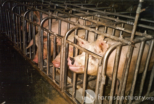
So the omega-6 content can cover a 10-fold range, 3% to 32%, with the highest omega-6 content in corn- and wheat-fed pigs who have been caged for fattening. Corn oil and wheat germ oil are 90% PUFA, and caging prevents exercise and thus inhibits the disposal of excess PUFA. Caging is a common practice in industrial food production; here is a picture of sows in gestation crates:

And here are some Chinese pigs in shipping cages for transport to market:

The Wall Street Journal reported Monday that McDonald’s, following Chipotle, has asked its pork suppliers to stop using gestation stalls, and the largest US hog producer, Smithfield Farms, has begun a 10-year plan to move pigs from small stalls into roomier “group housing systems.” So perhaps the omega-6 content of commercial pork will come down.

How much omega-6 are people actually getting from pork? In the Bridges database, the range in pork consumption across countries was 2 to 80 kg/yr, or 5 to 200 g/day. If this is from industrially raised pigs whose fat is 30% omega-6, then this works out to 0.25% to 10% of energy as omega-6 fats from pork. In most countries, pork is either the primary source of omega-6 fats or the second source after vegetable oils.

Moral of the story: If you’re going to eat a lot of pork, there are real benefits to finding a source of naturally raised pigs fed a healthy diet.

Aside: On a similar diet, human adipose tissue develops almost identical omega-6 levels to pig lard. The Finnish Mental Hospital Study [1] [2] [3], discussed in our book on pages 63-65, showed that on a normal dairy-rich hospital diet human adipose tissue is less than 10% omega-6, but on a soybean oil rich diet adipose tissue becomes 32% omega-6.

American diets have traversed this range in recent decades. Here is a plot of subcutaneous fat omega-6 levels from Stephan Guyenet:

But can omega-6 fats explain the remarkable correlation between pork consumption and liver cirrhosis mortality, hepatocellular carcinoma, and multiple sclerosis?

Polyunsaturated fats are usually a factor in liver diseases. As we discuss in the book (pp 57-58), polyunsaturated fats – either omega-6 or omega-3 – combined with alcohol or fructose are a recipe for fatty liver disease and metabolic syndrome, especially if micronutrient deficiencies figure in the mix. Two of the studies cited in the book:

  • Mice fed 27.5% of calories as alcohol developed severe liver disease and metabolic syndrome when given a corn oil diet (rich in omega-6), but no disease at all when given a cocoa butter diet (low in omega-6). (The first line of this paper reads, “The protective effect of dietary saturated fatty acids against the development of alcoholic liver disease has long been known”.) [4]
  • Scientists induced liver disease in mice by feeding alcohol plus corn oil.  They then substituted a saturated-fat rich mix based on beef tallow and coconut oil for 20%, 45%, and 67% of the corn oil. The more saturated fat, the healthier the liver. [5]

George Henderson, who got us started on this series, links to more papers connecting omega-6 fats to liver cirrhosis.

So: Pork can be a major source of omega-6 fats; and omega-6 fats are a cause of liver cirrhosis.

However, there are several reasons for thinking that omega-6 fats cannot be the primary reason pork raises mortality from our three diseases.

First, vegetable oil consumption seems to be largely uncorrelated with the pork-associated diseases. If omega-6 fats were the primary cause then vegetable oils should have been as strongly correlated as pork. Yet there are plenty of cases of high vegetable oil and low pork consumption (eg Israel), or low vegetable oil and high pork consumption. Disease rates track pork consumption only.

Second, high intake of omega-6 fats causes a mild elevation of risk for a wide range of diseases, much like obesity (which high omega-6 intake causes). Yet pork is associated with extreme elevation of three diseases, and little association with other diseases – not at all the pattern we would expect for omega-6 fats.

Overall, I think we can say that omega-6 fats are probably a contributing factor in liver disease and liver cancer, possibly in multiple sclerosis, but they are unlikely to be the primary factor in the high correlation between pork consumption and liver cirrhosis mortality, liver cancer mortality, and multiple sclerosis.

Processed Meat Toxins

In many countries, most pork consumption is in the form of processed meats. In the United States, about two-thirds of pork is processed. Here is a table (hat tip: Mary Lewis):

Smoked ham is 28% of US pork consumption, sausage is 13%, bacon 6%, processed lunchmeat 6%, and other forms of processed pork another 10%. Among fresh pork cuts, pork chops lead with 11% of US consumption.

In epidemiological studies, processed meat consumption is often associated with poor health. The strongest association is for colorectal cancer [6] and other cancers of the digestive tract, liver, and prostate.

The main types of processing are curing and smoking. Smoking introduces to the meat smoke toxins such as phenols, aldehydes, and polycyclic aromatic hydrocarbons. Curing uses salt, sugar, and nitrite, and while these are fairly benign on their own, various toxins can be formed from them, notably glycation products from the sugar and “N-nitroso compounds” such as nitrosamines from the nitrite.

Some people have concerns about the salt in processed pork. MScott provided evidence that the salt could promote peroxidation of omega-6 fats. Vladimir Heiskanen sent me a link to a blog post arguing that upsetting the sodium-potassium balance could be important:

Dr. Kublina also stressed that people must understand the massive impact that processing has on foods. She cites, for example, that 100 g of unprocessed pork contains 61 mg of sodium and 340 mg of potassium, but turning this into ham alters that ratio significantly, to yield a whopping 921 mg of sodium and, to boot, reduces the potassium content to 240 mg.

On the other hand, john linked to a paper showing that bacon protected against colon cancer, probably due to its salt content. Personally, I think salt is quite healthy, even at the levels contained in bacon, as long as one drinks water and eats vegetables for potassium.

Of all the toxins in processed pork, the most plausible causal agent for our three diseases are the N-nitroso compounds. These compounds are highly abundant in processed pork:

N-nitroso content of food items ranged from <0.01?g/100 g. to 142 ?g/100 g and the richest sources were sausage, smoked meats, bacon, and luncheon meats. [7]

The most common N-nitroso compound in pork products is N-nitrosodimethylamine (NDMA), followed by N-nitrosopiperidine (NPIP), N-nitrosodiethylamine (NDEA), N-nitrosopyrrolidine (NPYR), N-nitrosomorpholine, and N-nitrosothiazolidine (NTHZ).

Nitrosamine levels are increased by high-temperature cooking: “Frying of bacon and cured, smoked pork bellies led to substantially increased levels of NPYR.” [8] In general, high-temperature cooking of meats is a bad idea, as it can generate mutagenic and carcinogenic compounds even in fresh meat. [9]

N-nitroso compounds are known causal agents for liver cancer. Scientists commonly use N-nitrosodiethylamine (NDEA) to induce hepatocellular carcinoma in rats (669 citations, eg [10]). In primates, N-nitroso compounds specifically cause cancers of the liver:

Conversely, all except two of the N-nitroso compounds were carcinogenic. Diethylnitrosamine (DENA) was the most potent and predictable hepatocarcinogen in cynomolgus, rhesus, and African green monkeys. [11]

A Finnish study found an increased risk of colorectal cancer with exposure to N-nitrosodimethylamine (NDMA) from smoked and salted meats, mainly fish and pork [12]. In China, intake of N-nitroso compounds correlates with the incidence of esophageal cancer. [13]

So it seems like we have a likely causal agent here linking pork to liver cancer.

But not so fast!

Although N-nitroso compounds undoubtedly can cause liver cancer, there is a big obstacle to attributing the correlation of human liver cancer with pork consumption to the N-nitroso compounds in processed pork. This is that human liver cancer rates seem to be more strongly related to consumption of fresh pork than processed pork.

I’ve seen several studies showing this, and none showing the reverse. Here’s an example: “A prospective study of red and processed meat intake in relation to cancer risk” [14]. Remember, red meat includes pork, and pork is the most dangerous red meat; processed meat is mainly processed pork.

Here is the hazard ratio of various cancers for the top quintile versus bottom quintile of red meat intake:

Liver cancer has the highest hazard ratio, 1.61.

Here are the hazard ratios for processed meat:

.

Liver cancer is eleventh most likely among the cancers, and the hazard is insignificant.

Here’s another study, an analysis of colorectal cancer rates in the European Prospective Investigation into Cancer (EPIC), which also supports the idea that (a) pork is worse than beef and (b) fresh pork is worse than processed pork:

In analyses of subgroups of red meats, colorectal cancer risk was statistically significantly associated with intake of pork (for highest versus lowest intake, HR = 1.18, 95% CI = 0.95 to 1.48, Ptrend = .02) and lamb (HR = 1.22, 95% CI = 0.96 to 1.55, Ptrend = .03) but not with beef/veal (HR = 1.03, 95% CI = 0.86 to 1.24, Ptrend = .76). In analyses in which intake of each meat was mutually adjusted for intake of the other meats, only the trend for increased colorectal cancer risk with increased pork intake remained statistically significant (Ptrend = .03). Intakes of ham (for highest versus lowest intake, HR = 1.12, 95% CI = 0.90 to 1.37, Ptrend = .44), of bacon (HR = 0.96, 95% CI = 0.79 to 1.17, Ptrend = .34), and of other processed meats (mainly sausages) (HR = 1.05, 95% CI = 0.84 to 1.32, Ptrend =.22) were not independently related to colorectal cancer risk. [15]

Beef is harmless, lamb is not statistically significant after adjustment for pork intake, but pork was harmful in all analyses. However, processed pork had lower hazard ratios than fresh pork, and bacon even appeared protective!

Before I conclude this post, let me present one more fact. This is that fiber consumption is protective against pork-induced cancer. Here is representative data, from [15]:

Look at panel B: With high fiber intake there is essentially no additional cancer risk; but if fiber intake is low, then pork consumption is much more effective at elevating cancer rates.

Conclusion

So let’s add up the evidence and see where it leads:

  • First, the only potentially dangerous component of fresh natural pork, omega-6 fats, can’t account for the data.
  • Second, processed pork, which has other dangerous compounds like N-nitroso compounds, actually appears safer than fresh pork.
  • Third, fiber is protective against pork dangers.

To me these suggest that an infectious pathogen is the cause we are looking for.

Consider: Traditional methods of processing pork, such as salting, smoking, and curing, are antimicrobial. They were developed to help preserve pork from pathogens. So if processed pork is less risky than fresh pork, we should look for a pathogen that is reduced in number by processing.

If a pathogen is the cause, then it makes sense that fiber would be protective. Fiber increases gut bacterial populations. Gut bacteria get “first crack” at food and release proteases and other compounds that can kill pathogens. Also, a large gut bacterial population makes for a vigilant immune system at the gut barrier, making it more likely that pathogens will fail to enter the body. The gut flora are a valuable part of the gut’s immune defenses.

In my next post I’ll look at the pathogens that can infect both pigs and humans, and see (1) if there is a likely candidate for the association of pork consumption with liver cirrhosis, liver cancer, and multiple sclerosis, and (2) how we can best protect ourselves against this threat.

Related Posts

Posts in this series:

References

[1] Miettinen M et al. Effect of cholesterol-lowering diet on mortality from coronary heart-disease and other causes. A twelve-year clinical trial in men and women. Lancet. 1972 Oct 21;2(7782):835-8. http://pmid.us/4116551.

[2] Turpeinen O et al. Dietary prevention of coronary heart disease: the Finnish Mental Hospital Study. Int J Epidemiol. 1979 Jun;8(2):99-118. http://pmid.us/393644.

[3] Miettinen M et al. Dietary prevention of coronary heart disease in women: the Finnish mental hospital study. Int J Epidemiol. 1983 Mar;12(1):17-25. http://pmid.us/6840954.

[4] You M et al. Role of adiponectin in the protective action of dietary saturated fat against alcoholic fatty liver in mice. Hepatology. 2005 Sep;42(3):568-77. http://pmid.us/16108051.

[5] Ronis MJ et al. Dietary saturated fat reduces alcoholic hepatotoxicity in rats by altering fatty acid metabolism and membrane composition. J Nutr. 2004 Apr;134(4):904-12. http://pmid.us/15051845.

[6] Santarelli RL et al. Processed meat and colorectal cancer: a review of epidemiologic and experimental evidence. Nutr Cancer. 2008;60(2):131-44. http://pmid.us/18444144.

[7] Stuff JE et al. Construction of an N-nitroso database for assessing dietary intake. J Food Compost Anal. 2009 Dec 1;22(Suppl 1):S42-S47. http://pmid.us/20161416.

[8] Ellen G et al. N-nitrosamines and residual nitrite in cured meats from the Dutch market. Z Lebensm Unters Forsch. 1986 Jan;182(1):14-8.  http://pmid.us/3953157.

[9] Sinha R. An epidemiologic approach to studying heterocyclic amines. Mutat Res. 2002 Sep 30;506-507:197-204. http://pmid.us/12351159.

[10] Peto R et al. Effects on 4080 rats of chronic ingestion of N-nitrosodiethylamine or N-nitrosodimethylamine: a detailed dose-response study. Cancer Res. 1991 Dec 1;51(23 Pt 2):6415-51. http://pmid.us/1933906.

[11] Thorgeirsson UP et al. Tumor incidence in a chemical carcinogenesis study of nonhuman primates. Regul Toxicol Pharmacol. 1994 Apr;19(2):130-51. http://pmid.us/8041912.

[12] Knekt P et al. Risk of colorectal and other gastro-intestinal cancers after exposure to nitrate, nitrite and N-nitroso compounds: a follow-up study. Int J Cancer. 1999 Mar 15;80(6):852-6. http://pmid.us/10074917.

[13] Lin K et al. Dietary exposure and urinary excretion of total N-nitroso compounds, nitrosamino acids and volatile nitrosamine in inhabitants of high- and low-risk areas for esophageal cancer in southern China. Int J Cancer. 2002 Nov 20;102(3):207-11. http://pmid.us/12397637.

[14] Cross AJ et al. A prospective study of red and processed meat intake in relation to cancer risk. PLoS Med. 2007 Dec;4(12):e325. http://pmid.us/18076279.

[15] Norat T et al. Meat, fish, and colorectal cancer risk: the European Prospective Investigation into cancer and nutrition. J Natl Cancer Inst. 2005 Jun 15;97(12):906-16. http://pmid.us/15956652.

Pork: Did Leviticus 11:7 Have It Right?

If we were to rank popular meats by their healthfulness, the order would be (1) fish and shellfish, (2) ruminants (beef, lamb, goat), and (3) birds (duck, chicken, turkey). In last place would be pork.

Given the iconic place of bacon in the Paleo movement, it’s worth exploring the evidence against pork.  George Henderson has given us a great place to start:  “Nanji and Bridges identified possible problems with pork plus moderate alcohol in 1985 and other researchers have confirmed the pattern since.”

Pork Consumption and Liver Cirrhosis

Pork consumption has a strong epidemiological association with cirrhosis of the liver. Startlingly, pork may be even more strongly associated with alcoholic cirrhosis than alcohol itself!

The evidence was summarized by Francis Bridges in a recent (2009) paper [1], building on earlier work by Nanji and French [2]. A relation between pork consumption and cirrhosis of the liver is apparent across countries and has been consistently maintained for at least 40 years.

Here is the correlation between pork consumption and mortality from liver cirrhosis in 2003 [1]:

The correlation coefficient of 0.83 is extremely high – rarely seen in epidemiology. Correlation coefficients range from -1.0 to 1.0, and a coefficient of 1.0 would indicate that cirrhosis mortality was strictly proportional to pork consumption. The very low p-value confirms the statistical association.

Here is the relation between alcohol consumption and mortality from liver cirrhosis:

The correlation coefficient is lower than for pork consumption.

In epidemiological studies, beef, lamb, and pork are often grouped together as “red meat.” However, this may conceal differences between pork and the ruminant meats. Bridges found that beef actually appeared protective against cirrhosis:

In the present study using 2003 data, a significant negative association between dietary beef and rates of cirrhosis mortality was found…. [D]ietary beef may be a protective factor regarding the pathogenesis of alcoholic cirrhosis. [1]

This would be consistent with considerable evidence, discussed in our book (pp 57-58), showing that saturated fat is protective against liver disease, while polyunsaturated fat causes it. Epidemiological data confirms that saturated fat is protective; here is Bridges again [1]:

[A]nalysis of data from 17 countries indicated that diets high in cholesterol and saturated fat protected (i.e., inversely correlated) against alcoholic cirrhosis while polyunsaturated fats promoted (positively correlated) cirrhosis [8].

Beef is high in saturated fat, low in polyunsaturated fat. Pork is relatively high in polyunsaturated fat.

If the fat composition is playing a role, perhaps it is not that surprising that pork is more strongly related to cirrhosis than alcohol.

Either fructose or alcohol can react with polyunsaturated fat to produce liver disease. Sugar consumption, for example in soft drinks, may be just as likely to combine with pork to cause a cirrhotic liver as alcohol. But no other common dietary component can substitute for the role of polyunsaturated fat in causing liver disease.

Here Nanji and French summarize the correlation of pork with liver disease even in the absence of alcohol:

In countries with low alcohol consumption, no correlation was obtained between alcohol consumption and cirrhosis. However, a significant correlation was obtained between cirrhosis and pork. A similar relationship was seen in the ten Canadian provinces, where there was no correlation between cirrhosis mortality and alcohol consumption, but a significant correlation was obtained with pork. [2]

But fat composition is hardly likely to be the sole issue with pork. Most polyunsaturated fats in modern diets are derived from vegetable oils, not pork. It seems that there must be something else in pork besides polyunsaturated fat that is causing liver disease.

Pork and Liver Cancer

We would expect that if pork can cause liver cirrhosis it will also promote liver cancer, since injured and inflamed tissues are more likely to become cancerous.

Indeed, there is an association between pork consumption and the primary liver cancer, hepatocellular carcinoma. Nanji and French [3] write:

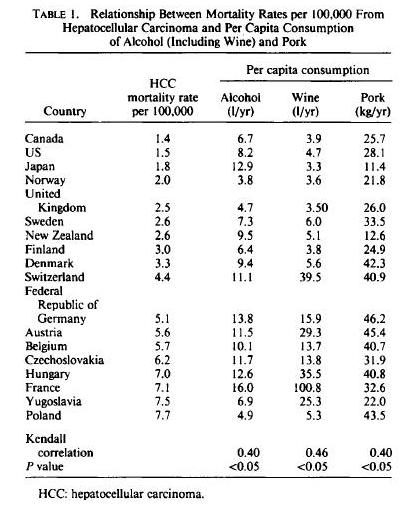
The authors investigated the possibility that dietary fat, meat, beef, and pork consumption might be factors that would, in addition to alcohol, correlate with mortality from hepatocellular carcinoma (HCC) in different countries….

The correlation between HCC and alcohol was 0.40 (p < 0.05); that with pork consumption was also 0.40 (p < 0.05). There was no correlation with total fat meat, beef, and cigarette and tobacco consumption.

Here is the raw data by country:

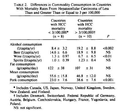
Another way of looking at the data is based on countries with low and high incidence of HCC. Countries with high incidence of HCC eat more pork and drink more alcohol, but actually eat less animal fat:

Pork and Multiple Sclerosis

Nanji and Norad [4] looked for other diseases that correlate with pork consumption, and hit upon multiple sclerosis. The connection is remarkable:

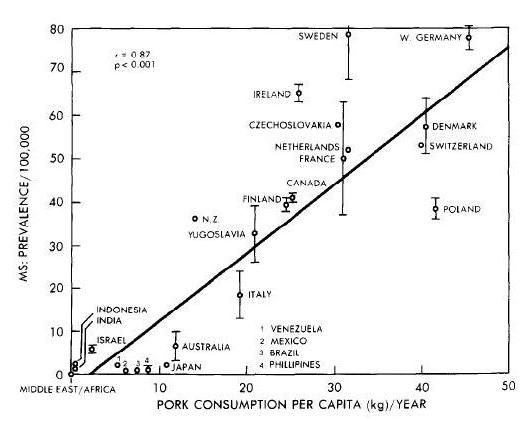
A significant correlation was obtained between prevalence of multiple sclerosis and … pork consumption (r = 0.87, p less than 0.001). There was no significant correlation with beef consumption. [4]

As noted earlier, a correlation coefficient of 0.87 is extremely high, and a p-value below 0.001 also shows a very strong relationship. MS is much more likely to befall pork eaters. Such a strong correlation makes it look like pork, or something found in pork, is the cause of MS.

Nanji and Norad further note that beef, the “other red meat,” is not associated with MS:

The correlation between pork consumption and MS prevalence was highly significant. Also, of major significance was the absence of a significant correlation between MS prevalence and beef consumption. This is consistent with the observations that MS is rare in countries where pork is forbidden by religious customs (e.g. Middle East) and has a low prevalence in countries where beef consumption far exceeds pork consumption (e.g. Brazil, Australia). [4]

The correlation between pork and MS may be seen here:

Lauer [5] verified the pork-MS link, but found it to be characteristic of processed pork:

When … quantitative data are taken into account, and a combined factor “smoked meat” or “smoked pork” is formed, the association is very high throughout. This factor is also compatible with the high risk of multiple sclerosis in Scotland and particularly in the Orkney and Shetland Islands and with the only transitorily high incidence in the Faroe Islands [6], whereas coffee can hardly explain both epidemiological features.

Arguments for the biological plausibility of some agents occurring in smoked and cured meat (in particular nitrophenol haptens and their protein conjugates) have been put forward [7]. There appears at present to be no plausibility for the factor “margarine”, which was also not compatible with the temporal pattern of multiple sclerosis in the Faroe Islands. [6]

Conclusion

There are remarkably strong correlations between pork consumption and liver disease, liver cancer, and multiple sclerosis.

What can be behind those relationships? The relatively high omega-6 fat content of pork may be a contributing factor, but it can’t be the whole story. It seems there is something else in pork that makes pork consumption risky.

What is it about pork that is so dangerous, and what does it mean for our dietary advice? That will be the topic of my next post.

Related Posts

Posts in this series:

References

[1] Bridges FS. Relationship between dietary beef, fat, and pork and alcoholic cirrhosis. Int J Environ Res Public Health. 2009 Sep;6(9):2417-25. http://pmid.us/19826553.

[2] Nanji AA, French SW. Relationship between pork consumption and cirrhosis.  Lancet. 1985 Mar 23;1(8430):681-3. http://pmid.us/2858627.

[3] Nanji AA, French SW. Hepatocellular carcinoma. Relationship to wine and pork consumption. Cancer. 1985 Dec 1;56(11):2711-2. http://pmid.us/2996744.

[4] Nanji AA, Narod S. Multiple sclerosis, latitude and dietary fat: is pork the missing link?  Med Hypotheses. 1986 Jul;20(3):279-82. http://pmid.us/3638477.

[5] Lauer K. The food pattern in geographical relation to the risk of multiple sclerosis in the Mediterranean and Near East region. J Epidemiol Community Health. 1991 Sep;45(3):251-2. http://pmid.us/1757770.

[6] Lauer K. Dietary changes in relation to multiple sclerosis in the Faroe Islands: an evaluation of literary sources. Neuroepidemiology. 1989;8(4):200-6. http://pmid.us/2755551.

[7] Lauer K. Environmental nitrophenols and autoimmunity. Mol Immunol. 1990 Jul;27(7):697-8. http://pmid.us/2395440.

[8] Nanji AA, French SW. Dietary factors and alcoholic cirrhosis. Alcohol Clin Exp Res. 1986 Jun;10(3):271-3. http://pmid.us/3526949.