



Choline Deficiency and Plant Oil Induced Diabetes

I’m going to deviate from my original plan for the “Dangers of a Zero-Carb Diet” series to discuss a topic that came up in the comments to the first post.

Leonie’s Diabetes and the Rose Corn Oil Trial

What prompted this diversion is Leonie’s interesting comment from Wednesday’s post:

I developed diabetes several years after being on a low carb diet. Continuing low carb to manage the diabetes did not halt its progress. It has taken about 18 months of adding more carbs (60 – 100 gr/day) to my diet to bring my fasting glucose down by a couple of mmol and eating more carbs has also lowered my Hba1c and post meal spikes significantly. I wonder if the liver is another organ that may be affected by carbohydrate deficiency.

I had not heard of such cases before, or so I thought, but Dr. Deans in the comments reminded us that Peter at Hyperlipid had noticed two similar cases in the Rose Corn Oil trial. [1] (The Rose Corn Oil trial, of course, figures prominently in our book’s discussion of PUFA toxicity.)

In the Rose Corn Oil trial, there were three arms – a normal diet arm, a high corn oil arm, and a high olive oil arm. The normal dieters were expected to eat “fried foods, fatty meat, sausages, … ice cream, cheese, … milk, eggs, and butter” while the oil arms were supposed to restrict these foods and replace them with corn or olive oil.

Here’s what happened:

Four patients were removed from the trial for other reasons. Two developed non-cardiac thromboembolism and were given anticoagulant therapy. The other two were removed because of diabetes mellitus. One of them already had mild diabetes, but glycosuria increased considerably soon after he started oil. Oil was stopped and glycosuria disappeared. Oil was restarted, but was stopped a month later because heavy glycosuria recurred. The other patient, not a previously recognized diabetic, developed glycosuria with a diabetic glucose-tolerance test a few weeks after starting oil. [1]

The patients who developed diabetes came one from the corn oil arm and one from the olive oil arm. Likewise, the patients who developed thromboembolisms came one from the corn oil arm and one from the olive oil arm. No such disasters occurred on the “fatty meat” arm.

Since all three diets were similarly fatty, it doesn’t appear to be the quantity of fat that was the issue. Rather it was the type of lipid, or some micronutrient that was present in the animal and dairy foods but lacking in the plant oils.

For insight into what the problem might be, let’s look at how scientists poison lab animals.

Insights from Diet Animal Poisoning Research

You have to pity diet researchers. It takes 60 years for bad diets to poison humans enough to significantly raise mortality rates. Yet a diet researcher is supposed to gain a Ph.D. in 4 years (or in 5 while simultaneously obtaining an MD!), do a postdoc in 2 years, win a grant in the first years of an entry-level position with PI status, and then demonstrate productive results within the term of a 2-to-5 year grant. Deadlines are pressing: A study needs to start rats or mice on two diets, and have one diet produce much better health than the other, in considerably less than a two-year time frame.

Just comparing McDonald’s fast food with a Mediterranean diet won’t do. Two years later both sets of mice will die happily of old age, with no significant differences between groups. Peer reviewers judge you to have discovered no new results. No new results means no paper, no grant, no job.

So “diet” researchers first have to become experts at quickly inducing disease in rats and mice. Find a diet that poisons animals in a few months, compare it to another diet that doesn’t, and you have a paper. Look for variations that slow or hasten the poisoning, and you have more papers. To be a highly productive scientist, one must be a skilled animal poisoner.

Various techniques have been developed for this purpose, including: knocking out some crucial gene; breeding a mutant strain that naturally develops disease; giving the animals poison with their food; or depriving them of crucial nutrients. Almost every study of diet in mice or rats uses one of these techniques.

If a missing nutrient can cause diabetes within a few years for Leonie and 12 to 18 months for the Rose Corn Oil trial volunteers, it’s likely to be pretty good at inducing disease in animals too. There’s a good chance diet animal poisoning researchers have already stumbled upon it in rats or mice.

Choline Deficiency Diseases

One of the most popular deficiency diets among researchers is the choline-deficient diet. A useful paper by Dutch scientists [2] gives a nice look at the impact of choline deficiency on rats.

Choline deficiency (CD) by itself induces metabolic syndrome (indicated by insulin resistance and elevated serum triglycerides and cholesterol) and obesity.

A combined methionine and choline deficiency (MCD) actually causes weight loss and reduces serum triglycerides and cholesterol, but induces more severe liver damage. The MCD diet prevents the body from manufacturing choline from methionine, vitamin B12, and folate, so MCD diets severely reduce choline levels; and without choline VLDL particles are not produced. Without VLDL particles, fats and cholesterol are trapped in the liver and never reach the blood and adipose cells.

Here is a measure of insulin resistance on the two diets:
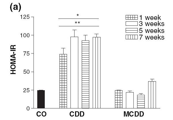
The induction of insulin resistance by the CD diet is very rapid, requiring less than a week.

Induction of insulin resistance is thought to be mediated by elevated TNF-alpha production by adipose cells and by hypertriglyceridemia. Since the MCD diet neither raised serum triglycerides nor caused obesity which induces TNF-alpha production in adipose cells, it did not cause insulin resistance.

What Does This Have to Do With Diabetes?

Insulin resistance is a key step in the development of diabetes:

  • Insulin resistance in the liver causes the liver to release more glucose into the blood (since insulin inhibits glucose release by the liver). This is discussed in a nice paper [3] found by LynMarie Daye and cited in the comments by CarbSane.
  • Peripheral insulin resistance means that the rest of the body is less sensitive to insulin. The pancreas has to produce more insulin to dispose of the excess glucose that the liver is releasing.

This elevation of insulin and glucose levels is a crucial step toward diabetes; it is “pre-diabetes.”

Persistently elevated glucose levels can then poison the beta cells of the pancreas, diminishing insulin secretion capability and causing diabetes. [4]

The Rose Corn Oil trial was not a low-carb diet, so postprandial glucose levels could easily have risen to toxic levels.

If a CD diet can cause insulin resistance in a week, it’s plausible that it might cause diabetes in 12 to 18 months, which is when the Rose Corn Oil trial patients developed it.

What About the Thromboembolism Cases?

MCD diets induce fibrinogenesis. In the blood, excess fibrin formation leads to clotting, and clots can block vessels to cause thromboembolisms. It may be that the thromboembolism cases in the Rose Corn Oil trial had methionine, folate, or B12 deficiencies to go with their choline deficiency.

Why Do Plant Oils Induce Diabetes But Not Animal Fats?

So why did diabetes develop in the corn and olive oil arms of the Rose Corn Oil trial but not the “fatty meat and dairy” arm?

Well, look at the choline content of these foods:

Choline content of one cup (~200 g) oil or fat or 227 g (1/2 lb) meat

Beef liver 968.0 mg
Cube steak (beef) 290.0 mg
Beef tallow 164.0 mg
Butter 42.7 mg
Olive oil 0.6 mg
Corn oil 0.4 mg

Source: http://nutritiondata.com.

Take away meat and dairy and replace them with plant oils, and it’s very easy to have a choline deficiency.

What Does This Have to Do With Zero-Carb Diets?

Maybe nothing … without carb consumption, postprandial glucose levels are not as high, and beta cell poisoning is less likely … but it may be that a zero-carb diet aggravates a choline deficiency in some fashion. I will leave this as a topic for further research.

UPDATE: Leonie in a new comment gives us more information: she has PCOS, goiter with nodules, and auto-antibodies. This suggests autoimmunity as a more likely explanation for her zero-carb diabetes.

Conclusion

In the book, we recommend the use of animal fats such as beef tallow for cooking, and recommend that pregnant women and vegetarians supplement with choline. We thought seriously about recommending that everyone supplement choline, but were reluctant to recommend too many supplements.

In retrospect, we should have recommended choline supplements for everyone who is overweight, has elevated blood glucose or lipids, or has elevated liver enzymes.

We have been using beef tallow as our cooking oil for several months now. It might be good practice for everyone to favor animal fats like beef tallow over plant oils for cooking.

References

[1] Rose GA et al. Corn oil in the treatment of ischaemic heart disease.  Br Med J. 1965 Jun 12;1(5449):1531-3. http://pmid.us/14288105.

[2] Veteläinen R et al. Essential pathogenic and metabolic differences in steatosis induced by choline or methione-choline deficient diets in a rat model. J Gastroenterol Hepatol. 2007 Sep;22(9):1526-33. http://pmid.us/17716355.

[3] Sonksen P, Sonksen J. Insulin: understanding its action in health and disease. Br J Anaesth. 2000 Jul;85(1):69-79. http://pmid.us/10927996.

[4] Leibowitz G et al. Glucose regulation of ?-cell stress in type 2 diabetes. Diabetes Obes Metab. 2010 Oct;12 Suppl 2:66-75. http://pmid.us/21029302.


Dangers of Zero-Carb Diets, I: Can There Be a Carbohydrate Deficiency?

It’s frequently said in the Paleo blogosphere that carbs are unnecessary. Here’s an example from Don Matesz, an outstanding blogger who eats a diet extremely close to ours:

Protein is essential, carbs are not…. You can only cut protein so much, but you can cut carbs dramatically.

Dr. Michael Eades has mocked the idea of a carbohydrate deficiency disease:

Are there carbohydrate deficiency diseases, Mr. Harper, that you know about that the rest of the nutritional world doesn’t?  I’ll clue you in: there aren’t.  But there are both fat and protein deficiency diseases written about in every internal medicine textbook.

Such statements made an impression on me when I first started eating Paleo five years ago. But several years and health problems later, I realized that this view was mistaken.

Why Aren’t Carbohydrate Deficiency Diseases Known?

How do doctors discover the existence of a nutrient deficiency disease?

It’s not as easy as you might think. For example, the existence of essential fatty acid deficiency diseases in humans was in doubt right up into the 1950s, even though omega-6 deficiency disease had been discovered and characterized in rats in the 1920s. [1] The reason is that omega-6 and omega-3 deficiencies can occur only on unnatural diets. It was infants fed fat-free formula in the 1940s and 1950s who ended up proving the existence of omega-6 deficiency disease in humans.

Two difficulties have made it challenging for science to recognize a carbohydrate deficiency syndrome:

  1. Lack of an animal model.
  2. The rarity of zero-carb diets among humans.

Until recently, few people save the Inuit ate very low-carb diets, and the Inuit didn’t leave good medical records. As a result, few or no humans developed recorded carbohydrate deficiency syndromes.

This wouldn’t be a problem if it were possible to induce carbohydrate deficiency in animals. However, it isn’t.

Animals don’t get carbohydrate deficiency diseases because they have small brains, meaning low glucose needs, and big livers, meaning high glucose manufacturing capacity. Animals can generate all the glucose they need from protein or from volatile acids like propionate produced by bacterial fermentation in their digestive tracts.

But, as we note in the book, humans are more fragile. We have small livers and big brains, and so the possibility of glucose deficiency is real.

Here is a comparison of brain, liver, and gut sizes in humans and other primates [2]:

Organ % body weight, humans % body weight, other primates
Brain 2.0 0.7
Liver 2.2 2.5
Gut 1.7 2.9

The brain is the biggest determinant of glucose needs.  While other primates need only about 7% of energy as glucose or ketones, humans need about 20%.

Compared to other primates, humans have a 12% smaller liver. This means we can’t manufacture as much glucose from protein as animals can. Humans also have a 40% smaller gut. This means we can’t manufacture many short-chain fatty acids, which supply ketones or glucogenic substrates, from plant fiber.

So, while animals can meet their tiny glucose needs (5% of calories) in their big livers, humans may not be able to meet our big glucose needs (20-30% of calories) from our small livers.

So any carbohydrate deficiency disease will strike humans only, not animals.

How Should We Look for a Carbohydrate Deficiency Disease?

To find a carbohydrate deficiency syndrome in humans, we should look at populations that eat very low-carb diets, such as:

  • The Inuit on their traditional hunting diet.
  • Epilepsy patients being treated with a ketogenic diet.
  • Optimal Dieters in Poland, who have been following a very low-carb diet for more than 20 years.
  • Very low-carb dieters in other countries, who took up low-carb dieting in the last 10 years as the Paleo movement gathered steam.

We should also have an idea what kind of symptoms we should be looking for. Major glucose-consuming parts of the body are:

  • Brain and nerves.
  • Immune system.
  • Gut.

The body goes to great lengths to assure that the brain and nerves receive sufficient energy, so shortfalls in glucose are most likely to show up in immune and gut function.

So, we’ve mapped our project. Over the coming week, or however long it takes before we get tired, we’ll investigate the evidence for carbohydrate deficiency conditions in humans.

Related Posts

Other posts in this series:

  1. Dangers of Zero-Carb Diets, II: Mucus Deficiency and Gastrointestinal Cancers A Nov 15, 2010.
  2. Danger of Zero-Carb Diets III: Scurvy Nov 20, 2010.
  3. Dangers of Zero-Carb Diets, IV: Kidney Stones Nov 23, 2010.

References

[1] Holman RT. The slow discovery of the importance of omega 3 essential fatty acids in human health. J Nutr. 1998 Feb;128(2 Suppl):427S-433S. http://pmid.us/9478042

[2] Aiello LC, Wheeler P. The expensive tissue hypothesis: the brain and the digestive system in human and primate evolution. Current Anthropology 1995(Apr); 36(2):199-211.

The Danger of Hospital Food

In the book we discuss how high blood sugar is a powerful predictor of poor outcomes in hospital patients.

Well, Abby’s grandmom is in the hospital with pneumonia. She is being fed with intravenous glucose, and has developed high blood sugar – 160 mg/dl – despite provision of insulin.

Abby says the doctors aren’t concerned. But they should be.

High Blood Sugar Causes Pneumonia

High blood glucose and high insulin both suppress immune function. As a result, pneumonia rates are much higher in hospital patients with high blood sugar. If hyperglycemia helps cause pneumonia, it’s surely a danger in someone who already has pneumonia.

A recent paper investigating the health effects of high blood sugar during total parenteral (meaning intravenous) nutrition gives us a quantitative assessment of the risks of elevated blood sugar. [1]

They report that pneumonia was the number one consequence of elevated blood sugar from intravenous feeding:

In multivariate analysis adjusting for age, sex, and history of diabetes, the blood glucose within 24 h of TPN >180 mg/dl was associated with increased risk of pneumonia (OR 3.6, 95% CI 1.6–8.4) and acute renal failure (2.2, 1.02–4.8 1) compared with patients with blood glucose <120 mg/dl. [1]

In other words: If 24 hours after intravenous feeding is started, blood glucose rises over 180 mg/dl, the likelihood of subsequently contracting pneumonia is increased 3.6 fold.

Abby’s grandma had blood glucose of 160 mg/dl, which is still at elevated risk [1]:

Here the measure is mortality, not pneumonia, but we can see that blood glucose in the range 150-180 mg/dl 24 hours after initiation of TPN is quite a bit more dangerous than blood glucose levels below 150 mg/dl.  Mortality rates are almost 50% higher.

This is not a new finding:

TPN therapy has been associated with increased risk for infections and mortality (2,1013). The increased risk of complications appears to be related, among other factors, to the development of hyperglycemia (4,14). Observational studies have reported a 33% mortality rate in TPN patients who developed hyperglycemia (15), as well as an increased risk of cardiac complications, infections, systemic sepsis, and acute renal failure (3,4,6). In agreement with these reports, we found a strong correlation between TPN-induced hyperglycemia and poor clinical outcome. [1]

Some of the reasons hyperglycemia is dangerous involve immune suppression:

The mechanisms underlying the detrimental effects of hyperglycemia relate to alterations in immune functions and inflammatory response (16,17). Hyperglycemia impairs leukocyte function, phagocytosis, and chemotaxis (18). Hyperglycemia also increases counterregulatory hormones, inflammatory cytokines, and oxidative stress (16,17), which can lead to endothelial dysfunction and cardiovascular complications (17). [1]

Significance for Treatment

Patients who need intravenous feeding, in pneumonia cases presumably due to high choking risk or need for oxygen, have great difficulty avoiding negative health consequences. But the risks of intravenous feeding are increased by suboptimal formulations that have too much sugar and too much omega-6 polyunsaturated fat. Such formulations can strongly suppress immune function, especially if they produce hyperglycemia.

I hesitate to second-guess doctors who are on the scene and privy to case knowledge, but I think the evidence is pretty strong for the dangers of hyperglycemia. If Abby’s grandma has enough muscle and fat, fasting and receiving parenteral water, vitamins, and minerals might be a better strategy. At a minimum, glucose provision should be reduced or insulin increased to reduce blood glucose levels.

Doctors are Enslaved to Faulty Practice Standards

The paper I cited is from 2010, but the dangers of hyperglycemia have been known for decades. Yet hospitals still commonly induce it.

Our current medical institutions seem to have left doctors terrified of deviating from standard practice, even if standard practice is known to be harmful. And new, better practices can’t be adopted until proven in costly clinical trials, even though the existing practices were adopted without such trials.

I wonder what Dr. House would do in Abby’s grandma’s case? And if Dr. House would do the right thing for the patient in defiance of standard practice, how long would he keep his medical license or his malpractice insurance?

References

[1] Pasquel FJ et al. Hyperglycemia during total parenteral nutrition: an important marker of poor outcome and mortality in hospitalized patients. Diabetes Care. 2010 Apr;33(4):739-41. http://www.ncbi.nlm.nih.gov/pubmed/20040658.

The Vanderbilt Protocol for Multiple Sclerosis

The antibiotic approach to MS therapy was developed at Vanderbilt by Drs. Stratton and Mitchell. They have patented multiple versions of their protocol, most recently in 2005. (Since patents are publicly available and the “Description of the Invention” is often an excellent overview of the science, we’ve put links to patents at the end of this post.)

Since we have some readers with MS, including Alexander, I thought I would post a summary of the Vanderbilt protocol. This was written by Dr. Stratton in 2009:

Treatment Protocol for Chronic Infections Caused by C. pneumoniae

As far as the Cpn Antimicrobial Regimen is concerned, my thoughts (as of 2009) are as follows:

First, as a general rule, the sicker a patient is, the slower they should go. This is why our early protocol started out with only one antibiotic and one dose, and then gradually adding the next dose/antibiotic as the reactions to each dose/antibiotic became apparent. These reactions appear to be caused by destruction of Chlamydia organisms as well as by the death of some of the infected host cells. Even destruction of elementary bodies by reducing agents such as N-acetyl-cysteine (NAC) can cause these reactions as chlamydial major outer membrane protein (MOMP) is released. MOMP is known to interact with Toll-like receptors (TLRs) and can thus induce the production of cytokines. Moreover, chlamydial cell wall contains LPS, which also interacts with TLRs and induces the production of cytokines. The reaction to anti-chlamydial therapy is sometimes referred to as “die-off” as presumable both chlamydial organisms and host cells are dying. These reactions can be delayed by days to weeks and may include “flu-like symptoms”, arthralgias and myalgias, “hangover-like symptoms (“brain-fog”, nausea, malaise), gastroenteritis (including diarrhea), and (rarely) fever. These reactions are akin to the “leprosy reaction” well described with the therapy of leprosy. The use of prednisone (10-20 mg per day) and/or pentoxifylline (Trental, 400 mg bid or tid), as done for the leprosy reaction, may be beneficial.

I think that all patients should start with supplements/vitamins before they start any antibiotics. Baseline lab studies, including CBC and liver function studies, should be done and these parameters followed every 3-4 months, more frequently (i.e., monthly) for sicker patients. As C. pneumoniae can infect white blood cells and liver cells, potential death of these cells should be monitored in sicker patients. Our initial protocol recommended this. I would add NAC to the supplements. We used amoxicillin, which is degraded in the body to penicillamine (a reducing agent) in our regimen as an anti-elementary body agent, but NAC seems to work equally well and may offer additional benefits in boosting the immune system as well as protecting the liver. As far as supplements/vitamins are concerned, I think Professor David Wheldon’s supplement/vitamin suggestions are very complete and should be the benchmark. Once antibiotics are ready to be started, I would start with a macrolide. We have used Azithromycin because it is easy to give and has become somewhat cheaper since it went off patent. Clarithromycin (500 mg twice a day) or Roxithromycin (300 mg once a day) can be used instead of Azithromycin. I would still give just one 250 mg azithromycin tablet and then wait two weeks to see if there is any reaction to it. Then I would give two tablets, one on Monday and one on Wednesday. Once again I would wait two weeks. I’d continue in this way, adding each dose until the patient was taking 250 mg of azithromycin MWF. If the patient has severe reactions (meaning they can’t work – most people are trying to work and take care of a family while they are on this therapy), I’d slow down the process. After the azithromycin, I’d add doxycycline – again doing this very slowly. Once the patient was taking both azithromycin (250 mg MWF) and doxycycline (100 mg twice a day), I’d start the metronidazole pulses – again, doing these slowly and working up to a once a month pulse of 7 days of metronidazole (500 mg twice a day). Once the patient could do the monthly pulse of metronidazole, I’d add rifampin (300 mg twice a day). Once this was tolerated, I would increase the metronidazole pulse to 14 days, doing so slowly. Eventually, the patient should be able to tolerate metronidazole (500 mg twice a day) on a daily basis. Once a patient could do this regimen without any reactions, I would continue it for at least a year and probably three years for MS patients. It might take a year or two (or longer) to get to the point where there is no reaction to the daily metronidazole, depending on the chlamydial load, followed by 1-3 years of therapy. This might be a 5-year program, but should allow the patient to continue to work with minimal disruption. Patients should also be gradually improving during this time. The sicker the patient is, the longer the therapy is going to be. There is no shortcut. Younger patients who have not been sick as long tend to respond more quickly. ?

With MS patients, due to the possible CNS damage that might occur by going slowly, I would move more quickly unless there were major reactions. This means compressing what might have taken a year into several months. 

The reactions patients have are varied – some are severe enough that they stop the antibiotics. That, of course, defeats the purpose of the therapy. It is very tricky and each patient has to learn his own limitations. When we started our protocol, we were thinking of a hotline to answer questions that are now easily and better answered via the internet (http://Cpnhelp.org). Finally, I don’t think this is the only regimen that will work nor do I think it will work better or faster. It is just what I do in 2009 when treating a patient.

I’ve been informed that the late stages of the protocol have been further refined in 2010. Now, once die-off reactions to the metronidazole subside, Dr. Stratton tries rifabutin or rifampin.

I would agree with Dr. Stratton that nutritional supplements should begin 3-4 months before antibiotics, but would add that our diet should be adopted 3-4 months before antibiotics begin. This is necessary to improve immune response and healing capacity; and to avoid unnecessary cell death and die-off toxicity when antibiotics begin. The eleven ways to enhance immunity, discussed in Step Four of the book, should be routinely practiced.

Simply introducing diet and nutrition alone can lead to significant die-off effects. This shows that the adoption of diet and nutritional supplements is therapeutic in its own right.

On a bad diet, antibiotics are dangerous, as they risk gut dysbiosis and introduction of new co-infections.

Like Dr. Stratton, I think being active at http://cpnhelp.org is essential for anyone on this protocol. Many people at cpnhelp.org have used this protocol, often for years, and their experience can be very helpful.

As Dr. Stratton notes, it takes years to cure MS. It is necessary to be patient and to balance speed of killing pathogens against allowing the body time to recover from die-off effects – toxic bacterial proteins and human cell death. Remember, “the dose makes the poison” – doubling the rate of pathogen killing may quadruple the toxicity effects, so the optimum course is not the one with highest rate of pathogen killing. Every patient has to progress at his own pace. Go as fast as you can but no faster.

There are steps that can be taken to reduce die-off toxicity, such as drinking lots of water and eating salt to help urine excretion, and taking “moppers” such as charcoal, bentonite clay, cholestyramine, or chlorella to help assure that toxins released from the liver through the bile are excreted in feces, not re-absorbed.

Conclusion

MS recovery is not impossible; indeed, many have recovered on this protocol. However, it is long and arduous. Optimizing diet will shorten time to recovery, but it will still take years. 

MS is not the only disease that may be caused by C. pneumoniae. Alzheimer’s dementia, atherosclerosis, stroke, rheumatoid arthritis, and rosacea are all associated with C. pneumoniae infections and may be treatable by Dr. Stratton’s protocol.

Links

“Multiple Sclerosis:  A Curable Infectious Disease?”, July 7, 2010, https://perfecthealthdiet.com/?p=157.

“Is Multiple Sclerosis an Autoimmune Disease?”, July 5, 2010, https://perfecthealthdiet.com/?p=151.

“Eleven Steps for Overcoming Alzheimer’s and Other Chronic Infectious Diseases,” July 1, 2010, https://perfecthealthdiet.com/?p=134.

A list of the Vanderbilt patents.

The 2005 patent, ID 7,094,397.